

IMG_7690.jpg
Diagnose this
analyse_media
Reading File
Finding Sources
Finding Sources
Searching the Web
acute paronychia vs blistering dactylitis finger purple swelling peeling diagnosis
Reading File
Reading File
Reading File
Let me also fetch the blistering dactylitis image from the textbook for visual comparison, and read more about the whitlow/felon differential.
Loading Image
Reading File
Now I have enough information to provide a thorough clinical analysis. Here is my assessment:
Clinical Assessment
Disclaimer: This is an AI-assisted clinical analysis for educational/supportive purposes only. It is not a substitute for in-person evaluation by a physician. Please seek medical care promptly, especially if there is increasing pain, spreading redness, fever, or pus.
Most Likely Diagnosis: Acute Paronychia with Abscess Formation (possibly a Whitlow/Felon)
Findings from the Image
| Feature | Finding |
|---|---|
| Nail fold | Markedly swollen, tense, deep reddish-purple discoloration extending over the dorsal distal phalanx |
| Skin | Circumferential peeling/desquamation around the swollen area (post-inflammatory or resolving blister roof) |
| Lateral nail fold | Yellowish-white focus consistent with a pustule or loculated pus |
| Nail plate | Yellowish-white discoloration at distal and lateral edge; dark subungual debris at lateral border |
| Overall | Tense, edematous, erythematous/violaceous distal finger - the appearance of a tensely swollen abscess |
Differential Diagnosis
1. Acute Paronychia (top diagnosis)
- Infection of the proximal/lateral nail fold, most commonly caused by S. aureus, with Streptococcus species also common
- Classic presentation: red, hot, tender, swollen nail fold that progresses to abscess if untreated
- The peeling skin suggests either a resolving bullous component or the blister roof of a superficial abscess that has partially ruptured
- Fitzpatrick's Dermatology: "S. aureus is the major infectious cause of acute paronychia, usually around the fingernails, often originating from a break in the skin, such as a hangnail. Clinically, skin and soft tissue of the proximal and lateral nailfold are red, hot, and tender, and can progress to abscess formation if not treated."
2. Felon (Whitlow)
- A purulent abscess involving the bulbous distal pulp/tip of the finger (rather than just the nail fold)
- The involvement here appears dorsal/periungual rather than purely volar, which is more consistent with paronychia
- However, if the swelling extends to the fingertip pulp, this distinction blurs
- Fitzpatrick's: "The finger bulb is red, hot, tender, and edematous, with possible abscess formation" - this can complicate or co-exist with paronychia
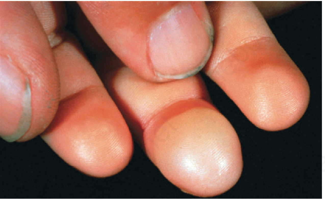
3. Blistering Distal Dactylitis (bulla repens)
- Caused by Group A Streptococcus or S. aureus, more common in children/adolescents
- Presents as a large tense blister on the volar fat pad; can extend to the nail folds
- The extensive peeling skin around the swelling is consistent with a ruptured or resolving bulla
- Andrews' Diseases of the Skin: "characterized by tense superficial blisters occurring on a tender erythematous base over the volar fat pad of the phalanx of a finger or thumb"
- This is the textbook image of blistering dactylitis for comparison:
Key Differentiating Points
| Feature | Paronychia | Felon | Blistering Dactylitis |
|---|---|---|---|
| Location | Nail fold (lateral/proximal) | Volar pulp | Volar fat pad, may extend to nail fold |
| Appearance | Erythema, swelling, pus at nail fold | Tense, throbbing pulp | Tense blister on erythematous base |
| Common pathogen | S. aureus | S. aureus | GAS, S. aureus |
| Age group | Any | Any | Children/adolescents (but not exclusively) |
| Peeling skin | Possible with resolving abscess | Rare | Yes - ruptured blister roof |
Recommended Management
- Urgent surgical/emergency evaluation - the degree of swelling, violaceous color, and pus suggests this likely needs incision and drainage (I&D)
- A digital nerve block may be required for the procedure
- Wound culture should be sent (especially given community-acquired MRSA rates)
- Antibiotics - systemic antibiotics are typically only indicated if there is significant surrounding cellulitis or the patient is immunocompromised; topical mupirocin may be used for mild cases
- Ensure tetanus prophylaxis is up to date
- If blistering dactylitis is confirmed: aspiration of the blister + oral penicillin/amoxicillin-clavulanate is appropriate
- Pfenninger & Fowler's Procedures for Primary Care: "As with all abscesses, it is best to incise and drain (I&D) them once a loculated area of purulence can be identified."
Bottom line: This finger shows a periungual bacterial abscess, most consistent with acute paronychia with abscess formation - possibly with a blistering dactylitis component given the extensive skin peeling. This requires prompt medical attention for drainage and likely antibiotic therapy. The deep purple/violaceous color indicates significant tissue tension from the collection of pus.
This is a shared conversation. Sign in to Orris to start your own chat.