
Ecg ko ache se samjho besic to advance clinical importance
Reading File
Finding Sources
Finding Sources
Finding Sources
Finding Sources
Reading File
Reading File
Finding Sources
Finding Sources
Reading File
Finding Sources
Finding Sources
Reading File
Loading Image
Searching Images
ECG ST elevation myocardial infarction STEMI changes
Reading File
Searching Images
ECG bundle branch block LBBB RBBB pattern QRS
Searching Images
ECG cardiac arrhythmia atrial fibrillation ventricular tachycardia
Ab main aapko ECG Basic se Advanced tak puri detail mein explain karta hoon — textbooks (Harrison's, Guyton, Braunwald's) ke saath.
📋 ECG (Electrocardiogram) — Basic se Advanced tak
⚡ ECG Kya Hai?
ECG ek aise recording hai jisme heart mein se nikalne wale electrical currents ko body surface ke electrodes ke through capture kiya jata hai. Jab cardiac impulse heart se guzarta hai, kuch current aas-paas ke tissues aur skin surface tak pahunch jaata hai — inhi potentials ko ECG paper par record kiya jaata hai.
(Guyton & Hall Textbook of Medical Physiology)
🔷 PART 1 — ECG KI WAVES AUR INTERVALS (Basics)

Waves Kya Represent Karti Hain?
| Wave / Segment | Physiological Event |
|---|---|
| P Wave | Atrial depolarization (SA node se signal) |
| QRS Complex | Ventricular depolarization (contraction se pehle) |
| T Wave | Ventricular repolarization (recovery) |
| U Wave | Late ventricular repolarization (Purkinje fibers) — hypokalemia mein prominent |
| ST Segment | Plateau phase of action potential — isoelectric hona chahiye |
| J Point | QRS ka end + ST ka start |
Yaad karo: P aur QRS = depolarization waves; T = repolarization wave
📏 Normal Intervals (Harrison's 22E)
| Interval | Normal Value | Clinical Importance |
|---|---|---|
| PR Interval | 120–200 ms (3–5 small boxes) | AV conduction time |
| QRS Duration | ≤ 110 ms (≤ 2.5 small boxes) | Ventricular depolarization speed |
| QT Interval | < 460 ms (women), < 450 ms (men) | Total repolarization time |
| RR Interval | Variable | Heart rate calculate karne ke liye |
ECG Paper ki Speed: 25 mm/sec
- 1 small box = 1 mm = 40 ms
- 1 large box = 5 mm = 200 ms
- 1 mV = 10 mm (standard calibration)
🧮 Heart Rate Calculate Karna
Method 1: 300 ÷ (large boxes between R-R)
Method 2: 1500 ÷ (small boxes between R-R)
- Normal: 60–100 bpm
- Bradycardia: < 60 bpm
- Tachycardia: > 100 bpm
🔷 PART 2 — 12 LEADS SYSTEM
6 Limb Leads (Frontal Plane)
- I, II, III — Standard bipolar leads
- aVR, aVL, aVF — Augmented unipolar leads
6 Precordial Leads (Horizontal Plane)
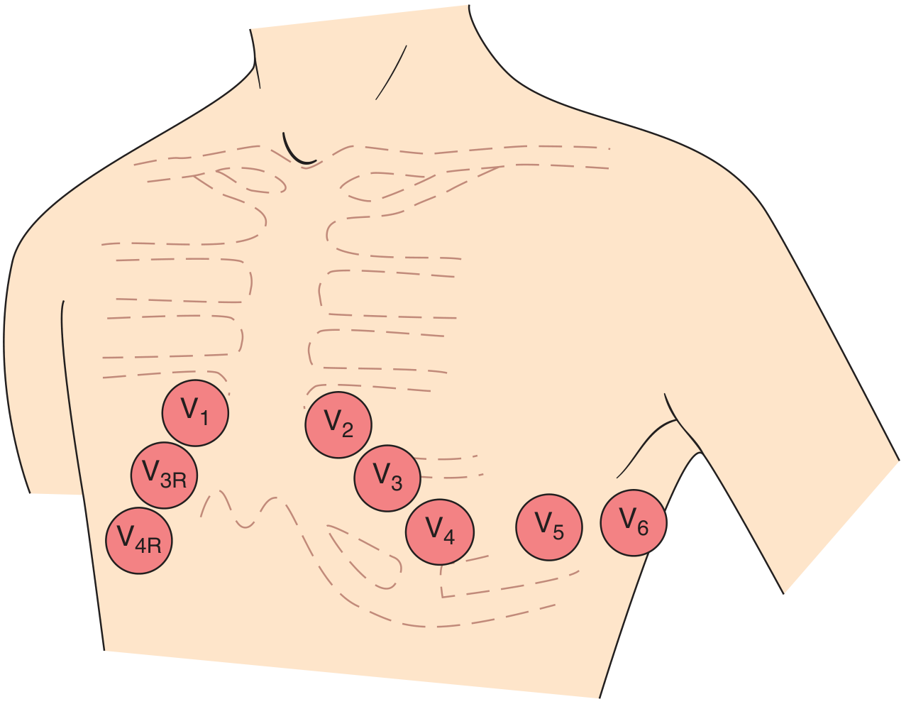
| Lead | Position |
|---|---|
| V1 | 4th intercostal space, right sternal border |
| V2 | 4th intercostal space, left sternal border |
| V3 | Between V2 and V4 |
| V4 | 5th intercostal space, mid-clavicular line |
| V5 | Anterior axillary line |
| V6 | Mid-axillary line |
Rule: Agar depolarization lead ke positive pole ki taraf jaye → positive (upright) deflection. Agar negative pole ki taraf → negative deflection.
🔷 PART 3 — QRS MORPHOLOGY & PROGRESSION
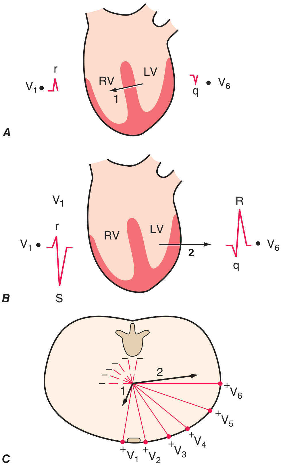
Ventricular depolarization 2 phases mein hoti hai:
- Phase 1 (Septal): Septum left se right depolarize hota hai → V1 mein small r wave, V6 mein small q wave
- Phase 2 (Main): Left ventricle dominate karta hai → V1 mein deep S, V6 mein tall R wave
Normal R Wave Progression:
- V1: rS pattern (small r, deep S)
- V3/V4: Transition zone (R = S)
- V5/V6: Rs pattern (tall R, small s)
🔷 PART 4 — CARDIAC AXIS
Hexaxial Diagram
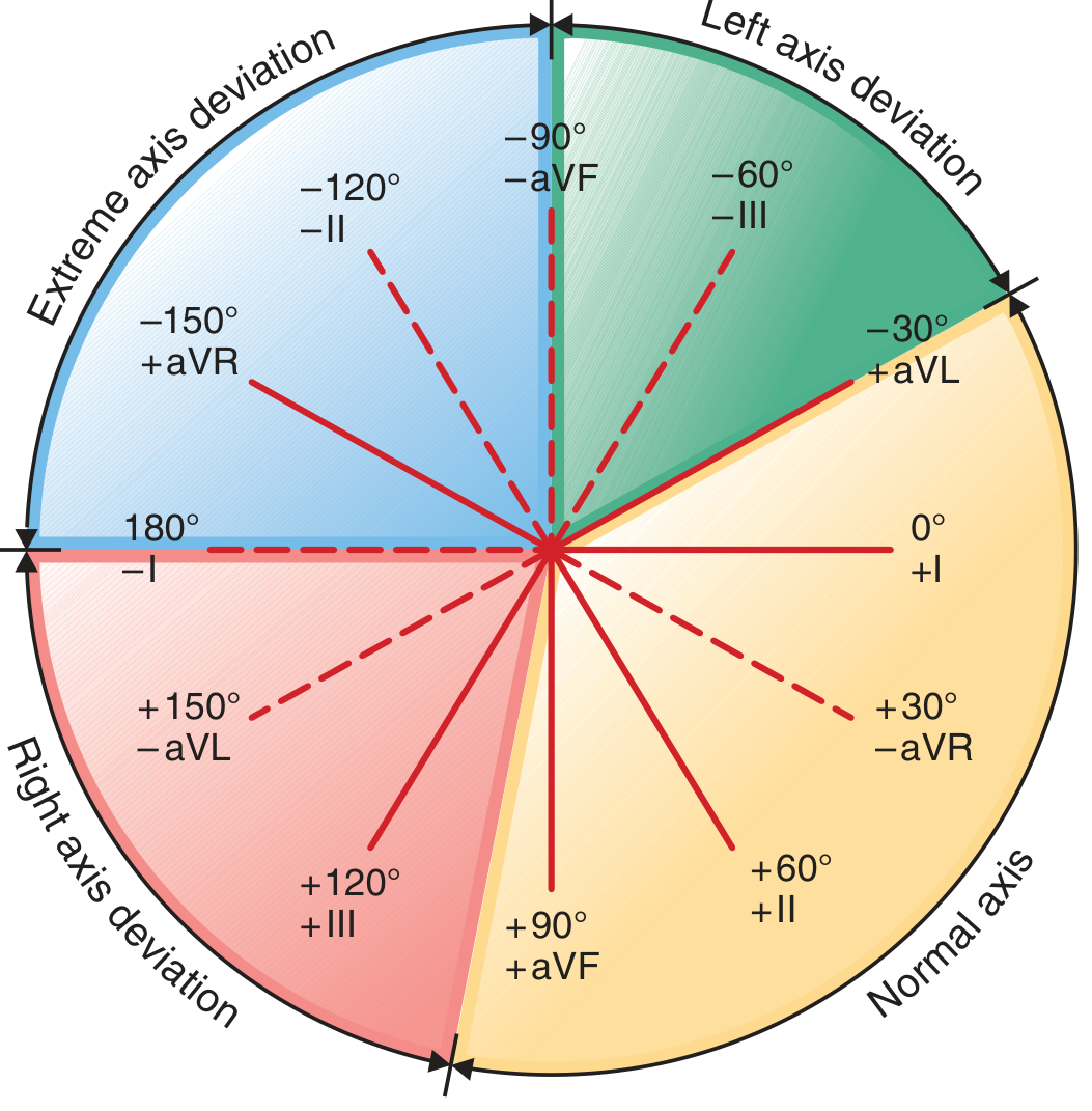
| Axis | Degree Range | Cause |
|---|---|---|
| Normal | -30° to +90° | Normal |
| Left Axis Deviation (LAD) | -30° to -90° | LBBB, inferior MI, LVH, left anterior fascicular block |
| Right Axis Deviation (RAD) | +90° to +180° | RVH, RBBB, lateral MI, pulmonary hypertension |
| Extreme/Northwest | -90° to -180° | Ventricular tachycardia, severe disease |
Quick Trick: Lead I aur aVF dekho:
- I (+), aVF (+) = Normal axis
- I (+), aVF (-) = LAD
- I (-), aVF (+) = RAD
- I (-), aVF (-) = Extreme axis
🔷 PART 5 — HYPERTROPHY PATTERNS
Left Ventricular Hypertrophy (LVH)
Sokolow-Lyon Criteria:
- S wave in V1 + R wave in V5 or V6 > 35 mm
Cornell Criteria:
- R in aVL + S in V3 > 28 mm (men), > 20 mm (women)
Clinical Importance: Hypertension, aortic stenosis, dilated cardiomyopathy — LVH increases risk of arrhythmias, heart failure, sudden cardiac death.
Right Ventricular Hypertrophy (RVH)
- Tall R wave in V1 (R > S in V1)
- Deep S wave in V5/V6
- Right axis deviation
- ST depression + T inversion in V1-V3
Causes: Pulmonary hypertension, pulmonary stenosis, cor pulmonale, mitral stenosis
🔷 PART 6 — BUNDLE BRANCH BLOCKS
Normal vs RBBB vs LBBB Pattern
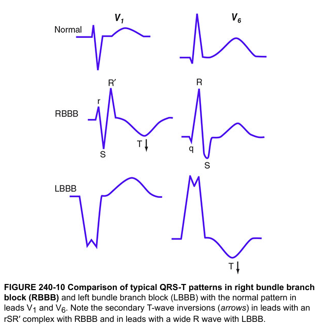
Right Bundle Branch Block (RBBB)
Pattern:
- V1: rSR' — "M shape" / "Rabbit ears"
- V6: Wide S wave
- QRS ≥ 120 ms
Complete vs Incomplete: Complete = QRS ≥ 120 ms; Incomplete = 110–120 ms
Clinical causes: Pulmonary embolism (acute), ASD, right heart strain, ischemia, normal variant (isolated RBBB)
Left Bundle Branch Block (LBBB)
Pattern:
- V1: Broad deep S wave (QS pattern)
- V5/V6: Broad notched R wave — "M shape" in lateral leads
- Absence of septal q wave in I, aVL, V5, V6
- QRS ≥ 120 ms
Clinical Importance (CRITICAL):
- New LBBB in chest pain = STEMI equivalent → treat as emergency
- LBBB masks ischemia changes on ECG
- Sgarbossa criteria se LBBB mein MI diagnose karo
🔷 PART 7 — ISCHEMIA, INJURY, INFARCTION (3 I's)
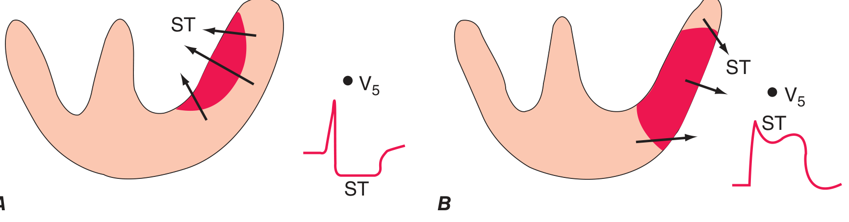
Phases of Myocardial Infarction on ECG:
| Time | ECG Finding | Represents |
|---|---|---|
| Minutes (Hyperacute) | Tall, peaked "hyperacute" T waves | Subendocardial ischemia |
| Hours | ST elevation (convex/tombstone) | Transmural injury — STEMI |
| Hours–Days | T wave inversion | Ischemia/early necrosis |
| Days–Weeks | Pathological Q waves | Myocardial necrosis |
| Chronic | Q waves persist, T may normalize | Old infarction |
ST Changes Kab Kahan?
- Subendocardial ischemia: ST depression (vector inward)
- Transmural injury: ST elevation (vector outward)
- Subendocardial ischemia → ST depression in precordial + ST elevation in aVR
🗺️ Localizing MI: Lead → Territory → Artery
| Leads with ST Elevation | Wall | Artery |
|---|---|---|
| V1–V4 | Anterior | LAD (Left Anterior Descending) |
| V5–V6, I, aVL | Lateral | LCX (Left Circumflex) |
| II, III, aVF | Inferior | RCA (Right Coronary Artery) |
| V1–V3 (ST depression) | Posterior | RCA or LCX |
| V3R–V4R | Right Ventricle | RCA (proximal) |
Anterior STEMI — LAD Occlusion
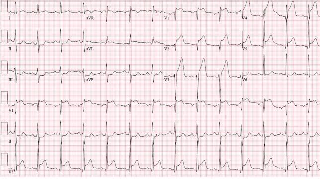
V1–V6 mein ST elevation + leads I, aVL → Anterior-lateral STEMI, LAD occlusion. Inferior leads mein reciprocal ST depression visible.
Inferior STEMI — RCA Occlusion
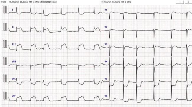
II, III, aVF mein ST elevation + I, aVL mein reciprocal changes → RCA territory.
Pathological Q Wave
- Width ≥ 40 ms (1 small box), Depth ≥ 25% of R wave height
- = Myocardial necrosis (dead tissue, no depolarization)
- Septal q wave (narrow, normal) se confuse mat karo
🔷 PART 8 — AV BLOCKS
| Type | PR Interval | QRS dropped? | Clinical Need |
|---|---|---|---|
| 1st Degree | >200 ms, constant | Never | No treatment usually |
| 2nd Degree Mobitz I (Wenckebach) | Progressively longer → then dropped | Yes (periodic) | Monitor |
| 2nd Degree Mobitz II | Constant → sudden drop | Yes (sudden) | Pacemaker risk |
| 3rd Degree (Complete) | P & QRS completely dissociated | All P waves dropped | Emergency pacemaker |
🔷 PART 9 — ARRHYTHMIAS
Atrial Fibrillation (AF)

Features:
- No distinct P waves → fibrillatory baseline
- Irregularly irregular R-R intervals
- Narrow QRS (unless aberrant conduction)
- Rate variable
Clinical: Stroke risk (CHA₂DS₂-VASc score) → anticoagulation decide karo
Supraventricular Tachycardia (SVT)
- Regular narrow complex tachycardia (150–250 bpm)
- Abrupt onset/offset
- P waves buried in QRS or after it
- Vagal maneuvers / Adenosine se terminate
Ventricular Tachycardia (VT)
- Wide complex tachycardia (QRS ≥ 120 ms)
- Rate 100–250 bpm
- AV dissociation, fusion beats, capture beats
- Emergency: defibrillation / amiodarone
Ventricular Fibrillation (VF)
- Chaotic irregular broad complexes
- No organized rhythm
- Cardiac arrest → immediate CPR + defibrillation
🔷 PART 10 — ELECTROLYTE CHANGES ON ECG
| Electrolyte | ECG Changes |
|---|---|
| Hyperkalemia | Peaked T waves → Wide QRS → Sine wave → VF |
| Hypokalemia | Flat T waves, prominent U wave, prolonged QT |
| Hypercalcemia | Short QT interval |
| Hypocalcemia | Prolonged QT interval |
| Digoxin toxicity | "Reverse tick" ST depression, various arrhythmias |
| Hypothermia | Osborn (J) waves |
🔷 PART 11 — CLINICAL PEARLS (Advanced)
Wellens Syndrome
- T wave inversion or biphasic T in V2-V3 without pain
- = Critical LAD stenosis → impending anterior MI → angiography urgent
De Winter T Waves
- ST depression + tall peaked T waves in precordial
- = LAD occlusion equivalent — no classic ST elevation
Brugada Pattern
- RBBB-like + ST elevation in V1-V3 (coved/saddleback)
- Risk of sudden cardiac death → ICD implant
Long QT Syndrome
- QTc > 450 ms (men), > 460 ms (women)
- Risk of Torsades de Pointes → VF → sudden death
- Causes: drugs (amiodarone, erythromycin, antipsychotics), hypokalemia, congenital
Pulmonary Embolism (PE) — S1Q3T3
- S wave in Lead I
- Q wave in Lead III
- T inversion in Lead III
-
- Sinus tachycardia + RBBB = PE until proven otherwise
Pericarditis
- Diffuse ST elevation (saddle-shaped) in multiple leads
- PR depression (pathognomonic)
- No reciprocal changes (unlike MI)
📌 ECG Padhne ka Systematic Approach (Always Follow)
1. Rate       → Normal / Brady / Tachy?
2. Rhythm     → Regular / Irregular? P before every QRS?
3. Axis       → Normal / LAD / RAD?
4. PR Interval → Normal / Prolonged / Variable?
5. QRS Width  → Narrow (<120ms) / Wide (≥120ms)?
6. ST-T Changes → Elevation / Depression / Inversion?
7. Q Waves    → Pathological?
8. QT Interval → Prolonged?
9. Hypertrophy? → Voltage criteria?
10. Specific Patterns → BBB, STEMI, Arrhythmia?
Sources: Harrison's Principles of Internal Medicine 22E, Guyton & Hall Medical Physiology, Braunwald's Heart Disease, Goldman-Cecil Medicine — Textbook of Medical Library---
Yeh tha ECG ka Basic se Advanced tak complete guide. Main neeche summary de raha hoon:
🎯 Kya Cover Kiya Gaya:
| Level | Topics |
|---|---|
| Basic | Waves (P, QRS, T, U), intervals (PR, QRS, QT), paper speed, leads |
| Intermediate | Cardiac axis, 12-lead system, R wave progression, hypertrophy (LVH/RVH) |
| Advanced | Bundle branch blocks, AV blocks, STEMI localization, arrhythmias |
| Clinical Pearls | Wellens, De Winter, Brugada, Long QT, PE (S1Q3T3), Pericarditis |
Key Points Yaad Rakhein:
- New LBBB + chest pain = STEMI equivalent — emergency
- ST elevation lead territory → artery → treat fast
- Peaked T waves = hyperkalemia tab tak jab tak aur prove na ho
- Irregularly irregular rhythm = Atrial Fibrillation — stroke risk
- ECG systematically padhein — Rate → Rhythm → Axis → Intervals → ST-T changes
Agar kisi specific topic par aur depth chahiye — jaise Brugada, Long QT, specific arrhythmias, ya practice MCQs — batao! 😊
Reload ache se samjho detailed mai
"Reload" se aap kya pooch rahe hain — thoda clarify karo:
Asking for Preferences
"Reload" se aap kiska matlab rakh rahe hain?
Reading File
Finding Sources
Finding Sources
Reading File
Reading File
Finding Sources
Reading File
Finding Sources
Finding Sources
Your credits are exhausted. Your daily credits reset tomorrow, or you can purchase a top-up for more.
This is a shared conversation. Sign in to Orris to start your own chat.