
Hodgkin lymphoma
I'll start by reading the medical library skill, then search for comprehensive Hodgkin lymphoma content in parallel.
Reading File
Now I'll run parallel searches across the library for Hodgkin lymphoma content.
Finding Sources
Finding Sources
Searching PubMed
"Hodgkin lymphoma"[MeSH Terms] AND treatment
Excellent - I found rich content across multiple textbooks. Now let me read the key sections in parallel - Robbins Pathology (most detailed pathology), Goldman-Cecil (clinical management), and Robbins Basic Pathology.
Reading File
Reading File
Reading File
Great content retrieved. Now let me get the detailed pathology/morphology from Robbins (subtypes) and the treatment section from Goldman-Cecil, plus fetch key histology images.
Reading File
Reading File
I have rich content. Let me now fetch the key histology image and read the treatment/staging sections from Goldman-Cecil.
Loading Image
Reading File
Excellent - rich content gathered. Let me read the treatment section from Goldman-Cecil and fetch the Reed-Sternberg cell image from Robbins.
Reading File
Finding Sources
I now have comprehensive content covering epidemiology, pathogenesis, morphology, classification, immunophenotype, staging, and treatment. Let me also fetch the imaging/staging figure and get the RS cell image from Robbins for completeness.
Loading Image
Finding Sources
Now let me read the treatment section from Goldman-Cecil for details on limited vs advanced stage treatment:
Reading File
I now have comprehensive information to compile a thorough answer. Let me deliver it.
Hodgkin Lymphoma
Definition and Overview
Hodgkin lymphoma (HL) is a B-cell lymphoid neoplasm defined by the presence of distinctive neoplastic giant cells - Reed-Sternberg (RS) cells - in a background of reactive inflammatory cells. It differs from non-Hodgkin lymphoma (NHL) in several key respects: it arises in a single lymph node or chain of nodes, spreads in a stepwise fashion to anatomically contiguous lymphoid tissues, and is curable in the majority of cases. It accounts for ~0.7% of all new cancers in the US, with approximately 9,000 new cases per year.
- Robbins, Cotran & Kumar Pathologic Basis of Disease, p. 573
- Goldman-Cecil Medicine, Chapter 172
Epidemiology
- Age distribution: Bimodal - an early peak in young adults (25-30 years) and a second peak in adults >55 years
- Sex: Slightly more common in men
- Race: More frequent in Whites than Blacks; much less frequent in Asian populations
- Incidence: ~2.7 per 100,000 per year in North America; approximately 30,000 new cases annually in North America and Europe combined
- Geography: Highest rates in the US, Canada, Switzerland, and northern Europe; much lower rates in eastern Asia
- In developing countries (e.g., Indian subcontinent), the age distribution shifts strongly into childhood
Classification (WHO)
Five subtypes are recognized, the first four grouped as Classic Hodgkin Lymphoma (cHL):
| Subtype | Frequency |
|---|---|
| Nodular sclerosis | ~60% |
| Mixed cellularity | ~9% |
| Lymphocyte rich | ~3% |
| Lymphocyte depleted | ~1% |
| Nodular lymphocyte-predominant HL (NLPHL) | ~8% |
| HL, not otherwise classifiable | ~19% |
- Goldman-Cecil Medicine, Table 172-1
Note: In the 2022 International Consensus Classification (ICC), nodular lymphocyte-predominant HL has been renamed "nodular lymphocyte-predominant B-cell lymphoma", dropping the Hodgkin designation entirely, because its RS cells carry a germinal center B-cell immunophenotype distinct from classic HL.
Pathogenesis
Cell of Origin
Elegant single-cell microdissection studies revealed that RS cells in classic HL carry clonal IGH gene rearrangements with the hallmarks of somatic hypermutation, establishing their origin as germinal center or post-germinal center B cells. Despite this B-cell origin, RS cells paradoxically fail to express most B-cell-specific genes (including immunoglobulin), due to widespread epigenetic reprogramming.
NF-κB Activation
Activation of NF-κB is the central oncogenic event, promoting RS cell growth and survival via several mechanisms:
- EBV+ tumors: The viral protein LMP-1 directly signals NF-κB upregulation
- EBV- tumors: Acquired loss-of-function mutations in IκB or TNF-α-induced protein 3 (both NF-κB inhibitors)
- Copy number gains in the REL proto-oncogene (chromosome 2p) also boost NF-κB activity
EBV Association
- EBV is present in RS cells in ~50% of classic HL cases overall (up to 70% in mixed-cellularity subtype)
- The integration site of EBV is identical in all RS cells in a given case, indicating infection preceded clonal expansion
- A history of infectious mononucleosis triples the lifetime risk of subsequent HL
Immune Evasion
RS cells escape immune destruction by:
- Loss of β2-microglobulin → failure to express class I MHC molecules → invisible to cytotoxic T cells
- High expression of PD-L1 and PD-L2 (from chromosome 9p copy number gains) - immune checkpoint ligands that suppress T-cell responses
Inflammatory Microenvironment
RS cells secrete multiple cytokines that recruit a reactive infiltrate (which paradoxically supports tumor survival):
- IL-5 → attracts eosinophils
- TGF-β → promotes fibrosis (nodular sclerosis subtype)
- IL-13 → may drive RS cell growth via autocrine loop
- IL-10, M-CSF, eotaxin → additional microenvironment modulation
The RS cells comprise only ~1-10% of total tumor cellularity; the remainder is reactive lymphocytes, macrophages, eosinophils, plasma cells, and fibroblasts.
Morphology
Reed-Sternberg Cell (Diagnostic)
The classic RS cell is large (~45 μm in diameter) with:
- Multiple nuclei (or a single nucleus with multiple lobes)
- Large "owl-eye" nucleoli (~5-7 μm, the size of a small lymphocyte)
- Abundant pale cytoplasm
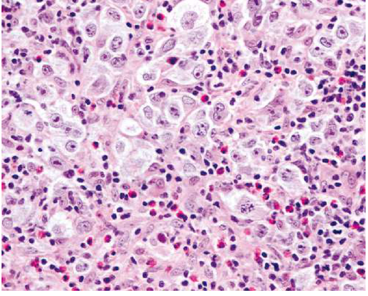
Histology of nodular sclerosing HL showing lacunar RS cells in a background of lymphocytes and eosinophils:
RS Cell Variants
| Variant | Appearance | Associated Subtype |
|---|---|---|
| Mononuclear (Hodgkin cell) | Single nucleus with prominent nucleolus | Classic HL |
| Lacunar cell | Folded/multilobate nucleus, pale cytoplasm that retracts in formalin (sits in a "lacuna") | Nodular sclerosis |
| Mummified cell | Pyknotic, shrunken cell death variant | Classic HL |
| Lymphohistiocytic (L&H / "popcorn cell") | Polypoid/multilobate nucleus, inconspicuous nucleolus | Nodular lymphocyte predominant |
Subtype Characteristics
1. Nodular Sclerosis (~60%): Most common; predominates in young women; mediastinal involvement typical; collagen bands divide tissue into nodules; lacunar cells present; birefringent collagen on polarized light
2. Mixed Cellularity (~9%): Classic RS cells in pleomorphic background; EBV+ in up to 70%; more common in males, older patients, HIV+ patients; intermediate prognosis
3. Lymphocyte Rich (~3%): Abundant small lymphocytes; few RS cells; excellent prognosis
4. Lymphocyte Depleted (~1%): Numerous RS cells, scant lymphocytes; most aggressive subtype; associated with HIV and older age
5. Nodular Lymphocyte Predominant (~8%): "Popcorn cells" (L&H cells) in a nodular background of B cells and follicular dendritic cells; distinct immunophenotype; rarely transforms to diffuse large B-cell lymphoma
Immunophenotype
| Marker | Classic HL | Nodular LP HL |
|---|---|---|
| CD30 | + (90-100%) | - |
| CD15 | + (75-85%) | - |
| PAX5/BSAP | + (dim, >90%) | + (strong) |
| CD20 | +/- (weak, ~40%) | ++ (strong) |
| CD79a | - | + |
| CD45 (LCA) | - | + |
| EBV (LMP-1) | + in ~50% of cHL | - |
This profile distinguishes HL from anaplastic large cell lymphoma (CD30+, CD15-, CD20-) and T-cell/histiocyte-rich B-cell lymphoma (resembles NLPHL but CD30-, IgD+ mantle cells present).
Clinical Features
Typical presentation:
- Painless cervical or supraclavicular lymphadenopathy (most common)
- Mediastinal adenopathy with or without cough/dyspnea (~60% have mediastinal involvement)
- Hepatosplenomegaly in advanced disease
B symptoms (present in ~30%, indicate worse prognosis):
- Unexplained fever >38°C (recurrent)
- Drenching night sweats
- Unexplained weight loss >10% body weight in 6 months
Alcohol-induced pain in involved lymph nodes - rare but pathognomonic
Pel-Ebstein fever: cyclical fever pattern (less common)
Staging - Modified Ann Arbor System (Cotswold Modification)
| Stage | Involvement |
|---|---|
| I | Single lymph node region (I) or one extralymphatic site (IE) |
| II | Two or more lymph node regions, same side of diaphragm (II), or local extralymphatic extension + lymph nodes, same side (IIE) |
| III | Lymph node regions on both sides of the diaphragm (III) ± local extranodal extension |
| IV | Diffuse involvement of one or more extralymphatic organs (bone marrow, liver, lung, bone) |
- Suffix A = no B symptoms; suffix B = B symptoms present
- Suffix X = bulky disease (largest diameter ≥10 cm)
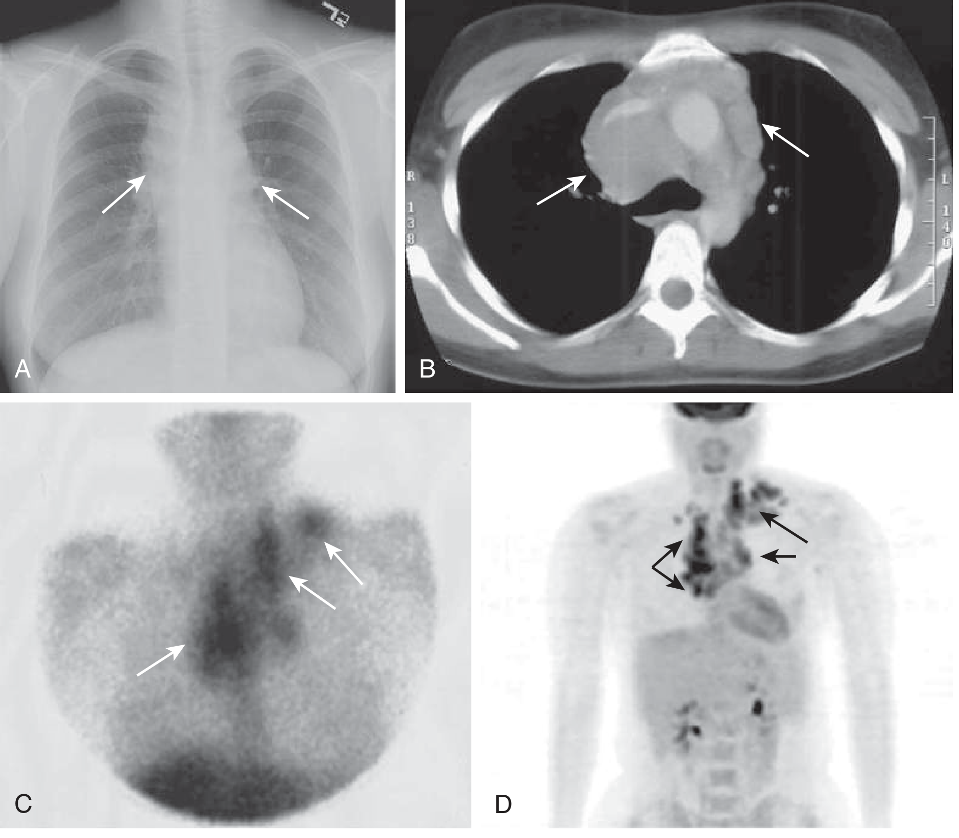
Staging Workup
- Complete history (B symptoms) + physical exam
- CBC, LDH, ESR, creatinine, alkaline phosphatase, albumin, serum protein electrophoresis
- Contrast-enhanced CT of neck, thorax, abdomen, pelvis
- FDG-PET/CT - now mandatory; replaces need for bone marrow biopsy in most cases
- Excisional lymph node biopsy for diagnosis
Treatment
Treatment is stratified by stage:
Limited Stage (IA or IIA, non-bulky)
- Standard: ABVD x 2 cycles + involved-region radiotherapy (IRRT)
- Alternative: ABVD x 3 cycles alone - if PET scan shows complete metabolic response, no further treatment; if partial response, add IRRT or checkpoint inhibitor x 8 doses + AVD x 4
- For unfavorable limited-stage (bulky mediastinum, extranodal extension, elevated ESR, ≥3 nodal areas): checkpoint inhibitors (nivolumab or pembrolizumab) combined with AVD showing >98% PFS in trials
Advanced Stage (IB, IIB, III, IV, or any stage with bulky disease)
- ABVD x 6 cycles (long-standing standard)
- A+AVD x 6 cycles (brentuximab vedotin + doxorubicin + vinblastine + dacarbazine) - preferred in many centers; omits bleomycin (reduces pulmonary toxicity)
- Escalated BEACOPP x 4-6 cycles (bleomycin, etoposide, doxorubicin, cyclophosphamide, vincristine, procarbazine, prednisone) - higher response rate but more toxicity
Regimen Summary
| Regimen | Drugs |
|---|---|
| ABVD | Adriamycin (doxorubicin) + Bleomycin + Vinblastine + Dacarbazine |
| A+AVD | Brentuximab vedotin + Doxorubicin + Vinblastine + Dacarbazine |
| BEACOPP | Bleomycin + Etoposide + Adriamycin + Cyclophosphamide + Oncovin + Procarbazine + Prednisone |
Relapsed/Refractory Disease
- Second-line chemotherapy (e.g., ICE, DHAP)
- Followed by high-dose chemoradiation + autologous hematopoietic cell transplantation (auto-HCT)
- Brentuximab vedotin (anti-CD30 antibody-drug conjugate) - active in relapsed cHL
- Checkpoint inhibitors: Nivolumab and pembrolizumab have shown significant activity in relapsed/refractory HL, exploiting the tumor's high PD-L1/L2 expression
Prognosis
Hodgkin lymphoma was the first human cancer to be successfully treated with radiation therapy and chemotherapy, and remains one of the most curable cancers:
- Early stage: ~90-95% cure rate
- Advanced stage: ~75-85% long-term disease-free survival
- The major clinical challenge today is minimizing long-term treatment toxicity (secondary malignancies, cardiac disease, pulmonary fibrosis from bleomycin) while maintaining cure rates
Adverse prognostic features for advanced disease (IPS - International Prognostic Score):
Albumin <4 g/dL, Hemoglobin <10.5 g/dL, Male sex, Stage IV, Age ≥45, WBC ≥15,000/μL, Lymphocyte count <600/μL or <8% of WBC - each factor subtracts ~7% from 5-year freedom from progression.
Sources: Robbins, Cotran & Kumar Pathologic Basis of Disease; Robbins & Kumar Basic Pathology; Goldman-Cecil Medicine, Chapter 172 (Connors & Savage); Henry's Clinical Diagnosis and Management by Laboratory Methods
This is a shared conversation. Sign in to Orris to start your own chat.