
Gist molecular pathology, diagnosis, targets therapy, sdh deficient gist
"gastrointestinal stromal tumor" AND "molecular pathology" AND "targeted therapy"
"SDH deficient GIST" AND "succinate dehydrogenase"
GIST gastrointestinal stromal tumor histology CD117 KIT mutation pathology
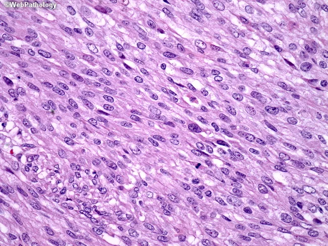
This is a high-magnification histopathology image of a gastrointestinal stromal tumor (GIST) showing a spindle cell-dominant morphology. Tissue is examined by light microscopy after Hematoxylin and Eosin staining, at ×400 magnification, highlighting elongated, fusiform cells arranged in intersecting fascicles with ovoid to elongated nuclei and eosinophilic cytoplasm. The stroma appears fibrous to lightly myxoid, with preserved cellular detail and minimal necrosis in this field. The spindle cell population is characteristic of many GISTs and supports a mesenchymal neoplasm of the gastrointestinal tract. In clinical practice, GISTs commonly harbor activating mutations in the c-KIT (CD117) gene in about 80% of cases or in PDGFRA (~8%), leading to constitutive tyrosine kinase signaling. Immunohistochemical CD117 positivity is a key diagnostic surrogate; DOG1 is frequently positive and may aid in challenging cases. Accurate molecular testing for c-KIT and PDGFRA mutations informs prognosis and eligibility for targeted therapy with tyrosine kinase inhibitors such as imatinib. This image serves as a reference for recognizing spindle cell GIST histology, differentiating it from smooth muscle or neural tumors, and illustrating how morphology guides ancillary testing, mutation profiling, and therapeutic decision-making in gastroenterology oncology and surgical pathology. This education-oriented caption emphasizes diagnostic workflow and targeted therapy implications for practice.
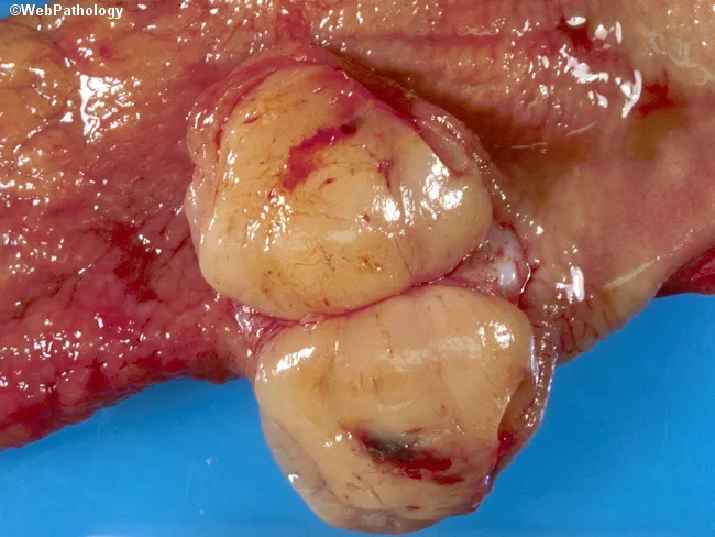
Gross pathology photograph of a resected gastric tumor illustrating the classic appearance of a gastrointestinal stromal tumor (GIST). The specimen demonstrates a solitary, well‑circumscribed, fleshy mass arising within the gastric wall, typically submucosal with a tan-yellow cut surface and occasional surface hemorrhage or focal necrosis. Gastric GISTs represent the most common mesenchymal neoplasms of the abdomen, with a predominance in the stomach (>50%), and most patients presenting in the sixth decade; pediatric cases are rare and may occur in the context of Carney triad. On gross examination, lesions are often exophytic or intraluminal and may appear as encapsulated nodules or plaque‑like thickenings. Definitive diagnosis requires microscopic evaluation and immunohistochemistry, including positivity for KIT (CD117) and DOG1, with variable spindle or epithelioid cell morphology and mitotic activity guiding risk stratification. The illustrated tumor’s solitary, well‑defined margins are consistent with a resectable, localized GIST, but differentiation from other submucosal spindle cell tumors (leiomyoma, leiomyosarcoma, schwannoma) relies on histology and immunophenotyping. Clinically, GISTs demand surgical excision and targeted therapy in select cases, and prognosis depends on size, mitotic rate, location, and rupture risk. This image is valuable for surgical pathology education, gross pathology training, and radiology–pathology correlation discussions.
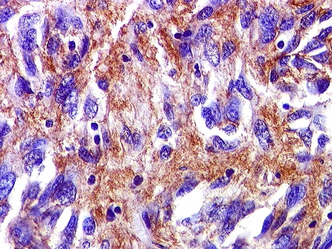
Immunohistochemical slide of a gastrointestinal stromal tumor (GIST) showing robust cytoplasmic/membranous brown staining on a hematoxylin counterstain background. The image likely represents a tissue section stained for CD117 (c-KIT) and/or DOG1, markers that confer high sensitivity and specificity for GIST. In this field, CD34 may be variably positive and SMA positivity can be focal, complicating differential diagnosis with leiomyosarcoma. The tissue is FFPE, sectioned at 4 μm, and stained with DAB chromogen to yield brown immunoreactivity. The tumor cells display spindle-cell morphology in fascicles embedded in a collagenous stroma; nuclei are ovoid to elongated with variable mitotic activity. The immunophenotype—predominant CD117 (c-KIT) and DOG1 positivity—supports the diagnosis of GIST, with DOG1 often providing superior specificity. The combined histology and IHC pattern differentiates GIST from smooth muscle and neural neoplasms. Clinically, this immunoprofile directs targeted therapy options (tyrosine kinase inhibitors such as imatinib or sunitinib) and informs mutational testing for KIT or PDGFRA to predict treatment response. This image is valuable for education in oncologic pathology, GI pathology, and diagnostic immunohistochemistry, illustrating how a mesenchymal GI tumor is identified by characteristic immunostains. It is suitable as a reference for immunophenotype, differential diagnosis, and molecular correlation in GIST research and teaching.
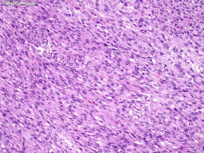
This is a light-m microscopy histopathology image of a gastrointestinal stromal tumor (GIST) in spindle cell form. The tissue shows spindle-shaped cells arranged in broad fascicles within a loose to moderate stroma. Nuclei are elongated with minimal pleomorphism and cytoplasm is eosinophilic to lightly basophilic; mitotic figures may be scarce in the depicted field. The overall pattern is classic for spindle cell GIST, which arises from interstitial cells of Cajal and can differentiate along smooth muscle or neural lines, though the image itself does not display immunohistochemical markers. Clinically, GISTs are most commonly located in the stomach or small intestine and require confirmation with immunostains such as KIT (CD117) and DOG1; CD34 is variably positive. The diagnostic significance lies in distinguishing GIST from other spindle cell GI tumors (e.g., leiomyosarcoma, schwannoma). Differential diagnoses include leiomyosarcoma, schwannoma, desmoid-type fibromatosis, and fibrosarcoma. Once confirmed, treatment typically involves surgical resection with adjuvant/targeted therapy guided by mutation status (e.g., imatinib for KIT/PDGFRA mutations). This image is relevant for educational discussions of GIST histology, spindle cell morphology, and differential diagnosis in GI pathology.
PMID: 39927693
PMID: 36980917
PMID: 42191879
SDH deficient GIST succinate dehydrogenase immunohistochemistry SDHB loss
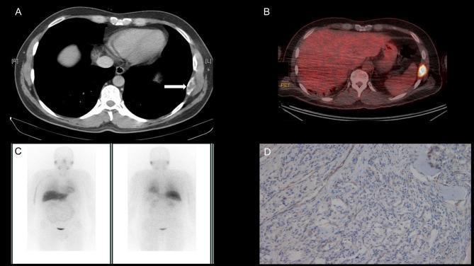
A composite clinical and diagnostic image set illustrating a case of metastatic paraganglioma with succinate dehydrogenase (SDH) deficiency. (A) Axial CT scan of the thorax shows an expansile, lytic-appearing metastatic deposit in the left 8th rib (indicated by a white arrow). (B) Axial 18F-FDG PET/CT fusion image demonstrates intense radiotracer uptake (avidity) at the site of the left rib lesion, indicating high glucose metabolism within the tumor. (C) Coronal I131-MIBG whole-body planar scans (anterior and posterior views) show a complete lack of tracer uptake at the rib metastasis site, highlighting the diagnostic limitation of MIBG for certain SDH-deficient neuroendocrine tumors. (D) Photomicrograph of tumor histology with SDHB immunohistochemistry (IHC) staining. The tumor cells show a diffuse loss of granular cytoplasmic staining (immunonegativity), while background stromal elements or internal controls show normal expression. This IHC pattern is a surrogate marker for deficiency in the SDH complex, specifically associated here with a germline SDHA mutation. The images demonstrate the multi-modal diagnostic approach and pathological correlation for evaluating malignant paragangliomas.
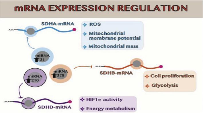
This pathophysiology diagram illustrates the post-transcriptional regulation of succinate dehydrogenase (SDH) subunits by specific microRNAs (miRNAs) in the context of cellular metabolism and cancer. The schematic identifies three primary regulatory axes: 1) miRNA-31 targets SDHA-mRNA, influencing reactive oxygen species (ROS) production, mitochondrial membrane potential, and mitochondrial mass. 2) miRNA-378 targets SDHB-mRNA, promoting cell proliferation and a metabolic shift toward glycolysis. 3) miRNA-210 inhibits SDHD-mRNA, leading to alterations in HIF1̧ activity and overall energy metabolism. Each mRNA is depicted with characteristic structural features including a 5' cap and a poly-A (AAA) tail. Upward-pointing arrows within the miRNA circles indicate overexpression, a common clinical finding in certain malignancies or post-radiation states. The diagram highlights how miRNA-mediated silencing of the SDH complex components disrupts the tricarboxylic acid (TCA) cycle and electron transport chain, contributing to metabolic reprogramming and tumor progression.
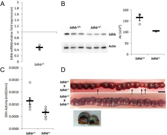
This composite educational image illustrates the molecular and developmental consequences of Sdhb (Succinate Dehydrogenase Complex Iron-Sulfur Subunit B) deletion in a rat model. Panel A: A scatter plot showing an approximately 50% reduction in Sdhb mRNA relative fold expression in heterozygous (Sdhb+/-) liver tissue compared to wild-type (Sdhb+/+), normalized to a baseline of 1. Panel B: An immunoblot (Western blot) and corresponding densitometric bar graph demonstrating reduced Sdhb protein expression in Sdhb+/- rats compared to Sdhb+/+ controls, using Actin as a loading control. Panel C: A scatter plot quantifying decreased SDH enzyme activity (mOD/min) in Sdhb+/- tissue. Panel D: A clinical macroscopic photograph of opened uteri from 12-day-old gravid rats. The top uterus (Sdhb+/- x Sdhb+/- cross) displays multiple developmentally stalled, smaller, and darker embryos (indicated by black arrows), illustrating the lethal effect of homozygous deletion. The bottom uterus (Sdhb+/+ x Sdhb+/+ cross) shows uniform, healthy embryonic development. This figure highlights the role of Sdhb in mitochondrial function and embryonic viability.
GIST: Molecular Pathology, Diagnosis, Targeted Therapy & SDH-Deficient GIST
1. Overview & Cell of Origin
- Stomach: ~60% of cases (most favorable prognosis)
- Small intestine: ~25-35%
- Colon, rectum, esophagus: rare
- Median age at diagnosis: ~60 years; slight male predominance
2. Molecular Pathology
KIT (c-KIT / CD117) Mutations (~75-85%)
| Exon | Domain | Frequency | Imatinib Response |
|---|---|---|---|
| 11 (most common) | Juxtamembrane | ~53-71% | High (~80-85% response) |
| 9 | Extracellular | ~8-9% | 40-50%; higher-dose imatinib needed |
| 13 | Kinase domain | ~1-4% | Moderate; often secondary resistance mutation |
| 17 | Activation loop | ~1% | Moderate; often secondary resistance mutation |
| 8 | Extracellular | rare | Variable |
PDGFRA Mutations (~5-16%)
Wild-Type / Triple-Negative GISTs (~10-17%)
3. Histology & Immunophenotype
| Feature | Detail |
|---|---|
| Spindle cell subtype | ~70% (fascicles of elongated cells) |
| Epithelioid subtype | ~20% |
| Mixed | ~10% |
| CD117 (KIT) | Positive in ~95%; strong diffuse cytoplasmic staining - key diagnostic marker |
| DOG1 | High sensitivity and specificity; positive in ~95%; critical in CD117-negative cases |
| CD34 | Variably positive (~70%) |
| SMA / S100 | Focal; helps differentiate from leiomyosarcoma/schwannoma |
Key Differential Diagnoses
4. Risk Stratification
- Tumor size (cm)
- Mitotic index (per 50 HPF or 5 mm²)
- Location (gastric vs. non-gastric; gastric carries lower risk for same size/mitotic count)
- Gastric GISTs ≤2 cm with mitotic index ≤5/50 HPF: near-zero risk of progression
- Tumors ≥10 cm with mitotic index >5/50 HPF: high risk
- Tumor rupture is an independent poor prognostic factor
5. Diagnosis
Imaging
- CT abdomen/pelvis (with IV and oral contrast): first-line imaging; identifies primary tumor and metastases
- Typical appearance: well-circumscribed submucosal mass; may be exophytic or intraluminal
- Metastases: liver and peritoneal cavity most common; lymph node metastasis is rare in conventional adult GIST (<5%)
- EUS: useful for biopsy guidance but may not yield adequate cellular material for genomic sequencing
Pathology Workup
- Morphology (H&E): spindle/epithelioid pattern
- IHC: CD117, DOG1 (both positive strongly supports GIST)
- Molecular testing (NGS): mandated for all resected GISTs to guide TKI selection - test for KIT exons 9, 11, 13, 17 and PDGFRA exons 12, 14, 18
- For KIT/PDGFRA-negative cases: SDHB IHC to identify SDH-deficient GIST (loss of granular cytoplasmic SDHB staining is a surrogate for SDH complex dysfunction)
6. Targeted Therapy
First-Line: Imatinib (Gleevec)
| Setting | Outcome |
|---|---|
| Metastatic disease | Median survival >5 years (vs. 18 months historical) |
| Adjuvant (1 year, ACOSOG Z9001 trial) | 1-year RFS 98% vs 83% (P<0.0001) |
| Adjuvant (3 years, SSG XVIII trial) | 5-year RFS 71% vs 52%; 5-year OS 92% vs 85% |
| Neoadjuvant | Can convert unresectable to resectable |
- KIT exon 11: most sensitive (~80-85% response)
- KIT exon 9: 40-50% response; higher-dose imatinib (800 mg/day) improves response
- Wild-type KIT/PDGFRA: up to 30% response
- PDGFRA mutations: generally resistant, especially D842V - essentially no response
- SDH-deficient: universally imatinib resistant
PDGFRA D842V: Avapritinib (First-Line)
Second-Line: Sunitinib
Third-Line: Regorafenib
Later Lines / Resistance: Ripretinib
7. SDH-Deficient GIST - Deep Dive
Definition & Epidemiology
- 10-15% of all GISTs
- ~5-10% of all gastric GISTs
- The majority of pediatric GISTs
- Virtually all GISTs in Carney triad and Carney-Stratakis syndrome
SDH Complex Biology
- Accumulation of succinate (oncometabolite)
- Competitive inhibition of alpha-ketoglutarate-dependent dioxygenases
- Genome-wide DNA hypermethylation (epigenetic silencing)
- HIF pathway activation (pseudohypoxia)
Mechanisms of SDH Loss
| Mechanism | Frequency |
|---|---|
| SDHC promoter hypermethylation (epimutation) | ~50% of SDH-deficient GISTs |
| SDHA somatic/germline mutation | ~30% (most common gene mutation) |
| SDHB, SDHC, SDHD mutations | ~20-30% combined; most are germline |
- The SDHC epimutation is a somatic event (not heritable); it silences SDHC transcription
- SDHA-mutant tumors typically require two hits (loss of wild-type allele + somatic second event)
- Most SDHx (B/C/D) mutations are germline, making genetic counseling essential
Clinical Features of SDH-Deficient GIST
| Feature | SDH-Deficient GIST | Conventional GIST |
|---|---|---|
| Age | Young adults, pediatric | Older adults (~60 yr) |
| Sex | Female predominance | Slight male predominance |
| Location | Stomach (predominantly) | Stomach > small intestine |
| Morphology | Epithelioid; multinodular/plexiform | Spindle cell (70%) |
| Multifocality | Common | Rare |
| Lymph node metastases | Common | Rare (<5%) |
| Clinical course | Indolent despite metastases | Behavior follows size/mitosis |
| Imatinib response | Universally resistant | Responsive (if KIT exon 11) |
| Risk stratification | Size/mitosis unreliable | Standard NIH/AFIP criteria |
Diagnosis of SDH-Deficient GIST
- Suspect when: KIT/PDGFRA negative, epithelioid morphology, young/female patient, multifocal gastric GIST
- SDHB IHC: Loss of granular cytoplasmic SDHB staining confirms SDH complex dysfunction (regardless of which subunit is mutated - SDHB protein is lost when any subunit is nonfunctional)
- SDHA IHC: If SDHA protein is also lost, this suggests SDHA mutation specifically
- Molecular testing: SDHx gene sequencing (somatic + germline) and SDHC promoter methylation analysis
- Germline testing: Recommended for ALL SDH-deficient GISTs regardless of which gene is affected - due to high germline prevalence
Associated Syndromes
| Syndrome | Features |
|---|---|
| Carney Triad | GIST + pulmonary chondromas + extra-adrenal paragangliomas; not heritable (SDHC epimutation) |
| Carney-Stratakis Syndrome | GIST + paraganglioma; autosomal dominant (germline SDHx mutations) |
| NF1-associated GIST | Multiple small intestinal GISTs; KIT overexpressed but no KIT/PDGFRA mutation; some SDH-deficient overlap |
| Familial GIST | Germline KIT or PDGFRA mutations; multifocal, indolent |
Emerging Targeted Therapy for SDH-Deficient GIST
- SDH loss causes genome-wide hypermethylation → disrupts genomic insulators → induces aberrant expression of FGF3 and FGF4 → activates an FGFR1 autocrine signaling loop
- Rogaratinib (pan-FGFR inhibitor) tested in 24 patients with advanced SDH-deficient GIST
- Objective response rate: 41.7% (10/24 partial responses)
- Median PFS: 31.0 months (95% CI: 20.2 - not reached)
- 1-year PFS: 77.4%
- Manageable toxicity: hyperphosphatemia (target engagement marker), fatigue, diarrhea
- This represents a proof-of-concept for targeting an epigenetically-activated oncogene pathway
8. Summary Algorithm
GIST Diagnosed (CD117+/DOG1+)
         |
    KIT / PDGFRA mutation testing
         |
   ┌─────┴──────────────────────────┐
KIT mutant               PDGFRA mutant          Wild-type (both negative)
   |                         |                          |
Imatinib                D842V?                    SDHB IHC
(exon 11: std dose;       Yes → Avapritinib      |            |
 exon 9: high dose)       No → Imatinib (poor)  Negative    Positive
                                                  (other WT)  = SDH-deficient
                                              BRAF/NTRK etc.    |
                                                          SDHx sequencing
                                                          Germline testing
                                                          FGF3/4 axis (rogaratinib)
- Quick Compendium of Clinical Pathology 5th Ed., p.404
- Sleisenger & Fordtran's GI and Liver Disease, p.481
- Harrison's Principles of Internal Medicine 22E, p.694
- Sabiston Textbook of Surgery 11e, p.1812
- Current Surgical Therapy 14e, p.154
- Henry's Clinical Diagnosis and Management, p.1848
- Florou et al., Cancer Med. 2025 - SDH-deficient GIST genomic workup algorithm (PMID: 39927693)
- Schipani et al., Genes (Basel). 2023 - SDHA germline mutations in SDH-deficient GIST (PMID: 36980917)
- Merriam et al., Nature Medicine. 2026 - Rogaratinib Phase 2 trial in SDH-deficient GIST (PMID: 42191879)