
Anion gap
Reading File
The anion gap (AG) is calculated as: Na+ - (Cl- + HCO3-). Normal range is roughly 8-12 mEq/L (or 10-14 if K+ is included). It represents unmeasured anions in plasma and is used primarily to classify metabolic acidosis.
Searching sources...
Finding Sources
Searching PubMed
"anion gap" AND "metabolic acidosis"
Reading File
Reading File
Reading File
Finding Sources
Reading File
Loading Image
Loading Image
Here is a thorough breakdown of the anion gap based on multiple authoritative textbooks.
Anion Gap
Definition and Formula
The anion gap (AG) is a calculated value representing the unmeasured anions in plasma. It arises from the principle of electrical neutrality: total cation charge must equal total anion charge. By looking only at the three most abundant measured electrolytes:
AG = [Na⁺] - ([Cl⁻] + [HCO₃⁻])
- Normal range: 8-12 mEq/L (most labs; some use 10-14 if K⁺ is included)
- The gap is "apparent" because it represents anions that are real but not routinely measured
The normal AG is composed primarily of anionic albumin and, to a lesser extent, phosphate, sulfate, urate, and organic acid anions such as lactate.
- National Kidney Foundation Primer on Kidney Diseases, 8e
Visual: Normal vs. High AG vs. Hyperchloremic Acidosis (Gamblegram)
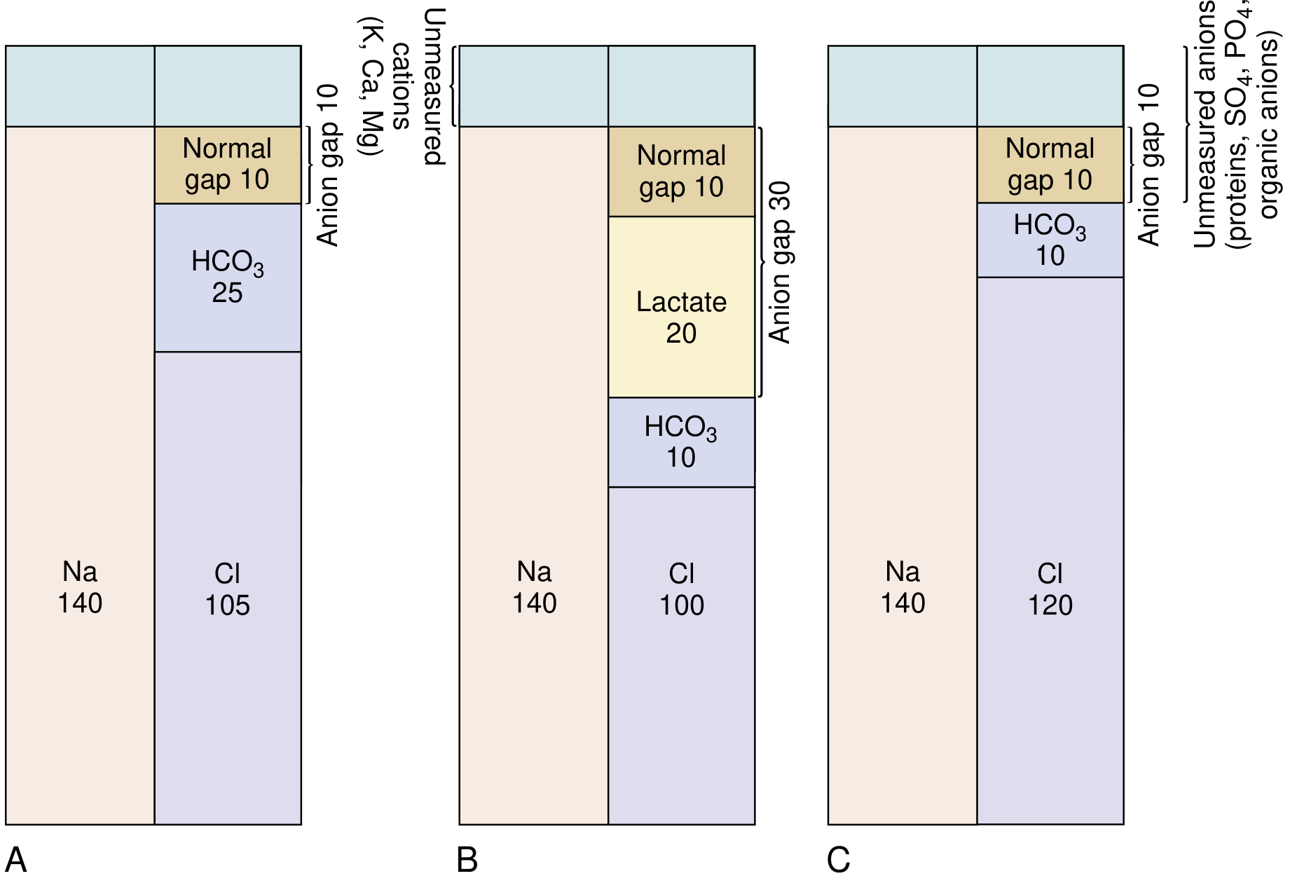
(A) Normal: Na 140, Cl 105, HCO₃ 25, AG = 10
(B) High AG acidosis (lactic): Na 140, Cl 100, HCO₃ 10, Lactate 20 - AG rises to 30
(C) Hyperchloremic/normal AG acidosis: Na 140, Cl 120, HCO₃ 10 - AG stays at 10
Why the Gap Exists
When an unmeasured anion (e.g., lactate, ketoacid) accumulates:
- It titrates HCO₃⁻, causing HCO₃⁻ to fall
- Cl⁻ stays the same (it is not the compensating anion)
- So AG = Na⁺ - (Cl⁻ + HCO₃⁻) increases
When HCO₃⁻ is lost and replaced by Cl⁻ (e.g., diarrhea, RTA):
- One measured anion replaces another
- AG stays normal (hyperchloremic acidosis)
Causes of Metabolic Acidosis by AG Type
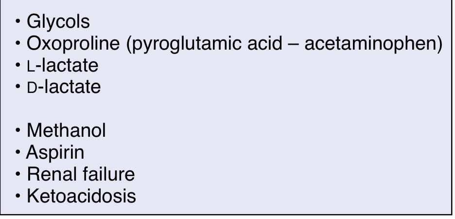
High AG Metabolic Acidosis - Mnemonic: GOLDMARK
| Category | Causes |
|---|---|
| Lactic acidosis | Type A (hypoperfusion, sepsis), Type B (metformin, liver failure), D-lactic acidosis |
| Ketoacidosis | Diabetic (DKA), alcoholic, starvation |
| Toxins/Drugs | Methanol, ethylene glycol (glycols), salicylates (aspirin), oxoproline (acetaminophen) |
| Renal failure | Accumulation of sulfate, phosphate, urate, hippurate |
- Costanzo Physiology, 7th Edition; National Kidney Foundation Primer, 8e
Normal AG Metabolic Acidosis (Hyperchloremic)
| Cause | Mechanism |
|---|---|
| Diarrhea | Loss of HCO₃⁻, replaced by Cl⁻ |
| Renal tubular acidosis (RTA) | Failure to excrete acid or retain HCO₃⁻ |
| Carbonic anhydrase inhibitors | Urinary HCO₃⁻ wasting |
| Saline infusion | Dilutional, Cl⁻ load |
| Early renal failure | - |
Albumin Correction (Critical Concept)
The normal AG is largely anionic albumin. In hypoalbuminemia (common in critically ill patients), the AG is falsely low and may mask a true AG acidosis.
Corrected AG (AGc) = AG + 2.5 × (4 - measured albumin [g/dL])
For every 1 g/dL drop in albumin below 4, add 2.5 mEq/L to the AG.
- National Kidney Foundation Primer, 8e
The Delta-Delta Ratio (Δ/Δ)
Once a high AG acidosis is confirmed, the delta-delta ratio detects concurrent metabolic disturbances:
Δ/Δ = (Measured AG - Normal AG) / (Normal HCO₃⁻ - Measured HCO₃⁻) Using: Normal AG = 12, Normal HCO₃⁻ = 24
| Delta Ratio | Interpretation |
|---|---|
| < 0.4 | Hyperchloremic (normal AG) acidosis predominates |
| 0.4 - 1.0 | Mixed high AG + hyperchloremic acidosis |
| 1.0 - 2.0 | Pure high AG metabolic acidosis (expected) |
| > 2.0 | High AG acidosis + concurrent metabolic alkalosis (or pre-existing elevated HCO₃⁻) |
- Miller's Anesthesia, 10e; Barash Clinical Anesthesia, 9e
Osmolar Gap (Special Consideration)
In methanol and ethylene glycol poisoning, a high AG acidosis is accompanied by an osmolar gap (measured osmolality > calculated osmolality by >10 mOsm/kg). This is because these low-molecular-weight toxins directly increase measured osmolality but are not included in the standard osmolality formula. Ketoacids and lactate do NOT typically cause a significant osmolar gap due to their higher molecular weights.
Clinical Approach Summary
- Calculate AG = Na⁺ - (Cl⁻ + HCO₃⁻)
- Correct for albumin if hypoalbuminemic
- If AG > 12: identify cause using GOLDMARK; check for osmolar gap if toxin suspected
- If AG normal: evaluate for hyperchloremic causes (diarrhea, RTA)
- In high AG acidosis: calculate Δ/Δ to unmask mixed disorders
This is a shared conversation. Sign in to Orris to start your own chat.