Ammonorrhoea

Reading File
Finding Sources
Searching PubMed

amenorrhea diagnosis management

Reading File
Reading File
Reading File
Reading File
Reading File
Reading File
Searching Images

amenorrhea diagnostic algorithm flowchart evaluation

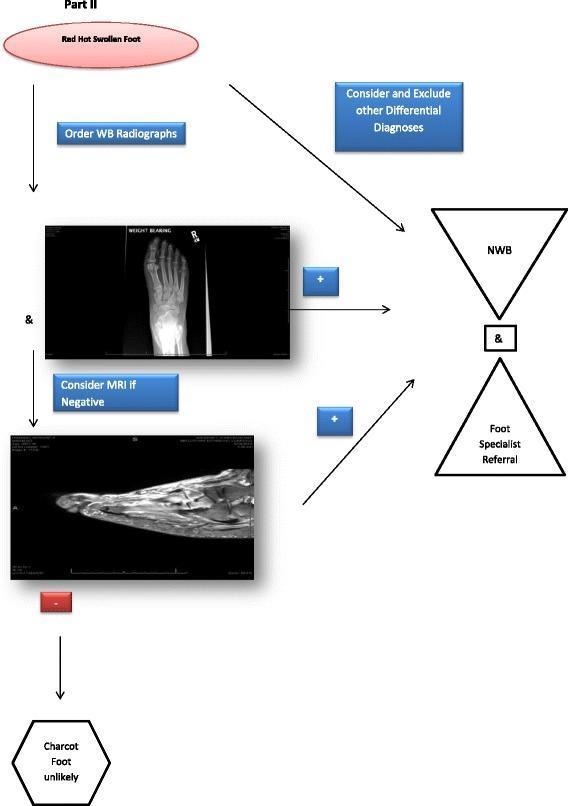
A medical diagnostic flowchart titled 'CN Stage 0 Evaluation Algorithm Part II', outlining the clinical pathway for suspected Charcot Neuroarthropathy (CN). The algorithm begins with the clinical presentation of a 'Red Hot Swollen Foot'. The primary diagnostic step involves ordering weight-bearing (WB) radiographs, shown as an anteroposterior X-ray image of the foot. Simultaneously, the clinician should consider and exclude other differential diagnoses. If the radiograph is positive (+) for CN signs, or if radiographs are negative and a subsequent MRI (shown as a sagittal T1-weighted image) is positive, the patient is transitioned to a non-weight-bearing (NWB) status and referred to a foot specialist. If both imaging modalities are negative (-) for diagnostic features of CN, the algorithm concludes that Charcot foot is unlikely. This educational resource is designed for podiatrists and orthopedic specialists to assist in the early identification and management of Charcot Neuroarthropathy Stage 0 to prevent permanent structural foot deformity.

A medical diagnostic flowchart titled 'CN Stage 0 Evaluation Algorithm Part II', outlining the clinical pathway for suspected Charcot Neuroarthropathy (CN). The algorithm begins with the clinical presentation of a 'Red Hot Swollen Foot'. The primary diagnostic step involves ordering weight-bearing (WB) radiographs, shown as an anteroposterior X-ray image of the foot. Simultaneously, the clinician should consider and exclude other differential diagnoses. If the radiograph is positive (+) for CN signs, or if radiographs are negative and a subsequent MRI (shown as a sagittal T1-weighted image) is positive, the patient is transitioned to a non-weight-bearing (NWB) status and referred to a foot specialist. If both imaging modalities are negative (-) for diagnostic features of CN, the algorithm concludes that Charcot foot is unlikely. This educational resource is designed for podiatrists and orthopedic specialists to assist in the early identification and management of Charcot Neuroarthropathy Stage 0 to prevent permanent structural foot deformity.

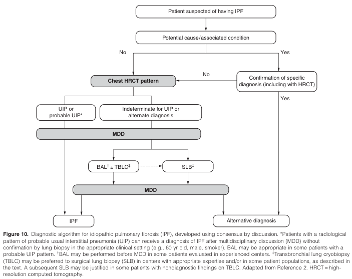
Summary : This flowchart presents a diagnostic algorithm for idiopathic pulmonary fibrosis (IPF), detailing the stepwise evaluation of patients suspected of having IPF, including assessment of potential causes, imaging patterns, multidisciplinary discussion, and biopsy options.

flowchart:
# Nodes :
  • Patient suspected of having IPF (rectangle)
  • Potential cause/associated condition (rectangle)
  • Confirmation of specific diagnosis (including with HRCT) (rectangle)
  • Chest HRCT pattern (rounded rectangle)
  • UIP or probable UIP* (rectangle)
  • Indeterminate for UIP or alternate diagnosis (rectangle)
  • MDD (multidisciplinary discussion) (rounded rectangle, appears twice)
  • BAL† ± TBLC‡ (rectangle)
  • SLB‡ (rectangle)
  • IPF (rectangle)
  • Alternative diagnosis (rectangle)

# Connectors :
  • Patient suspected of having IPF → Potential cause/associated condition (down arrow)
  • Potential cause/associated condition → Confirmation of specific diagnosis (right arrow, labeled "Yes")
  • Confirmation of specific diagnosis → Alternative diagnosis (down arrow, labeled "Yes")
  • Potential cause/associated condition → Chest HRCT pattern (down arrow, labeled "No")
  • Chest HRCT pattern → UIP or probable UIP* (left branch)
  • Chest HRCT pattern → Indeterminate for UIP or alternate diagnosis (right branch)
  • UIP or probable UIP* → MDD (down arrow)
  • Indeterminate for UIP or alternate diagnosis → MDD (down arrow)
  • MDD → BAL† ± TBLC‡ (left branch)
  • MDD → SLB‡ (right branch)
  • BAL† ± TBLC‡ → MDD (dashed arrow to right, indicating possible subsequent step)
  • SLB‡ → MDD (down arrow)
  • MDD (second instance) → IPF (left arrow)
  • MDD (second instance) → Alternative diagnosis (right arrow)

# Layout :
  • The flowchart is organized top-to-bottom, with initial patient assessment at the top.
  • Branching occurs based on presence of potential cause/associated condition.
  • Imaging (HRCT) and diagnostic pattern assessment are central.
  • Multidisciplinary discussion (MDD) is a key decision node, leading to biopsy options.
  • Final outcomes are IPF or alternative diagnosis at the bottom.

# Analysis :
  • The algorithm emphasizes exclusion of known causes before considering IPF.
  • HRCT imaging pattern is pivotal in guiding further workup.
  • Multidisciplinary discussion is required for diagnosis, especially in cases with probable UIP pattern.
  • Biopsy options (BAL, TBLC, SLB) are considered for indeterminate cases, with iterative discussion.
  • The process is designed to minimize unnecessary invasive procedures and ensure accurate diagnosis.

Summary : This flowchart presents a diagnostic algorithm for idiopathic pulmonary fibrosis (IPF), detailing the stepwise evaluation of patients suspected of having IPF, including assessment of potential causes, imaging patterns, multidisciplinary discussion, and biopsy options. flowchart: # Nodes : • Patient suspected of having IPF (rectangle) • Potential cause/associated condition (rectangle) • Confirmation of specific diagnosis (including with HRCT) (rectangle) • Chest HRCT pattern (rounded rectangle) • UIP or probable UIP* (rectangle) • Indeterminate for UIP or alternate diagnosis (rectangle) • MDD (multidisciplinary discussion) (rounded rectangle, appears twice) • BAL† ± TBLC‡ (rectangle) • SLB‡ (rectangle) • IPF (rectangle) • Alternative diagnosis (rectangle) # Connectors : • Patient suspected of having IPF → Potential cause/associated condition (down arrow) • Potential cause/associated condition → Confirmation of specific diagnosis (right arrow, labeled "Yes") • Confirmation of specific diagnosis → Alternative diagnosis (down arrow, labeled "Yes") • Potential cause/associated condition → Chest HRCT pattern (down arrow, labeled "No") • Chest HRCT pattern → UIP or probable UIP* (left branch) • Chest HRCT pattern → Indeterminate for UIP or alternate diagnosis (right branch) • UIP or probable UIP* → MDD (down arrow) • Indeterminate for UIP or alternate diagnosis → MDD (down arrow) • MDD → BAL† ± TBLC‡ (left branch) • MDD → SLB‡ (right branch) • BAL† ± TBLC‡ → MDD (dashed arrow to right, indicating possible subsequent step) • SLB‡ → MDD (down arrow) • MDD (second instance) → IPF (left arrow) • MDD (second instance) → Alternative diagnosis (right arrow) # Layout : • The flowchart is organized top-to-bottom, with initial patient assessment at the top. • Branching occurs based on presence of potential cause/associated condition. • Imaging (HRCT) and diagnostic pattern assessment are central. • Multidisciplinary discussion (MDD) is a key decision node, leading to biopsy options. • Final outcomes are IPF or alternative diagnosis at the bottom. # Analysis : • The algorithm emphasizes exclusion of known causes before considering IPF. • HRCT imaging pattern is pivotal in guiding further workup. • Multidisciplinary discussion is required for diagnosis, especially in cases with probable UIP pattern. • Biopsy options (BAL, TBLC, SLB) are considered for indeterminate cases, with iterative discussion. • The process is designed to minimize unnecessary invasive procedures and ensure accurate diagnosis.

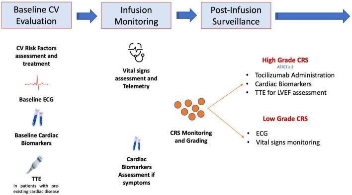
This flowchart delineates a diagnostic and management strategy for cardiovascular (CV) complications in patients undergoing CAR-T cell therapy. The clinical algorithm is organized into three sequential phases: Baseline CV Evaluation, Infusion Monitoring, and Post-Infusion Surveillance. The baseline phase involves CV risk factor assessment, ECG, baseline cardiac biomarkers (e.g., troponin, NT-proBNP), and transthoracic echocardiography (TTE) for patients with pre-existing heart disease. The infusion phase focuses on vital sign assessment, telemetry, monitoring for Cytokine Release Syndrome (CRS), and symptom-driven biomarker assessment. Post-infusion surveillance is risk-stratified based on American Society for Transplantation and Cellular Therapy (ASTCT) CRS grading. High-grade CRS (ASTCT ≥2) triggers tocilizumab administration, repeat cardiac biomarkers, and TTE for Left Ventricular Ejection Fraction (LVEF) assessment. Low-grade CRS requires continued ECG and vital signs monitoring. This infographic serves as a clinical guideline for oncologists and cardiologists to manage cardiotoxicity and systemic inflammatory responses during advanced immunotherapy.

This flowchart delineates a diagnostic and management strategy for cardiovascular (CV) complications in patients undergoing CAR-T cell therapy. The clinical algorithm is organized into three sequential phases: Baseline CV Evaluation, Infusion Monitoring, and Post-Infusion Surveillance. The baseline phase involves CV risk factor assessment, ECG, baseline cardiac biomarkers (e.g., troponin, NT-proBNP), and transthoracic echocardiography (TTE) for patients with pre-existing heart disease. The infusion phase focuses on vital sign assessment, telemetry, monitoring for Cytokine Release Syndrome (CRS), and symptom-driven biomarker assessment. Post-infusion surveillance is risk-stratified based on American Society for Transplantation and Cellular Therapy (ASTCT) CRS grading. High-grade CRS (ASTCT ≥2) triggers tocilizumab administration, repeat cardiac biomarkers, and TTE for Left Ventricular Ejection Fraction (LVEF) assessment. Low-grade CRS requires continued ECG and vital signs monitoring. This infographic serves as a clinical guideline for oncologists and cardiologists to manage cardiotoxicity and systemic inflammatory responses during advanced immunotherapy.

No relevant amenorrhea flowchart found in the image library. I have excellent textbook content to synthesize a thorough answer now.

Amenorrhoea

Definition

Amenorrhoea is the absence of menstruation. Normal menstruation requires an intact, coordinated interplay between the hypothalamus (pulsatile GnRH), pituitary (FSH, LH), ovary (oestrogen, progesterone), uterus, and outflow tract. Failure at any level produces amenorrhoea.

Classification

Primary Amenorrhoea

  • No secondary sexual characteristics by age 13 (i.e., no breast development)
  • Normal secondary sexual characteristics but no menses by age 15
These thresholds represent two standard deviations above the mean age of puberty onset. The absence of secondary sexual characteristics by 13 always warrants investigation.

Secondary Amenorrhoea

  • Absence of menstruation for 3 months in a woman with previously regular cycles
  • Women with fewer than 9 cycles per year or cycle length > 35 days should be evaluated
Note: The distinction between primary and secondary amenorrhoea is not absolute - many causes are shared between the two categories.

Physiology of Normal Menstruation

For menstruation to occur:
  1. Hypothalamus secretes pulsatile GnRH (modulated by neurotransmitters and hormones)
  2. GnRH stimulates pituitary to release FSH and LH
  3. FSH promotes ovarian follicular development; the follicle secretes oestrogen
  4. LH surge triggers ovulation and corpus luteum formation
  5. Corpus luteum secretes progesterone + oestrogen, causing endometrial maturation
  6. Withdrawal of hormones (if no pregnancy) triggers menstruation
Disruption at any step - hypothalamic, pituitary, ovarian, uterine/outflow tract - causes amenorrhoea.

WHO Classification of Amenorrhoea

GroupCharacteristicsExample
Group I - Hypogonadotropic hypogonadismNo endogenous oestrogen, normal/low FSH, normal prolactin, no H-P lesionAnorexia, exercise-induced
Group II - Normogonadotropic anovulationEvidence of oestrogen, normal FSH and prolactinPCOS
Group III - Hypergonadotropic hypogonadismElevated FSH (gonadal insufficiency/failure)Turner syndrome, POI
(Added) Group IV - Hyperprolactinaemic anovulationAnovulation specifically due to hyperprolactinaemiaProlactinoma

Causes

Primary Amenorrhoea

A. Without secondary sexual characteristics (no oestrogen exposure)
Hypergonadotropic hypogonadism (high FSH):
  • Turner syndrome (45,X) and gonadal dysgenesis variants
  • XX gonadal dysgenesis
  • 17-hydroxylase deficiency (ovaries and adrenal glands)
Hypogonadotropic hypogonadism (low FSH/LH):
  • Kallmann syndrome (GnRH deficiency + anosmia)
  • Hypopituitarism
  • Constitutional delay of puberty (physiologic)
  • Nutritional disorders / eating disorders
B. With secondary sexual characteristics + anatomic abnormality
  • Mayer-Rokitansky-Kuster-Hauser (MRKH) syndrome - Mullerian agenesis (absent uterus/upper vagina, normal ovaries)
  • Imperforate hymen (commonest outflow obstruction - causes cyclic pain, haematocolpos)
  • Congenital vaginal atresia
  • Androgen insensitivity syndrome (46,XY - blind vaginal pouch, absent uterus, testes)
  • Transverse vaginal septum
C. With secondary sexual characteristics + normal anatomy - see secondary amenorrhoea causes below

Secondary Amenorrhoea (common causes)

CategoryExamples
PhysiologicPregnancy, lactation, menopause
HypothalamicFunctional hypothalamic amenorrhoea (stress, weight loss, excessive exercise, eating disorders - especially anorexia nervosa), tumours, infiltrative disease
PituitaryHyperprolactinaemia (prolactinoma), Sheehan syndrome, empty sella, pituitary apoplexy, craniopharyngioma
OvarianPCOS, primary ovarian insufficiency (POI/premature ovarian failure before age 40), ovarian tumours, resistant ovary syndrome
Uterine/OutflowAsherman syndrome (intrauterine adhesions post-D&C or infection)
AdrenalCushing syndrome, late-onset congenital adrenal hyperplasia, virilising adrenal tumours, adrenocortical insufficiency
ThyroidHypothyroidism, hyperthyroidism
DrugsAntipsychotics (phenothiazines, haloperidol, clozapine), antidepressants (TCAs, MAOIs), antihypertensives (methyldopa, reserpine, Ca channel blockers), cytotoxic agents, OCP, marijuana

Evaluation

Step 1 - Always Rule Out First

  • Pregnancy test (serum/urine beta-hCG) - must exclude before any further workup

Step 2 - History

  • Menstrual pattern, weight changes, exercise habits, dietary history
  • Galactorrhoea, hot flushes, symptoms of hypothyroidism/hyperthyroidism
  • Hirsutism, acne (androgen excess)
  • Medications, prior pelvic/uterine surgery, stress
  • Cyclic pelvic pain (suggests outflow obstruction)

Step 3 - Physical Examination

  • Secondary sexual characteristics (breast development, pubic/axillary hair)
  • Visual fields (pituitary tumour)
  • Thyroid size
  • Signs of Cushing (cushingoid appearance, striae)
  • Galactorrhoea, signs of androgen excess
  • Pelvic exam: vaginal patency, uterine/ovarian masses, clitoral enlargement

Step 4 - Initial Laboratory Tests

TestPurpose
beta-hCGRule out pregnancy
ProlactinScreen for hyperprolactinaemia
TSHScreen for thyroid disease
FSH + LHDifferentiate hypergonadotropic vs hypogonadotropic
Oestradiol (E2)Assess oestrogen status
AMHMay help assess ovarian reserve (especially in POI workup)

Step 5 - Localising with FSH

FSH LevelInterpretation
High (> 40 IU/L)Primary ovarian insufficiency / gonadal failure
Low or normalHypothalamic or pituitary cause

Step 6 - Progesterone Challenge Test

  • Give progestin (e.g., medroxyprogesterone 10 mg x 10 days)
  • Withdrawal bleed occurs: Confirms oestrogen production is sufficient; likely PCOS, stress, exercise, medication
  • No withdrawal bleed: Suggests either (a) inadequate oestrogen (check FSH/E2) or (b) outflow obstruction (Asherman syndrome, anatomic)

Step 7 - Additional Tests as Indicated

FindingNext Step
Elevated FSH, age < 25 or short statureKaryotype (rule out Turner, Y chromosome)
POI in young womenScreen for fragile X premutation (FMR1), autoimmune antibodies (21-hydroxylase, thyroid)
Suspected pituitary/hypothalamic diseaseMRI of the sella turcica
Gonadotropin deficiencyGnRH stimulation test (differentiate hypothalamic from pituitary)
Suspected Cushing24-hr urine cortisol or overnight dexamethasone suppression test
Outflow tract suspectedPelvic USS or hysteroscopy
PCOS suspectedTestosterone, LH:FSH ratio, pelvic USS

Differential Diagnosis by Hormone Pattern (Secondary Amenorrhoea)

CauseFSHLHOestrogenBleed after Progesterone
Hypothalamic dysfunction (stress/exercise/weight loss)↓ or N↓ or N↓ or N±
Anorexia nervosaNegative
Hypothalamic failure (lesion)Negative
PCOSN or ↑↑ (often)N (oestrone ↑)Positive
Primary ovarian insufficiency↑↑↑↑Negative
Hyperprolactinaemia↓ or N↓ or N±
Asherman syndromeNNNNegative

Management

Management is directed at the underlying cause:

Specific Treatments

CauseTreatment
HypothyroidismLevothyroxine
ProlactinomaDopamine agonists (cabergoline, bromocriptine)
PCOSWeight loss, OCP, metformin, clomiphene (if fertility desired)
Asherman syndromeHysteroscopic adhesiolysis
Imperforate hymen / vaginal atresiaSurgical correction
Kallmann syndromeGnRH pulsatile therapy or HRT
Cushing syndromeTreat the underlying cause

Hormone Therapy (for hypo-oestrogenic states)

  • Oestrogen + progestogen replacement is used to:
    • Initiate and maintain secondary sexual characteristics
    • Provide symptomatic relief (hot flushes, vaginal atrophy)
    • Protect bone density (low oestrogen = ↑ fracture risk)
  • Calcium and Vitamin D supplementation for women with low oestrogen

Fertility Management

  • Ovulation induction: clomiphene citrate, gonadotropins, pulsatile GnRH
  • Egg donation for women with POI

Functional Hypothalamic Amenorrhoea

  • Correct the underlying behavioural cause: weight restoration, reduce exercise intensity, manage stress
  • Multidisciplinary approach (nutritionist, psychiatrist if eating disorder)
  • Dietary and lifestyle management review (2024) confirms nutritional rehabilitation as the primary intervention

Special Considerations

  • Bone health: Any cause of prolonged hypooestrogenaemia carries risk of osteoporosis - assess bone mineral density (DEXA) in long-standing cases
  • POI and autoimmunity: 20-40% of POI cases are autoimmune; screen for adrenal (21-hydroxylase antibodies), thyroid antibodies
  • Young women with POI: Karyotype is mandatory if < 25 years or < 5 feet tall (exclude Y chromosome material - gonadoblastoma risk)
  • Fragile X premutation: Must screen in spontaneous POI due to FMR1 premutation association
  • Gonadal dysgenesis with Y chromosome: Gonads must be surgically removed (gonadoblastoma risk)

Sources: Berek & Novak's Gynecology, Ch. 34 | Tietz Textbook of Laboratory Medicine, 7th Ed., Ch. 58 | Swanson's Family Medicine Review
This is a shared conversation. Sign in to Orris to start your own chat.