
Acute pyelonephritis
"acute pyelonephritis"[MeSH Terms]
Acute Pyelonephritis
Definition
- Robbins Basic Pathology, p. 514 | Tintinalli's Emergency Medicine, p. 620
Pathogenesis & Routes of Infection
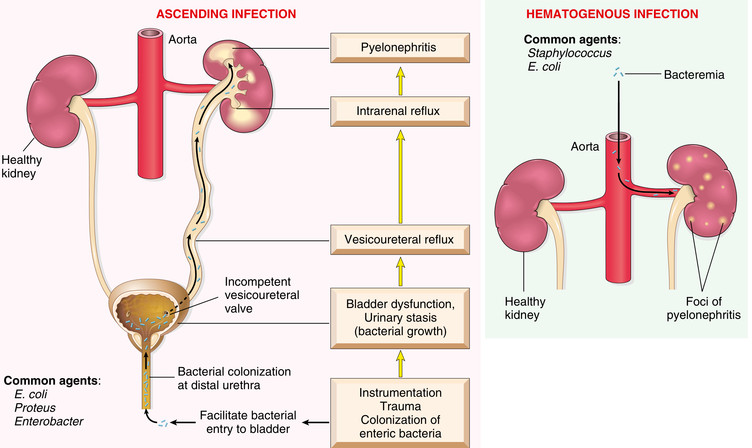
- Enteric bacteria colonize the distal urethra/introitus → ascend to bladder → travel up ureters → infect renal pelvis and parenchyma
- Women are particularly susceptible because of the short urethra and its proximity to the rectum
- Enhanced by: urethral instrumentation, catheterization, cystoscopy
- Bacteremia (e.g., from bacterial endocarditis or sepsis) seeds the kidney
- More common in: diabetics, immunocompromised patients, people who inject drugs
| Factor | Mechanism |
|---|---|
| Urinary outflow obstruction (BPH, calculi, uterine prolapse) | Urinary stasis allows bacterial growth |
| Vesicoureteral reflux (VUR) | Present in 20-40% of children with UTI; incompetent ureterovesical valve allows infected urine to reflux into renal pelvis |
| Diabetes mellitus | Increased infection susceptibility + neurogenic bladder → stasis |
| Pregnancy | Uterine pressure causes stasis; progesterone causes ureteral smooth muscle relaxation and dilatation |
| Immunosuppression | Reduced host defense |
| Pre-existing renal scarring | Intraparenchymal obstruction |
- Robbins Basic Pathology, p. 514-515 | Bailey and Love's Surgery, p. 3635
Causative Organisms
| Organism | Notes |
|---|---|
| E. coli | By far the most common (75%+ of uncomplicated cases) |
| Klebsiella pneumoniae | Second most common |
| Proteus mirabilis | Urease producer; associated with struvite stones |
| Enterobacter spp. | |
| Pseudomonas aeruginosa | More common in complicated/nosocomial cases |
| Staphylococcus aureus | Hematogenous route; in people who inject drugs, endocarditis |
| Enterococcus faecalis | Particularly in complicated/catheter-associated UTI |
- Robbins Basic Pathology, p. 514 | Tintinalli's Emergency Medicine, p. 3145
Pathology / Morphology
- One or both kidneys may be affected (usually unilateral)
- Kidney may be enlarged
- Discrete yellowish-white raised abscesses visible on the cortical surface
- If obstruction is prominent → pus fills the renal pelvis, calyces, and ureter = pyonephrosis
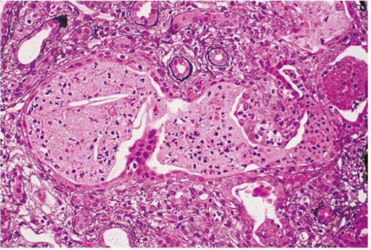
- Neutrophil-rich inflammation initially limited to tubules, later spreading to the interstitium
- Neutrophils extend from tubules into collecting ducts → WBC casts in urine
- Glomeruli are typically spared
- Tubular damage and interstitial edema
-
Papillary necrosis - ischemic + suppurative necrosis of the tips of renal papillae; seen with diabetes, urinary obstruction, or sickle cell anemia
-
Pyonephrosis - pus-filled, obstructed collecting system
-
Renal/perirenal abscess - may result from coalescence of microabscesses
-
Robbins Basic Pathology, p. 515 | Comprehensive Clinical Nephrology 7e, p. 762
Clinical Features
- Fever ≥38°C with chills/rigors
- Flank pain / costovertebral angle (CVA) tenderness
- Lower urinary tract symptoms (dysuria, frequency, urgency) - variably present
-
Onset is usually sudden
-
Symptoms range from mild illness to septic shock with renal failure (~10-19% of sepsis cases originate from GU tract)
-
Missed pyelonephritis can lead to untreated sepsis - a more dangerous missed diagnosis than cystitis
-
Presentation may be subtle in spinal cord injury patients, immunocompromised, and the elderly
-
Absence of pyuria in a patient with pyelonephritis suggests obstructed urinary tract
-
Tintinalli's Emergency Medicine, p. 3159 | Bailey & Love's Surgery, p. 3636 | Comprehensive Clinical Nephrology 7e, p. 762
Investigations
Urinalysis
- Pyuria - almost always present (WBCs in urine)
- WBC casts - highly specific for pyelonephritis (infrequent but diagnostic)
- Bacteriuria (positive Gram stain of urine sediment can differentiate gram-positive vs. gram-negative - guides empiric therapy)
- Urine culture: ≥10⁴ CFU/mL (some labs require ≥10⁵)
Urine Culture
- Should be performed in all patients with acute pyelonephritis
- Positive in nearly all true pyelonephritis cases
Blood Cultures
- Indicated in severe or hospitalized cases; ~15-20% positive
Imaging
- Not required in straightforward uncomplicated cases
- Indications for imaging: failure to respond to antibiotics (48-72 hrs), atypical presentation, suspicion of obstruction/abscess
- Renal ultrasound - first-line modality when imaging needed; detects hydronephrosis, pyonephrosis, abscess
- Contrast-enhanced CT (gold standard) - shows decreased patchy, wedge-shaped, or linear opacification of affected parenchyma
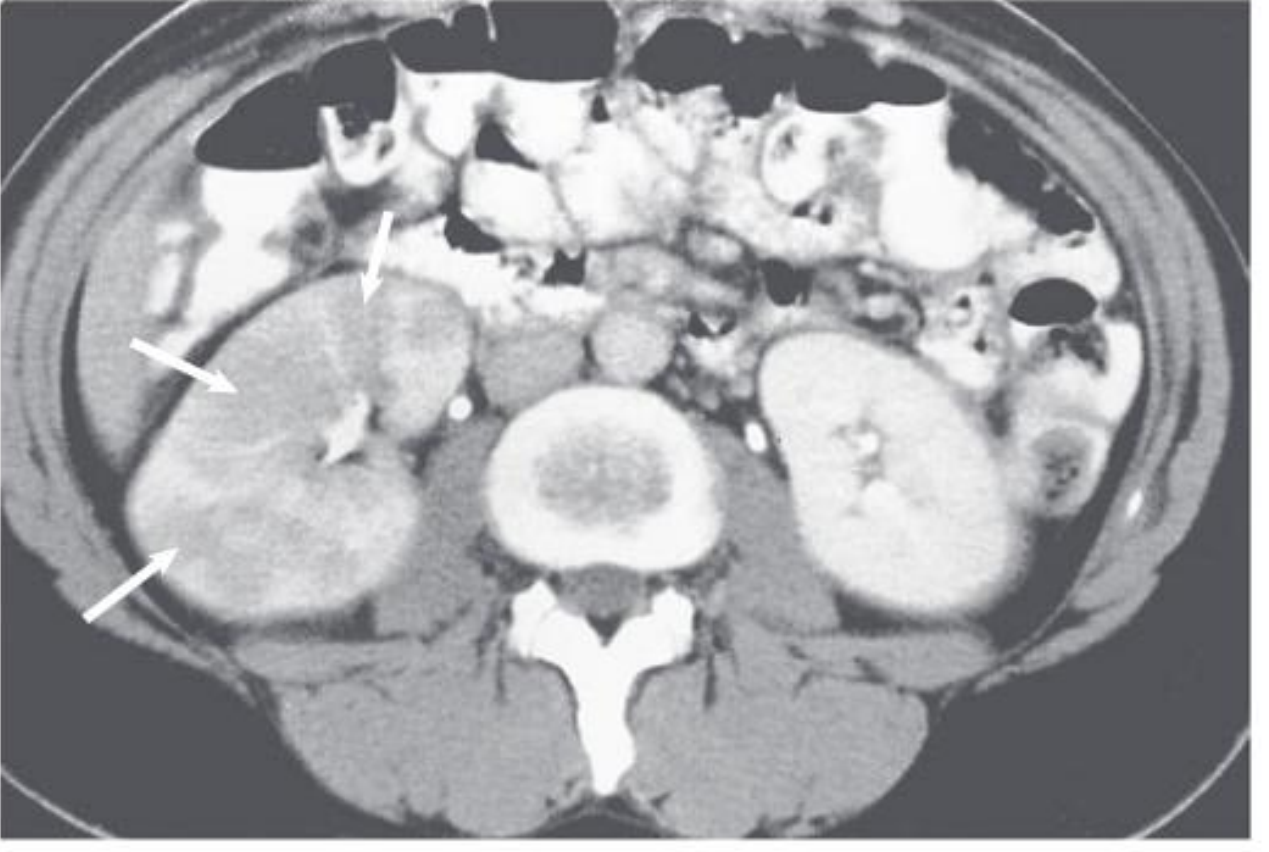
- Bailey & Love's Surgery, p. 3639-3640 | Comprehensive Clinical Nephrology 7e, p. 762
Management
Decision: Inpatient vs. Outpatient
- Inability to maintain oral hydration or take medications
- Uncertain social situation / compliance concerns
- Diagnostic uncertainty
- Severe illness: high fever, severe pain, marked debility
- Signs of sepsis or septic shock
- Immunocompromised or pregnant
Only ~7% of adult females with acute pyelonephritis require hospitalization in population-based studies. - Comprehensive Clinical Nephrology 7e
Antibiotic Therapy
| Drug | Dose / Duration | Notes |
|---|---|---|
| Ciprofloxacin (first-line if susceptibility confirmed) | 500 mg BID x 5-7 days | Fluoroquinolones achieve excellent tissue levels |
| Levofloxacin | 750 mg OD x 5-7 days | |
| Trimethoprim-sulfamethoxazole | DS BID x 14 days | Use only if local E. coli resistance <20% |
| Cefixime (oral) | 400 mg OD x 7-14 days | After initial parenteral dose |
| Drug | Notes |
|---|---|
| Aminoglycosides (gentamicin) | Initial IV; associated with better outcomes; preferred in areas with high resistance |
| Ceftriaxone (IV) | Broad-spectrum; step down to oral cephalosporin |
| Piperacillin-tazobactam | 4.5 g q8h x 7 days for complicated/resistant cases |
| Meropenem | 1 g q8h x 4-5 days for ESBL/resistant organisms |
| Plazomicin | 15 mg/kg once daily for complicated/resistant cases |
- IV-to-oral transition is appropriate at 24-48 hours if clinical improvement
- Nitrofurantoin and fosfomycin are NOT appropriate for pyelonephritis (insufficient tissue levels in renal parenchyma)
- Total treatment duration: 7-14 days (5-7 days adequate for fluoroquinolones)
In Pregnancy
-
Hospital admission is standard practice for pregnant women with pyelonephritis
-
First-line: penicillins or cephalosporins (safe for fetus)
-
Avoid: fluoroquinolones, aminoglycosides (if possible), trimethoprim (first trimester)
-
Prophylactic antibiotics until delivery to prevent recurrence and preterm labor risk
-
Progesterone-induced ureteral dilatation + urine stasis make pregnant women especially susceptible; pyelonephritis is one of the most common causes of septic shock in pregnancy
-
Goldman-Cecil Medicine, p. 2979 | Comprehensive Clinical Nephrology 7e, p. 762-763 | Bailey & Love's Surgery, p. 3647-3648
Upper UTI Spectrum / Complications
- Acute bacterial nephritis (lobar nephronia) - localized, non-liquefied inflammation without frank abscess
- Renal abscess - localized collection of pus; diagnosed by CT; may require percutaneous drainage
- Emphysematous pyelonephritis - gas-forming infection (diabetics, immunocompromised); life-threatening; requires emergent nephrectomy or drainage
- Pyonephrosis - pus-filled obstructed collecting system; requires urgent nephrostomy drainage
- Papillary necrosis - seen with diabetes, obstruction, sickle cell anemia; associated with worse prognosis
- Tintinalli's Emergency Medicine, p. 3095
Differential Diagnosis
- Acute cystitis (no systemic features, no CVA tenderness)
- Renal/ureteric calculus (colicky pain, hematuria)
- Appendicitis (right-sided pain)
- Ovarian pathology (females)
- Lumbar musculoskeletal pain
- Pneumonia (basal, right-sided)
- Psoas abscess
Prognosis
-
Uncomplicated acute pyelonephritis follows a benign course; symptoms resolve within a few days of appropriate antibiotic therapy. Bacteria may persist in urine or recurrence with new serotypes can occur.
-
When predisposing factors are present, disease may become recurrent or chronic, leading to chronic pyelonephritis, irregular cortical scarring (especially at poles), and eventually chronic kidney disease.
-
The development of papillary necrosis is associated with a worse prognosis.
-
Bilateral involvement may cause renal failure.
-
Robbins Basic Pathology, p. 515 | Comprehensive Clinical Nephrology 7e
/generatehandwrittenimage downsyndrome
Handwritten medical student notes on lined paper about Down Syndrome (Trisomy 21). The notes are written in blue and black ink in a neat handwriting style with headings underlined. Include the following sections clearly written out: DOWN SYNDROME (TRISOMY 21) Incidence: 1 in 700 live births — most common chromosomal disorder GENETICS: • Trisomy 21 (95%) — meiotic nondisjunction, karyotype 47,XX,+21 • Translocation (4%) — long arm of chr 21 to chr 14 or 22 (often familial, Robertsonian) • Mosaic (1%) — mitotic nondisjunction; milder phenotype • Maternal age effect: risk ↑ with age (1:1550 at <20 yrs → 1:25 at >45 yrs) • Extra chromosome is of maternal origin in 95% of cases CLINICAL FEATURES: • Flat facial profile, oblique palpebral fissures, epicanthic folds • Simian (single palmar) crease • Hypotonia, short neck, protruding tongue, low-set ears • Intellectual disability (IQ 25–50 in 80%), delayed milestones • Short stature COMPLICATIONS: ❤ Congenital heart disease (40%) — endocardial cushion defects, ASD, VSD 🔬 Leukemia — 10–20× increased risk (ALL + AML) 🧠 Alzheimer disease — ALL patients >40 yrs develop Alzheimer changes; 30% develop dementia by age 60 🦠 Abnormal immune responses — recurrent lung infections, thyroid autoimmunity ⚡ Epilepsy, hypothyroidism (↑ risk) 🦴 Atlantoaxial instability (10–20%) GI: Hirschsprung disease, duodenal atresia Eyes: cataracts, refractive errors DIAGNOSIS: • Prenatal: maternal serum β-hCG ↑, PAPP-A ↓, nuchal fold thickening • Cell-free fetal DNA (liquid biopsy) — sensitive & specific • Confirmed by amniocentesis / karyotype TREATMENT: • No specific therapy • Manage complications: cardiac surgery, leukemia treatment, thyroid screening, spine precautions • Supportive education and environment
400, message: Got more than 4194304 bytes when reading: b'data: {"candidates": [{"content": {"role": "model","parts": [{"inlineData": {"mimeType": "image/png"...'.
We encountered an error while processing your request. Please try again or reach out to us for help.
/generatehandwrittenimage /generatehandwrittenimage downsyndrome
Handwritten medical student notes on lined white paper, written in blue and black ballpoint pen, neat cursive and print mix handwriting style, with headings underlined in red pen. The notes read: DOWN SYNDROME (TRISOMY 21) Incidence: 1 in 700 live births — most common chromosomal disorder GENETICS: - Trisomy 21 (95%) — meiotic nondisjunction → karyotype 47,XX,+21 - Translocation (4%) — chr 21 long arm fused to chr 14 or 22; familial (Robertsonian) - Mosaic (1%) — mitotic nondisjunction; milder phenotype - Maternal age effect: risk rises sharply with age (1:1550 at age <20 yrs vs 1:25 at age >45 yrs) - Extra chr 21 is maternal in origin in 95% of cases CLINICAL FEATURES: - Flat facial profile, oblique palpebral fissures, epicanthic folds - Single palmar (simian) crease - Hypotonia, short neck, protruding tongue - Intellectual disability (IQ 25–50 in ~80%) - Short stature, delayed milestones ASSOCIATIONS / COMPLICATIONS: - Congenital heart disease (40%) — ASD, VSD, endocardial cushion defects - Leukemia risk x10–20 (ALL and AML) - Alzheimer disease — ALL pts >40 yrs develop plaques/tangles - Hypothyroidism, epilepsy (increased risk) - Atlantoaxial instability (10–20%) - GI: duodenal atresia, Hirschsprung disease - Eyes: cataracts, refractive errors - Recurrent infections (T-cell dysfunction) PRENATAL DIAGNOSIS: - Serum: beta-hCG ↑, PAPP-A ↓ - Ultrasound: nuchal fold thickening - Cell-free fetal DNA (liquid biopsy) — highly sensitive - Confirmed by amniocentesis + karyotype MANAGEMENT: - No disease-specific therapy - Treat complications: cardiac surgery, leukemia Rx, thyroid screen - Cervical spine precautions before anaesthesia - Supportive education + multidisciplinary care
400, message: Got more than 4194304 bytes when reading: b'data: {"candidates": [{"content": {"role": "model","parts": [{"inlineData": {"mimeType": "image/png"...'.
We encountered an error while processing your request. Please try again or reach out to us for help.