
Reddish blisters on legs with pedal edema cause
Reading File
Now let me search the medical library for this clinical presentation - reddish blisters on legs with pedal edema.
Finding Sources
Finding Sources
Finding Sources
I found a relevant result in Harrison's. Let me read that section and also search for more specific conditions.
Reading File
Finding Sources
Finding Sources
Reading File
Finding Sources
Finding Sources
Finding Sources
Finding Sources
Searching the Web
reddish blisters on legs with pedal edema causes differential diagnosis
Now let me fetch the image from Harrison's to display it, and look for the Dermatology textbook content on this topic.
Loading Image
Finding Sources
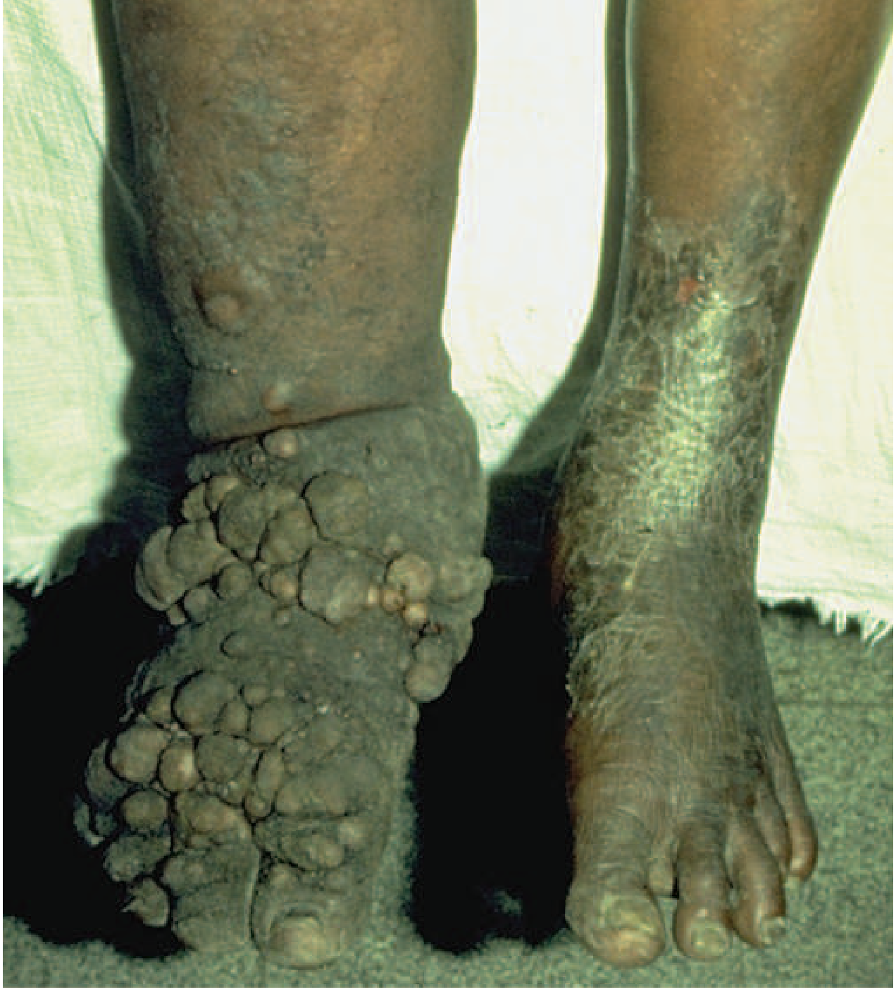
I now have comprehensive information to provide a detailed answer. The image from Harrison's shows filarial elephantiasis (not quite what was asked), and the Fitzpatrick's result confirms lipodermatosclerosis as a key diagnosis. Let me compose the full answer.
Reddish Blisters on Legs + Pedal Edema: Causes & Differential Diagnosis
This combination - erythematous (reddish) blisters/bullae on the legs together with pedal edema - is an important clinical presentation with several possible causes. Here is a structured differential:
Most Common Causes
1. Chronic Venous Insufficiency (CVI) / Varicose Veins
The most frequent cause in outpatient settings. Increased venous pressure leads to:
- Pitting pedal/ankle edema
- Varicose veins
- Red-brown skin discoloration (hemosiderin staining)
- Stasis dermatitis - erythematous, weeping, eczematous patches
- Tension blisters (bullae) form when edema is severe and skin is overstretched
- Eventually leads to lipodermatosclerosis (fibrosis and hyperpigmentation, "champagne bottle" shape)
2. Cellulitis / Erysipelas
- Unilateral red, warm, tender, swollen leg
- Systemic features: fever, chills
- Blisters/bullae may form in severe cases
- Often mistaken for venous disease - and vice versa
- Key: Acute onset, unilateral, systemic illness (Nottinghamshire APC Guidance on inflamed lower limbs)
3. Lymphedema
- Non-pitting edema of lower legs/feet
- Skin becomes thickened, rubbery, hyperkeratotic
- Blisters may form from lymph fluid leaking through skin (lymphorrhea)
- Filariasis (Wuchereria bancrofti, Brugia malayi) - a parasitic cause of lymphedema: brawny edema, vesicles, ulcers, hyperpigmentation, and papular/blister lesions on legs. The image below shows advanced filarial elephantiasis:
4. Bullous Pemphigoid
- Autoimmune blistering disorder
- Tense, fluid-filled bullae on erythematous base on legs/trunk
- More common in elderly
- Can occur on edematous skin
5. Contact Dermatitis (Allergic or Irritant)
- Erythema, vesicles, blisters on legs
- From topical treatments, compression stockings, rubber in footwear
- Often associated with underlying edematous skin
Systemic Causes of the Edema Component (with secondary skin changes)
| Cause | Edema Character | Key Features |
|---|---|---|
| Congestive Heart Failure | Bilateral, pitting, worsens at day-end | Dyspnea, JVD, orthopnea |
| Nephrotic Syndrome | Periorbital + pedal, pitting | Proteinuria, hypoalbuminemia |
| Liver Cirrhosis | Bilateral + ascites | Jaundice, splenomegaly |
| Hypothyroidism (myxedema) | Non-pitting, doughy | Cold intolerance, fatigue |
| Deep Vein Thrombosis | Unilateral, acute | Pain, warmth, Homan's sign |
In systemic causes, the skin becomes chronically edematous and fragile, and blisters ("tension blisters") can form from skin stretching.
6. Necrotizing Fasciitis (Emergency - Do Not Miss)
- Rapidly spreading, dark mottled skin + edema + multiple bullae
- Limb feels "heavy," crepitus possible
- Systemic sepsis features
- Surgical emergency - requires urgent debridement (Textbook of Family Medicine, 9e)
Key Distinguishing Features
| Feature | Venous/Stasis | Cellulitis | Lymphedema | Bullous Pemphigoid |
|---|---|---|---|---|
| Bilateral | Usually | Rarely | Can be | Can be |
| Fever | No | Yes | No | No |
| Blister type | Tension/weeping | Flaccid/hemorrhagic | Lymphorrhea | Tense, turgid |
| Skin color | Red-brown, pigmented | Bright red | Skin thickened | Erythematous |
| Pitting | Yes | Yes (early) | No | Variable |
Approach
- History: Duration, bilateral vs unilateral, trauma, systemic symptoms, medications (CCBs, steroids, NSAIDs are common culprits for edema)
- Labs: CBC, CMP, albumin, TSH, urinalysis, LFTs
- Doppler ultrasound: Rule out DVT, assess venous insufficiency
- Wound swab if infective etiology suspected
- Skin biopsy if bullous pemphigoid or vasculitis suspected
Red flags requiring urgent assessment: Rapid spread of redness, fever, pain disproportionate to appearance, crepitus, or skin necrosis - these suggest necrotizing infection.
Sources: Harrison's Principles of Internal Medicine 22e; Fitzpatrick's Dermatology 5e; Textbook of Family Medicine 9e; Wound Care Today - Red Legs
This is a shared conversation. Sign in to Orris to start your own chat.