
Is spo2 n po2 same
No, SpO2 and PO2 are not the same thing. Here's a quick breakdown:
- SpO2 (peripheral oxygen saturation) is the percentage of hemoglobin molecules in the blood that are carrying oxygen. Measured non-invasively by pulse oximetry.
- PO2 (partial pressure of oxygen) is the pressure exerted by dissolved oxygen molecules in the blood, measured in mmHg. Requires an arterial blood gas (ABG) test.
They are related but distinct measurements, and the relationship between them is non-linear (described by the oxyhemoglobin dissociation curve).
Searching sources...
Reading File
Finding Sources
Finding Sources
Reading File
Finding Sources
Searching Images
oxyhemoglobin dissociation curve SpO2 PO2
Loading Image
Reading File
No, SpO2 and PO2 are not the same. They measure two completely different things, though they are related to each other through the oxyhemoglobin dissociation curve.
SpO2 vs PO2 - Key Differences
| Feature | SpO2 | PO2 |
|---|---|---|
| Full name | Peripheral oxygen saturation | Partial pressure of oxygen |
| What it measures | % of hemoglobin molecules carrying O2 | Pressure of dissolved O2 in blood (mmHg) |
| Unit | Percentage (%) | mmHg |
| How measured | Pulse oximeter (non-invasive) | Arterial blood gas - ABG (invasive) |
| Normal arterial value | 95-100% | ~95 mmHg (arterial) |
| Normal venous value | ~75% | ~40 mmHg (venous) |
How They Are Related
They are linked by the oxyhemoglobin dissociation curve - a sigmoid (S-shaped) curve. The curve plots PO2 (x-axis) against saturation % (y-axis):
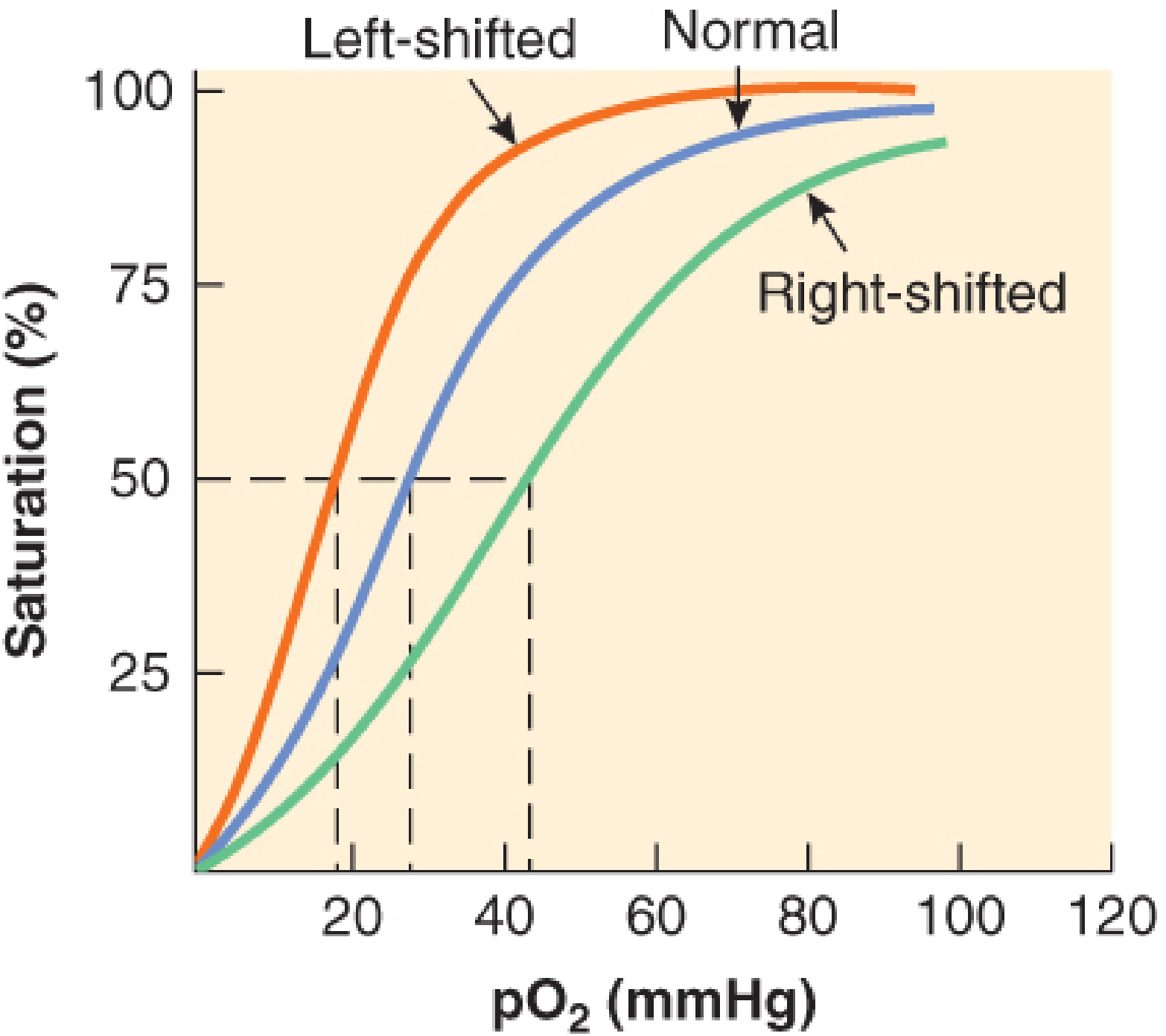
Key points from the curve (sourced from Barash's Clinical Anesthesia and Guyton & Hall Medical Physiology):
- Steep part of the curve (PO2 < 60 mmHg): A small drop in PO2 causes a large fall in SpO2. Here, SpO2 is a good indicator of how hypoxic the patient is.
- Flat/plateau part of the curve (PO2 > 75 mmHg): SpO2 stays high (~97-100%) even as PO2 continues to rise. So a patient with PO2 of 90 mmHg and PO2 of 150 mmHg may both show SpO2 ~99%. SpO2 cannot detect hyperoxia.
- Normal values: Arterial blood has PO2 ~95 mmHg → SpO2 ~97%. Venous blood has PO2 ~40 mmHg → SpO2 ~75%.
Why the Distinction Matters Clinically
-
SpO2 misses hyperoxia: A patient on high-flow O2 may have a dangerously elevated PO2 (e.g., 300-400 mmHg) but SpO2 still reads 100%. You need an ABG to detect this.
-
Curve shifts change the relationship:
- Right shift (fever, acidosis, high CO2, 2,3-DPG): same PO2 gives lower saturation - O2 offloads more easily to tissues.
- Left shift (alkalosis, hypothermia, CO poisoning, low 2,3-DPG): same PO2 gives higher saturation - O2 is held more tightly by hemoglobin.
-
CO poisoning: Pulse oximetry reads SpO2 as falsely normal (carboxyhemoglobin absorbs light similarly to oxyhemoglobin), but the actual PO2 and true O2 content are dangerously low. ABG is needed.
-
P50 is the PO2 at which hemoglobin is exactly 50% saturated. Normal P50 is ~26-27 mmHg. It shifts with the curve and reflects O2 affinity of hemoglobin.
Bottom line: SpO2 tells you how loaded the hemoglobin is with oxygen (in %). PO2 tells you the driving pressure of oxygen in blood (in mmHg). They are related by the oxyhemoglobin dissociation curve, but they are not interchangeable, and there are important clinical situations where one can be normal while the other is not.
- Barash, Cullen & Stoelting's Clinical Anesthesia, 9e, Monitoring of Arterial Oxygenation by Pulse Oximetry
- Guyton and Hall Textbook of Medical Physiology, Reversible Combination of O2 With Hemoglobin
Is po2 same with pao2
PO2 is a general term for the partial pressure of oxygen in any body fluid or gas. PaO2 is a specific form - the partial pressure of oxygen in arterial blood. So PaO2 is a type of PO2, but PO2 is not always PaO2.
Searching sources...
Finding Sources
Finding Sources
Finding Sources
No, PO2 and PaO2 are not exactly the same - PaO2 is a specific type of PO2. Here is the full breakdown:
PO2 vs PaO2 - The Difference
PO2 is a generic term meaning "partial pressure of oxygen." It can refer to oxygen tension in any compartment. The subscript letter tells you which compartment:
| Notation | Full Name | Location | Normal Value |
|---|---|---|---|
| PaO2 | Partial pressure of O2 in arterial blood | Systemic arteries | ~95 mmHg |
| PvO2 | Partial pressure of O2 in venous blood | Mixed venous blood | ~40 mmHg |
| PAO2 | Partial pressure of O2 in the alveoli | Alveolar gas | ~100 mmHg |
| PO2 (generic) | Any partial pressure of O2 | Depends on context | Varies |
So when someone says "PO2" without a subscript, they usually mean arterial PO2 = PaO2 by convention - but this is loose usage. Technically they are not identical.
The Important A-a Gradient (PAO2 vs PaO2)
One clinically very important distinction is between PAO2 (alveolar) and PaO2 (arterial). These are NOT the same:
-
PAO2 is calculated using the alveolar gas equation:PAO2 = (FiO2 × [PB - PH2O]) - (PaCO2 / R) On room air: PAO2 ≈ 100 mmHg
-
PaO2 is directly measured from an arterial blood gas (ABG): ~95 mmHg normally.
The difference between them is called the A-a gradient (A-a O2 difference):
A-a gradient = PAO2 - PaO2 ≈ 5-15 mmHg (normal, breathing room air)
A widened A-a gradient (>20-25 mmHg) indicates a problem with gas exchange - such as:
- V/Q mismatch (e.g., pulmonary embolism, pneumonia)
- Diffusion impairment (e.g., pulmonary fibrosis)
- Intracardiac or intrapulmonary shunt
As Barash's Clinical Anesthesia states: "Systemic arterial partial pressure of oxygen (PaO2) is the best indicator of pulmonary oxygen transfer from alveolar gas to pulmonary capillary blood."
Quick Summary
- PO2 = general term (partial pressure of O2 anywhere)
- PaO2 = PO2 in arterial blood (measured by ABG)
- PAO2 = PO2 in the alveoli (calculated)
- PvO2 = PO2 in venous blood
- In everyday clinical speech, "PO2" often loosely means PaO2 - but when reading ABG results or literature, always check the subscript. The capital "A" (PAO2) vs lowercase "a" (PaO2) distinction is especially important.
This is a shared conversation. Sign in to Orris to start your own chat.