
Incisional hernia management
I'll read the medical library skill first, then search for incisional hernia management content.
Reading File
Now I'll run parallel searches across the library and PubMed simultaneously.
Finding Sources
Searching PubMed
"incisional hernia"[MeSH Terms] AND repair
Excellent. Now reading the key sections from Bailey & Love and Fischer's Mastery in parallel.
Reading File
Reading File
Finding Sources
Now reading the management/repair sections from Bailey & Love (prevention + treatment) and Fischer's operative sections in parallel.
Reading File
Reading File
Reading File
Now reading the mesh repair and laparoscopic sections, plus prevention details from Bailey & Love.
Reading File
Reading File
Finding Sources
Excellent. Now reading the rest of Bailey & Love's repair section (open + laparoscopic details) and getting the ICAP mesh placement content from Fischer's.
Reading File
Reading File
I now have comprehensive material. Let me fetch the key clinical images and also quickly search for the classification system (EHS/Chevrel).
Loading Image
Finding Sources
Finding Sources
I now have comprehensive data to write a thorough response. Let me also fetch the mesh planes diagram.
Loading Image
I have all the material needed. Here is the comprehensive response:
Incisional Hernia - Management
Definition
An incisional hernia arises through a defect in the musculofascial layers of the abdominal wall at the site of a postoperative scar, and can occur anywhere a laparotomy has been made.
- Bailey and Love's Short Practice of Surgery, 28th Ed.
Incidence and Aetiology
- Reported in 10-50% of laparotomy incisions and 1-5% of laparoscopic port-site incisions
- Predisposing factors fall into three groups:
| Category | Factors |
|---|---|
| Patient | Genetic collagen disorders, obesity, malnutrition, immunosuppression/steroids, chronic cough, cancer |
| Wound | Poor tissue quality, wound tension, wound infection |
| Surgical | Inappropriate suture material, poor closure technique |
Pathologically, the hernia begins as disruption of musculofascial layers in the early postoperative period. If the skin heals, the defect goes unnoticed and a visible swelling may not appear for weeks, months or years.
Clinical Features
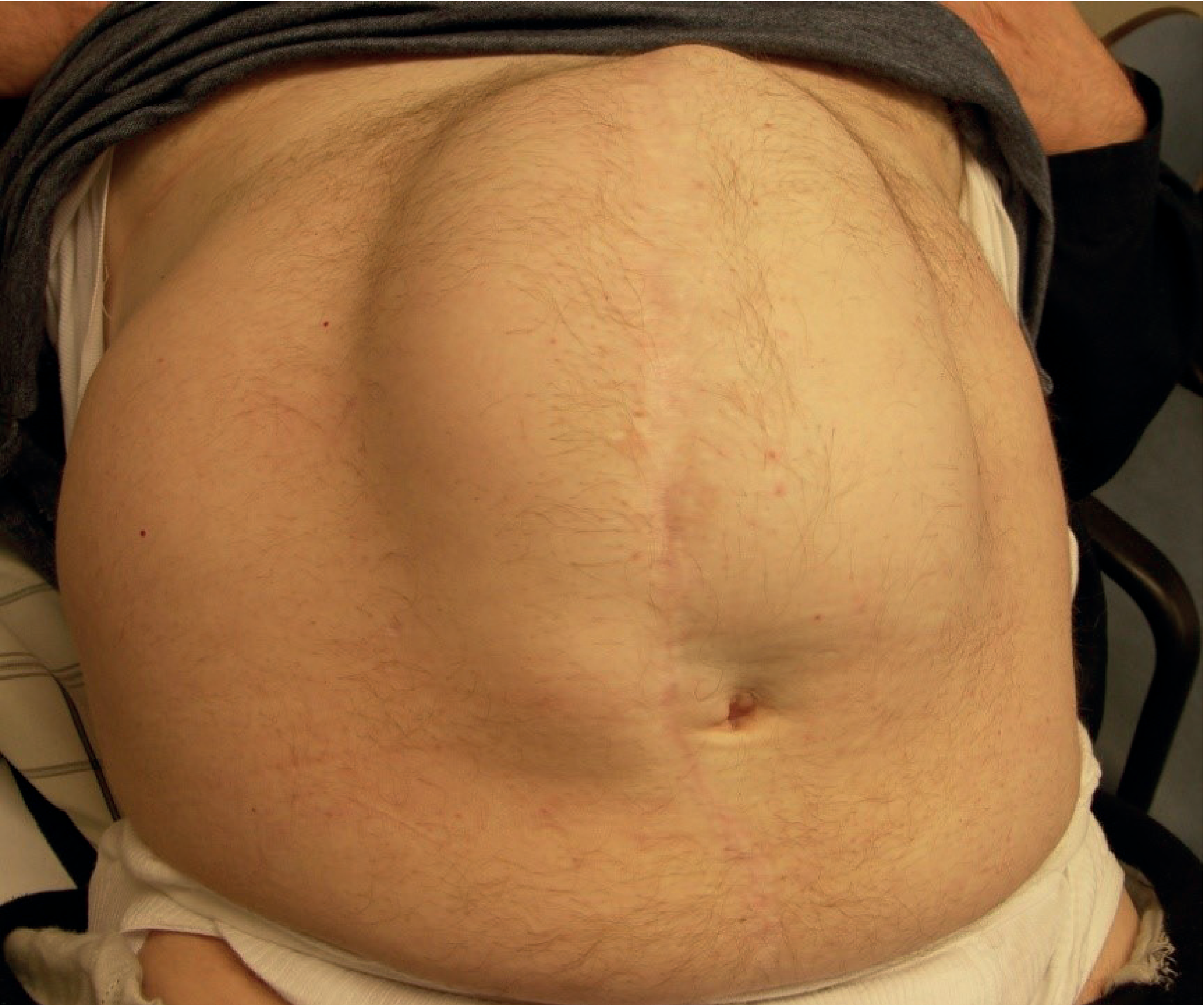
- Localised swelling at the scar, or diffuse bulging of the entire incision length
- Multiple discrete defects along the incision are common - clinically apparent single hernias frequently have unsuspected additional defects at operation
- Tend to increase steadily in size; overlying skin may become thin, atrophic, and may ulcerate
- Obstruction is common due to internal adhesions
- Strangulation is less frequent because most incisional hernias are shallow and wide-necked (risk is highest when the fibrous defect is small and the sac is large)
Classification (EHS)
The European Hernia Society classifies incisional hernias by:
- Location: midline (subxiphoid, epigastric, umbilical, infraumbilical, suprapubic) vs. lateral (subcostal, flank, iliac, lumbar)
- Width of defect: W1 (<4 cm), W2 (4-10 cm), W3 (>10 cm)
- Recurrent vs. primary
Prevention
Prevention is the priority and targets modifiable risk factors:
Preoperative optimisation:
- Smoking cessation
- Weight loss (obesity)
- Correction of malnutrition
- Optimise immunosuppression where possible
Surgical technique - the STITCH trial principles:
- Small bites: 5 mm apart and 5-8 mm back from the wound edge (compared to the older 1 cm/1 cm rule)
- Incorporate fascia only in suture bites - no fat or muscle, which becomes ischaemic and causes suture slack, allowing fascial edges to separate
- 2/0 slowly resorbable monofilament suture (e.g., PDS or Monocryl) is preferred over heavier or non-absorbable materials
- Suture-to-wound-length ratio must be at least 4:1; a ratio less than this indicates bites are too far apart or too tight
- Drains must exit through separate stab incisions, never through the laparotomy wound itself
- Continuous or interrupted technique are equivalent; if continuous, avoid tight pulls that cut out
- In high-risk wound infections (abdominal sepsis): consider partial skin closure or negative pressure wound therapy
Prophylactic mesh:
- Evidence supports prophylactic mesh in high-risk patients (e.g., bariatric surgery, stoma formation)
- A large-pore polypropylene mesh in the retromuscular plane at the time of stoma formation reduces parastomal hernia rates significantly (baseline rate >50%)
Treatment
Conservative Management
Asymptomatic incisional hernias in patients with significant comorbidities may not require surgery. An abdominal binder/belt provides symptomatic relief and may slow enlargement. The decision to operate must involve shared decision-making weighing surgical risk vs. benefit.
Pre-operative Optimisation ("Prehabilitation")
Before elective repair, patients should undergo:
- Weight loss if obese: 7% of total bodyweight achieves significant metabolic improvement; every 5 kg of weight lost creates approximately 1 extra litre of intra-abdominal space in men (0.5 L in women)
- Smoking cessation
- Fitness and core strength training
- Multidisciplinary team assessment (becoming standard of care)
Surgical Repair - Principles
Three core principles apply regardless of technique:
- The repair must cover the entire length of the previous incision (multiple unsuspected defects are common)
- Musculofascial approximation should be performed with minimal tension
- Prosthetic mesh must be used to reduce recurrence - simple suture repair (Mayo overlapping technique, da Silva layered closure) carries an unacceptable recurrence rate
Mesh is contraindicated in a grossly contaminated field (e.g., perforated strangulated bowel), but may still be used in a clean-contaminated field (e.g., elective bowel resection with prophylactic antibiotics).
Mesh must augment primary fascial closure - it should not bridge a gap between fascial edges, as unsupported central mesh will inevitably bulge and fail ("pseudo-recurrence").
Surgical Techniques
1. Open Primary Repair (Small Defects <2 cm)
- Primary suture closure is acceptable for defects <2 cm
- Apply small-bite technique: 5 mm bites, 5 mm spacing, 2-0 slowly resorbable monofilament on a 30-40 mm needle
- The suture length used must be at least 4x the incision length
- Mesh use based on shared decision-making considering infection risk and recurrence risk
For wide midline defects where running suture causes undue tension:
- Interrupted figure-of-eight sutures with slowly absorbable monofilament are placed loosely along the wound edges, then cinched together simultaneously - distributes tension more evenly
2. Open Mesh Repair
Procedure:
- Incision extended past the hernia defect
- Complete adhesiolysis from the anterior abdominal wall is performed
- Bowel inspected for injury
- All prior intraperitoneal mesh should be removed
- Mesh repair then proceeds after fascial preparation
Mesh Positioning (ICAP 2019 Classification):
| Position | Anatomical Plane | Notes |
|---|---|---|
| Onlay | Subcutaneous, on top of anterior rectus sheath/external oblique aponeurosis | Simplest; higher SSI risk |
| Inlay | Between muscles as interposition (bridges gap) | Avoid electively - poor outcomes; acceptable only in emergencies |
| Retrorectus (sublay) | Between rectus abdominis and posterior rectus sheath | Gold standard for midline hernias |
| Preperitoneal (sublay) | Between transversalis fascia and peritoneum | |
| IPOM (Intraperitoneal Onlay Mesh) | Inside abdomen on peritoneum | Requires anti-adhesion (barrier-coated) mesh; used in laparoscopic repair |
3. Component Separation Techniques
Used when the defect is large and primary fascial closure would create undue tension. Goal: allow fascial closure with wide mesh overlap.
Anterior Component Separation (ACS) - Ramirez technique (1990):
- Skin/subcutaneous flaps raised over anterior rectus sheath lateral to the rectus
- Posterior rectus sheath incised just lateral to linea alba; rectus separated from posterior sheath
- External oblique aponeurosis incised just lateral to linea semilunaris, from costal margin to iliac crest
- External oblique separated from underlying internal oblique
- Each step provides additional medial advancement; midline fascia then closed
Transversus Abdominis Release (TAR) - Novitsky (2012):
- Extension of the retrorectus repair
- After creating the retrorectus space, the posterior lamella of the internal oblique is incised just medial to the neurovascular bundles and linea semilunaris
- Transversus abdominis (TA) muscle is divided to reveal peritoneum and transversalis fascia
- Lateral dissection into the retroperitoneum to the lateral border of the psoas
- Provides maximal fascial advancement with wide posterior mesh space
Caution: the TAR incision must be medial to neurovascular bundles to prevent rectus denervation and avoid inadvertently cutting anterior lamella of internal oblique/external oblique (which creates a new lateral wall hernia).
4. Laparoscopic Repair (IPOM)
- Adhesiolysis performed laparoscopically; hernia contents reduced
- Fibrous defect margins exposed (falciform ligament and median umbilical fold are often divided)
- Options: close fascial defect(s) with sutures before mesh reinforcement (preferred for larger defects), or bridge the defect with mesh alone (only safe for small hernias)
- Large defects must have the fascia closed first - bridging large defects leads to mesh bulging ("pseudo-recurrence")
- Mesh placed as IPOM: directly on peritoneum, fixed with tissue glue, sutures, or tacks
- Anti-adhesion (tissue-separating/barrier-coated) mesh is mandatory for IPOM - these are expensive
- Risk: in dense adhesions, bowel injury may occur and go unrecognised, leading to postoperative peritonitis
5. Botulinum Toxin A (Chemical Component Separation)
Preoperative BTX-A injection into the lateral abdominal wall muscles causes temporary paralysis and lengthening of the muscles, facilitating medialization and primary fascial closure in complex hernias. Systematic review evidence exists (PMID 37801164), though some collaborative registry data has not shown improved fascial closure rates vs. standard care.
Special Situations
Parastomal Hernia
- Occurs in >50% of patients with a stoma
- Creates stoma management difficulties (appliance leakage, obstruction, prolapse)
- Options:
- Stomal reversal (ideal if feasible)
- Resiting the stoma (not recommended - parastomal hernia recurs at same rate at new site)
- Mesh repair: retromuscular placement preferred; intraperitoneal (Sugarbaker technique) also used
- Laparoscopic: modified Sugarbaker or keyhole technique (large mesh with central hole)
- Prophylactic mesh at time of stoma formation (large-pore polypropylene in retromuscular plane, bowel through central hole) significantly reduces parastomal hernia rates
EHS Classification of Parastomal Hernia: Small (<5 cm) vs. Large (>5 cm), with or without concomitant incisional hernia.
Loss of Domain
- Very large hernias where abdominal contents have "lost domain" and cannot be safely reduced
- Requires progressive pneumoperitoneum and/or BTX-A preoperatively to expand abdominal cavity
- Bridging mesh (biologic or biosynthetic) may be used as a damage control strategy in the emergent setting to preserve native tissue planes for future reconstruction
Outcomes and Recurrence
- Suture repair alone: recurrence rate 25-63%
- Mesh reinforced repair: significantly lower recurrence rates
- Retrorectus/TAR with mesh overlap: lowest reported recurrence rates for large/complex hernias
- Laparoscopic IPOM: comparable recurrence to open with shorter hospital stay, but higher mesh bulging rates if fascia not closed
Sources
- Bailey and Love's Short Practice of Surgery, 28th Edition (Chapter 64)
- Fischer's Mastery of Surgery, 8th Edition (Chapters 213, 216)
- Current Surgical Therapy, 14th Edition (Chapter 10)
- Recent systematic review on BTX-A in complex incisional hernia repair: Barretto et al., Hernia 2024 (PMID 37801164)
This is a shared conversation. Sign in to Orris to start your own chat.