


26727.jpg
+2 more
Explain each question and explain further
I'll analyze all three images and then explain each question in detail. Let me read the medical library skill first and analyze the images simultaneously.
Reading File
analyse_media
analyse_media
analyse_media
I now have all the questions extracted. Let me provide a thorough explanation of all 20 MCQs and the short/long answer questions.
KMTC/QP-08/EPS - Full Exam Explanation
SECTION 1: MULTIPLE CHOICE QUESTIONS
Q1. Which one of the following is true about natural killer (NK) cells?
✅ Correct Answer: B - They kill the invading microorganism by lysis
Explanation:
Natural Killer (NK) cells are large granular lymphocytes that are part of the innate immune system. They do NOT need prior sensitization to act.
- How they kill: NK cells release perforin (punches holes in the target cell membrane) and granzymes (enzymes that trigger apoptosis/programmed cell death inside the target). This process is called lysis.
- They target virus-infected cells and tumor (cancer) cells - not just bacteria, so option D is wrong.
- They do not engulf pathogens (that is phagocytosis, done by neutrophils and macrophages) - so option A is wrong.
- They definitely DO attack microbes/infected cells - so option C is wrong.
Key point: NK cells kill by lysis (perforin/granzyme mechanism), without needing antibodies or MHC presentation.
Q2. The disease that causes large lips, nose, and tongue is called:
✅ Correct Answer: C - Acromegaly
Explanation:
Acromegaly is a hormonal disorder caused by excess Growth Hormone (GH), usually from a pituitary adenoma (benign tumor of the pituitary gland).
- GH causes soft tissue overgrowth - hence enlarged lips, tongue (macroglossia), nose, hands, and feet.
- Other features: protruding jaw (prognathism), large hands/feet, coarse facial features, joint pain, headaches.
- It differs from gigantism (same cause but happens before growth plates close, so the person grows very tall).
- Parkinson's and Alzheimer's are neurological diseases - they don't cause physical enlargement of features.
- Cancer can cause mass effects locally but not this characteristic pattern.
Q3. The following are side effects of opioids EXCEPT:
✅ Correct Answer: B - Cough
Explanation:
Opioids (morphine, codeine, fentanyl, tramadol) act on opioid receptors (mu, kappa, delta) throughout the body.
Known opioid side effects include:
- Constipation ✔ - opioids slow gut motility (very common, doesn't go away with tolerance)
- Sedation ✔ - CNS depression
- Respiratory depression ✔ - the most dangerous/life-threatening effect; opioids suppress the brainstem respiratory centers
- Miosis (pin-point pupils), nausea, vomiting, urinary retention, euphoria
Cough is NOT a side effect - in fact, opioids suppress cough (codeine and pholcodine are used as antitussives/cough suppressants). So cough itself is not caused by opioids; it is suppressed.
Q4. The following is NOT a route of exposure to organophosphate poisoning:
✅ Correct Answer: D - Injection
Explanation:
Organophosphates are chemicals found in pesticides/insecticides (e.g., malathion, parathion) and nerve agents. They inhibit acetylcholinesterase, causing a buildup of acetylcholine.
Routes of exposure:
- Inhalation ✔ - breathing in sprayed pesticides
- Ingestion ✔ - swallowing (common in intentional poisoning/suicide attempts)
- Skin/mucous membrane contact ✔ - absorbed through the skin (especially in farm workers)
Injection is NOT a naturally occurring route of exposure for organophosphates. These are agricultural/environmental chemicals, not injectable drugs.
SLUDGE/DUMBELS - mnemonic for organophosphate toxicity symptoms:
Salivation, Lacrimation, Urination, Defecation, GI distress, Emesis + Bradycardia, Bronchoconstriction, Miosis.
Q5. The emerging infectious disease is:
✅ Correct Answer: D - COVID-19
Explanation:
An emerging infectious disease is one that has newly appeared in a population, or has existed but is rapidly increasing in incidence or geographic range.
- HIV/AIDS - emerged in the 1980s; now well-established, classified as an established disease
- TB - ancient disease, re-emerging in some contexts but not "newly emerging"
- Cancer - not an infectious disease
- COVID-19 - caused by SARS-CoV-2, first identified in Wuhan, China in December 2019, declared a global pandemic by WHO in March 2020. This is the classic example of an emerging infectious disease.
Q6. The following is NOT a clinical feature of Diabetic Ketoacidosis (DKA):
✅ Correct Answer: D - Hematuria
Explanation:
DKA occurs in Type 1 Diabetes (mainly) when there is insufficient insulin. Fat is broken down, producing ketone bodies, leading to metabolic acidosis.
Classic features of DKA (the "3 Polys" + more):
- Polyuria ✔ - excess urination due to osmotic diuresis from high blood glucose
- Polydipsia ✔ - excessive thirst
- Polyphagia ✔ - excessive hunger (cells starving despite high blood glucose)
- Also: Kussmaul breathing (deep rapid breaths), fruity/acetone breath, nausea, vomiting, abdominal pain, altered consciousness
Hematuria (blood in urine) is NOT a feature of DKA. It suggests urinary tract pathology (infection, stones, bladder cancer) - a completely different process.
Q7. Which one of the following is the link between innate and adaptive immunity?
✅ Correct Answer: C - Natural Killer cells
Explanation:
The immune system has two arms:
- Innate immunity - fast, non-specific (NK cells, neutrophils, macrophages)
- Adaptive immunity - slow, specific (T cells, B cells, antibodies)
NK cells bridge both:
- They act immediately like innate cells (no prior sensitization needed)
- But they can be activated and regulated by adaptive immune cytokines (like IL-2, IL-12, IFN-γ from T-cells)
- NK cells also interact with antibodies via ADCC (Antibody-Dependent Cell-Mediated Cytotoxicity) - using antibodies from the adaptive system to target cells
- They communicate with dendritic cells, which present antigens to T-cells (adaptive system)
Note: Some textbooks cite dendritic cells as the bridge, but among the choices given, NK cells fit best as they functionally operate in both arms.
Q8. The following infections are caused by Staphylococcus aureus EXCEPT:
✅ Correct Answer: B - Sinusitis (from context) / D - Headache
(Note: The image cuts off at B. Sinusitis. The remaining options C and D are visible on the next page as C. Endocarditis and D. Headache.)
Full question options:
- A. Furuncles (boils)
- B. Sinusitis
- C. Endocarditis
- D. Headache
✅ Correct Answer: D - Headache
Explanation:
Staphylococcus aureus is a gram-positive cocci that causes many infections:
- Furuncles (boils) ✔ - classic staph skin infection
- Sinusitis ✔ - S. aureus can cause sinusitis
- Endocarditis ✔ - S. aureus is the most common cause of acute bacterial endocarditis (infects heart valves, especially in IV drug users)
Headache is a symptom, not an infection - it is not "caused by" S. aureus specifically. It can be a symptom of any infection but is not a named disease entity caused by S. aureus.
Q9. Which among the following is NOT a congenital heart disease?
✅ Correct Answer: D - Cerebrovascular disease
Explanation:
Congenital heart diseases (CHD) are structural heart defects present from birth.
- Septal defect ✔ - ASD (Atrial Septal Defect) or VSD (Ventricular Septal Defect) - holes in the heart walls - classic CHD
- Obstruction defect ✔ - e.g., pulmonary stenosis, aortic stenosis, coarctation of aorta - CHD
- Cyanotic heart disease ✔ - e.g., Tetralogy of Fallot, Transposition of Great Arteries - CHD causing blue discoloration
Cerebrovascular disease (stroke, TIA, cerebral hemorrhage) affects blood vessels of the brain, NOT the heart. It is an acquired disease, not a congenital heart defect. It can be a complication of heart disease but is not itself a CHD.
Q10. The following is NOT an example of an NSAID:
✅ Correct Answer: D - Amoxicillin
Explanation:
NSAIDs (Non-Steroidal Anti-Inflammatory Drugs) work by inhibiting COX-1 and COX-2 enzymes, reducing prostaglandin synthesis. They have anti-inflammatory, analgesic, and antipyretic effects.
NSAIDs include:
- Ibuprofen ✔ - classic NSAID
- Aspirin ✔ - also an NSAID (though unique in its irreversible COX inhibition)
- Diclofenac ✔ - NSAID
- Others: naproxen, indomethacin, celecoxib
Amoxicillin is a penicillin-type antibiotic (beta-lactam) - it kills bacteria. It has NO anti-inflammatory mechanism and is not an NSAID.
Q11. Type of antibody that assumes a major role in bloodborne and tissue infections:
✅ Correct Answer: A - IgG
Explanation:
There are 5 classes of immunoglobulins (antibodies): IgG, IgA, IgM, IgE, IgD.
IgG is the most abundant antibody in blood and tissue fluids (~75% of all serum antibodies):
- It is the only antibody that crosses the placenta (providing passive immunity to newborns)
- It plays the major role in secondary immune responses (long-term memory)
- It opsonizes pathogens (coats them for phagocytosis)
- Effective against bloodborne and tissue infections (bacteria, viruses, toxins)
Other antibodies:
- IgA - found in secretions (saliva, tears, breast milk, gut) - mucosal immunity
- IgM - first antibody produced in a new infection (primary response); also activates complement
- IgE - allergic reactions and combating parasites
- IgD - B-cell receptor; function not fully understood
Q12. The following is supportive management of DKA before referral:
✅ Correct Answer: C - Do ABC and fluid administration
Explanation:
DKA is a medical emergency. Before transferring to a higher facility, the priority is stabilization:
- ABC = Airway, Breathing, Circulation - the foundation of emergency management
- Fluid resuscitation - DKA patients are severely dehydrated due to osmotic diuresis. IV normal saline (0.9% NaCl) is started immediately.
Why the others are wrong:
- Inject antibiotics - only if there's a known infection precipitating DKA, not routine
- Send to lab - labs are important but NOT before stabilizing the patient
- Administer painkillers - not a priority in DKA management
First-line management: ABC + IV fluids + insulin (in definitive care) + correct electrolytes (especially potassium).
Q13. Which type of antibody is responsible for combating parasitic infections?
✅ Correct Answer: B - IgE
Explanation:
IgE is specialized for fighting parasites (helminths/worms) and mediating allergic reactions:
- When IgE binds to a parasite, it activates mast cells and eosinophils (via Fc-epsilon receptors)
- Eosinophils release toxic granules that kill parasites
- Elevated IgE levels are a clue for parasitic infections or atopic conditions (asthma, eczema, allergic rhinitis)
Memory trick: IgE = Eosinophils = parasitEs and allErgiEs
Q14. Which one of the following cells is responsible for humoral immunity?
✅ Correct Answer: B - B-lymphocytes
Explanation:
The adaptive immune system has two arms:
- Humoral immunity - mediated by antibodies (immunoglobulins)
- Cell-mediated immunity - mediated by T-cells
B-lymphocytes (B-cells) produce antibodies. When activated (with help from Helper T-cells), B-cells differentiate into plasma cells that secrete large quantities of antibodies. Some become memory B-cells for long-term immunity.
- Helper T-cells - assist B-cells and cytotoxic T-cells; they don't directly produce antibodies
- Killer T-cells (Cytotoxic T-cells) - destroy infected or cancerous cells directly (cell-mediated)
- T-lymphocytes - cell-mediated immunity, NOT humoral
Q15. Which type of condition is best known as an endocrine disorder?
✅ Correct Answer: B - Hyperthyroidism
Explanation:
The endocrine system involves glands that secrete hormones (thyroid, pituitary, adrenal, pancreas, gonads, etc.). An endocrine disorder = hormonal imbalance.
Hyperthyroidism - excess production of thyroid hormones (T3, T4) by the thyroid gland. Classic example of an endocrine disorder. Causes: Graves' disease (most common), toxic multinodular goiter, toxic adenoma. Features: weight loss, heat intolerance, palpitations, sweating, tremor, exophthalmos (Graves').
- Cystic fibrosis - genetic disease affecting chloride channels (CFTR gene); primarily a respiratory and GI disease
- Heterochromia - different colored irises; a genetic pigmentation variation, not endocrine
- Chimerism - having two genetically distinct cell populations in one body; genetic, not endocrine
Q16. Which one of the following innate cells is responsible for pus formation?
✅ Correct Answer: D - Neutrophils
(Options complete: A. Mast cell, B. Monocytes, C. Macrophages, D. Neutrophils)
Explanation:
Pus is a thick yellowish-white fluid that forms at sites of infection. It consists of:
- Dead and dying neutrophils
- Dead bacteria
- Cellular debris
- Tissue fluid
Neutrophils are the first responders to bacterial infection. They migrate to the site of infection, engulf and kill bacteria through phagocytosis, and release enzymes and reactive oxygen species. As they die in large numbers, they accumulate as pus. This is why pus = neutrophils.
- Mast cells - involved in allergic reactions and releasing histamine
- Monocytes - circulate in blood, mature into macrophages in tissue
- Macrophages - long-term cleanup cells, but neutrophils make the acute pus
Q17. The following is NOT a risk factor for COPD:
✅ Correct Answer: B - Drinking dirty water
Explanation:
COPD (Chronic Obstructive Pulmonary Disease) is a lung disease causing airflow obstruction. Risk factors are primarily things that damage the airways/lungs:
- Cigarette smoking ✔ - #1 cause of COPD worldwide
- Mosquito coil ✔ - burning mosquito coils indoors produces particulate matter and chemicals that damage airways (biomass smoke is a major COPD risk factor in developing countries)
- Biogas fuel ✔ - indoor burning of biomass/solid fuels (wood, dung, crop waste) for cooking is a major COPD risk in low-income countries
Drinking dirty water is associated with gastrointestinal diseases (cholera, typhoid, dysentery), NOT lung disease. Water quality does not affect airway function.
Q18. The following is NOT a factor affecting drug absorption:
✅ Correct Answer: B - Dosages
Explanation:
Drug absorption refers to how a drug moves from the site of administration into the bloodstream.
Factors affecting drug absorption:
- Drug solubility ✔ - lipid-soluble drugs cross membranes better; poorly soluble drugs are poorly absorbed
- pH ✔ - affects drug ionization; weak acids are better absorbed in acidic environments (stomach), weak bases in alkaline (intestine)
- Presence of food ✔ - food can increase or decrease absorption depending on the drug (some drugs must be taken on an empty stomach)
- Also: blood flow to absorption site, surface area, gastrointestinal motility, drug formulation
Dosage determines how much drug you give - it affects the amount that needs to be absorbed, but it is not a factor that determines the rate or extent of absorption. A factor affecting absorption refers to properties that change how efficiently the drug gets into the bloodstream.
Q19. The following are fungal infections EXCEPT:
✅ Correct Answer: C - Bordetella pertussis
Explanation:
- Valley fever (Coccidioidomycosis) ✔ - caused by the fungus Coccidioides immitis; affects the lungs
- Athlete's foot (Tinea pedis) ✔ - caused by dermatophyte fungi (Trichophyton spp.); affects feet
- Ringworm (Tinea corporis) ✔ - caused by dermatophyte fungi; despite the name, no worm involved - it's fungal
Bordetella pertussis is a bacterium (gram-negative coccobacillus) that causes whooping cough (pertussis) - a respiratory bacterial infection. It is NOT a fungus.
Q20. The following is a WORRYING complication of NSAIDs:
✅ Correct Answer: B - Gastrointestinal bleeding
Explanation:
NSAIDs inhibit COX-1, which normally produces prostaglandins that protect the stomach lining (stimulate mucus and bicarbonate secretion, maintain mucosal blood flow).
When COX-1 is inhibited:
- The stomach lining loses its protection
- This leads to peptic ulcers and gastrointestinal (GI) bleeding - the most serious/worrying complication
- GI bleeding can be life-threatening (hematemesis, melena)
The other options (cough, vomiting, headache) are minor side effects. GI bleeding is the one that kills patients and requires hospitalization. This is why proton pump inhibitors (PPIs) like omeprazole are co-prescribed with NSAIDs in at-risk patients.
SECTION 2: SHORT ANSWER QUESTIONS
Q21. List the bacterial infections of the skin (5 Marks)
- Impetigo - Staphylococcus aureus or Streptococcus pyogenes; honey-colored crusting lesions
- Furuncles (Boils) - S. aureus; painful hair follicle infections
- Carbuncles - Multiple interconnected furuncles
- Cellulitis - S. aureus or S. pyogenes; deep skin/subcutaneous tissue infection
- Erysipelas - S. pyogenes; superficial skin infection with sharp borders
- Folliculitis - S. aureus; hair follicle inflammation
- Leprosy - Mycobacterium leprae; chronic skin/nerve disease
Q22. Supportive management of organophosphate poisoning (5 Marks)
- Remove from exposure - Remove contaminated clothing, wash skin with soap and water
- Airway management - Ensure open airway; suction secretions; consider intubation if needed
- Oxygen administration - High-flow O2 (organophosphates cause bronchoconstriction and respiratory failure)
- IV fluid administration - Maintain circulation
- Atropine - Antidote; blocks muscarinic effects (SLUDGE); give until secretions dry up
- Pralidoxime (2-PAM) - Reactivates acetylcholinesterase if given early
- Refer to higher facility immediately after stabilization
Q23. Clinical signs and symptoms of coronavirus (COVID-19) infection (5 Marks)
- Fever (most common)
- Dry cough
- Fatigue/malaise
- Shortness of breath / difficulty breathing
- Loss of taste (ageusia) and loss of smell (anosmia) - hallmark features
- Headache
- Sore throat
- Muscle aches (myalgia)
- Diarrhea (in some patients)
- Severe: pneumonia, ARDS (Acute Respiratory Distress Syndrome), multi-organ failure
Q24. Risk factors for hypertension (5 Marks)
- Age - risk increases with age
- Obesity/overweight - excess weight increases cardiac workload
- Physical inactivity - sedentary lifestyle
- High salt (sodium) diet - increases fluid retention and blood pressure
- Smoking and alcohol - damage blood vessels
- Diabetes mellitus - damages blood vessels
- Family history/genetics
- Stress
- Kidney disease - secondary hypertension
Q25. List 5 examples of opioids (5 Marks)
- Morphine - gold standard for severe pain
- Codeine - mild-moderate pain; also antitussive
- Fentanyl - very potent; used in anesthesia and chronic pain patches
- Tramadol - synthetic opioid; moderate pain
- Pethidine (Meperidine) - used in labor pain
- Methadone - used in opioid dependence treatment
- Oxycodone - moderate-severe pain
Q26. Classification of antimicrobial agents (5 Marks)
| Class | Examples |
|---|---|
| Beta-lactams (Penicillins, Cephalosporins) | Amoxicillin, Ceftriaxone |
| Macrolides | Erythromycin, Azithromycin |
| Fluoroquinolones | Ciprofloxacin, Levofloxacin |
| Aminoglycosides | Gentamicin, Streptomycin |
| Tetracyclines | Doxycycline, Tetracycline |
| Antifungals | Fluconazole, Amphotericin B |
| Antivirals | Acyclovir, Oseltamivir |
| Antiparasitics | Metronidazole, Albendazole |
Q27. List 5 indications of muscle relaxants (5 Marks)
- Surgical anesthesia - to facilitate intubation and surgery
- Muscle spasms - back pain, neck pain, spasms from injury
- Spasticity - in multiple sclerosis, cerebral palsy, stroke
- Tetanus - to manage severe muscle rigidity
- Electroconvulsive therapy (ECT) - to prevent fractures during seizures
- Facilitation of mechanical ventilation
Q28. Risk factors for hypoglycemia (5 Marks)
- Excessive insulin dose - in diabetic patients
- Skipping meals / inadequate food intake
- Strenuous exercise - increases glucose consumption
- Alcohol use - inhibits hepatic gluconeogenesis
- Oral hypoglycemic drugs (e.g., sulfonylureas like glibenclamide)
- Liver disease - impaired glycogen stores
- Kidney disease - reduced insulin clearance
- Insulinoma - insulin-secreting tumor
SECTION 3: LONG ESSAY QUESTIONS
Q29. Diabetic Ketoacidosis (DKA) - 20 Marks
Definition: DKA is a life-threatening metabolic emergency characterized by the triad of hyperglycemia (blood glucose >11 mmol/L), ketonemia/ketonuria, and metabolic acidosis (pH <7.3), occurring mainly in Type 1 Diabetes.
Causes/Precipitating factors:
- Missed insulin dose - most common
- Infection (pneumonia, UTI)
- New-onset Type 1 Diabetes (first presentation)
- Trauma, surgery, myocardial infarction, stroke
Pathophysiology: Insulin deficiency → cells can't use glucose → fat breakdown (lipolysis) → fatty acids → ketone bodies (acetoacetate, beta-hydroxybutyrate, acetone) → metabolic acidosis + osmotic diuresis from hyperglycemia → dehydration, electrolyte imbalance
Clinical Features:
- The "3 Polys": Polyuria, Polydipsia, Polyphagia
- Nausea, vomiting, abdominal pain
- Kussmaul breathing (deep, rapid)
- Fruity/acetone breath
- Dehydration (dry mucous membranes, sunken eyes)
- Altered consciousness, coma in severe cases
Investigations:
- Blood glucose (elevated >11 mmol/L)
- Blood gases (low pH, low bicarbonate)
- Urine dipstick (ketones +++)
- Serum electrolytes (low K+ initially; total body K+ depleted)
- Full Blood Count (look for infection)
- ECG (K+ changes affect the heart)
Management:
- IV fluids (0.9% NaCl)
- Insulin infusion
- Potassium replacement (critically important - serum K+ drops as insulin drives K+ into cells)
- Monitor and treat precipitating cause
- Antibiotics if infection confirmed
Q30. Life Cycle of Plasmodium falciparum (Malaria) - 20 Marks
P. falciparum causes the most severe form of malaria. Its life cycle involves two hosts: mosquito (sexual cycle) and human (asexual cycle).
In the Human (asexual cycle):
- Infected female Anopheles mosquito bites → injects sporozoites into bloodstream
- Sporozoites travel to liver → infect hepatocytes → multiply → form merozoites (hepatic/pre-erythrocytic stage, ~5-7 days)
- Merozoites released into blood → infect red blood cells (RBCs)
- Inside RBCs: ring stage → trophozoite → schizont → rupture releases more merozoites (every 48 hours for P. falciparum - causing tertian fever pattern)
- Some merozoites develop into gametocytes (male/female forms)
In the Mosquito (sexual cycle):
- Mosquito bites infected human → ingests gametocytes
- In the mosquito gut: gametocytes mature → fertilization → zygote → ookinete → oocyst
- Oocyst ruptures → releases sporozoites → migrate to salivary glands
- Mosquito bites next human → cycle repeats
Why P. falciparum is most dangerous:
- Infects all ages of RBCs (others prefer young or old RBCs only)
- Causes cytoadherence - infected RBCs stick to blood vessel walls → blockage → cerebral malaria, organ failure
- No dormant liver stage (unlike P. vivax and P. ovale)
This covers all 30 questions from your exam paper. Let me know if you'd like any specific topic explained in even more detail!
General medicine question and answers examined by KMTC explain further respectively
Finding Sources
Finding Sources
Finding Sources
Finding Sources
Finding Sources
Finding Sources
Finding Sources
Finding Sources
Finding Sources
Finding Sources
Reading File
Reading File
Reading File
Loading Image
Here is a full, deeply expanded explanation of every question in your KMTC General Medicine exam paper, backed by authoritative medical textbooks.
KMTC/QP-08/EPS - General Medicine
Complete Exam Explanation (All 30 Questions)
SECTION 1: MULTIPLE CHOICE QUESTIONS
Q1. Which one of the following is true about natural killer (NK) cells?
✅ Answer: B - They kill the invading microorganism by lysis
What are NK cells?
Natural Killer cells are large granular lymphocytes that belong to the innate immune system. The term "natural killer" comes from the fact that they can kill without prior sensitization - they do not need to have "seen" the target before to destroy it, unlike T-cells which need prior activation.
How NK cells kill - the lysis mechanism:
NK cells use two main killing strategies:
- Perforin-granzyme pathway: NK cells release perforin, a protein that punches circular pores (holes) into the membrane of the target cell. Through these pores, granzymes (proteolytic enzymes) enter the target cell and activate caspases, triggering apoptosis (programmed cell death from inside the cell).
- Death receptor pathway: NK cells express FasL on their surface, which binds to Fas on the target cell surface and directly triggers apoptosis.
What NK cells target:
- Virus-infected cells - NK cells detect cells that have LOST MHC class I molecules (viruses often downregulate MHC-I to hide from cytotoxic T-cells; NK cells exploit this "missing self" recognition)
- Tumor/cancer cells - which also often lose MHC-I expression
- They are NOT limited to bacteria only (option D is wrong)
- They do NOT engulf pathogens - phagocytosis is done by neutrophils and macrophages (option A is wrong)
Why option C is wrong: NK cells absolutely DO attack and destroy target cells - they are one of the body's critical first lines of defense.
Source: Katzung's Basic and Clinical Pharmacology, 16th Edition; Roitt's Essential Immunology
Q2. The disease that causes large lips, nose, and tongue is called:
✅ Answer: C - Acromegaly
What is Acromegaly?
Acromegaly is a chronic hormonal disorder caused by excess secretion of Growth Hormone (GH) from the anterior pituitary gland, almost always due to a benign pituitary adenoma (tumor of the somatotrope cells).
Why do facial features enlarge?
Growth hormone stimulates the liver and other tissues to produce Insulin-Like Growth Factor 1 (IGF-1). IGF-1 drives growth of soft tissues and bones. In adults (after growth plates have closed), bones cannot elongate but they can widen, and soft tissues continue to grow, causing:
- Enlarged, coarsened facial features
- Macroglossia (large tongue)
- Enlarged lips and nose
- Protruding jaw (prognathism)
- Prominent forehead (frontal bossing)
- Enlarged hands and feet (patients notice rings no longer fit, shoe size increases)
- Enlarged internal organs (heart, liver - organomegaly)
Acromegaly vs. Gigantism:
- Gigantism = excess GH occurs BEFORE puberty (growth plates still open) → person grows extremely tall
- Acromegaly = excess GH occurs AFTER puberty (growth plates closed) → features enlarge but height does not increase much
Associated complications:
- Diabetes mellitus (GH is anti-insulin / diabetogenic)
- Hypertension and cardiomegaly
- Sleep apnea (enlarged tongue/soft tissues obstruct airway)
- Arthritis (joint changes)
- Hyperprolactinemia in 20-40% of cases (co-secretion with prolactin)
Treatment:
- Surgical removal of pituitary adenoma (transsphenoidal surgery)
- Somatostatin analogues (octreotide, lanreotide) - suppress GH secretion
- GH receptor antagonist (pegvisomant) for resistant cases
Source: Ganong's Review of Medical Physiology, 26th Ed.
Q3. The following are side effects of opioids EXCEPT:
✅ Answer: B - Cough
How do opioids work?
Opioids bind to mu (μ), kappa (κ), and delta (δ) opioid receptors throughout the CNS and peripheral tissues. Mu receptors are most important for analgesia, but also mediate most side effects.
Opioid side effects (confirmed by multiple textbooks):
| Side Effect | Mechanism |
|---|---|
| Constipation | Opioids bind μ-receptors in the gut → reduced peristalsis → delayed gastric emptying → constipation. This NEVER goes away with tolerance. |
| Sedation | CNS depression via μ-receptors in the brain |
| Respiratory depression | Most dangerous effect - suppresses the brainstem respiratory center (pre-Botzinger complex); can be fatal in overdose |
| Nausea & vomiting | Stimulates the chemoreceptor trigger zone (CTZ) |
| Miosis (pin-point pupils) | Constriction of the pupil; classic sign of opioid toxicity |
| Urinary retention | Relaxes detrusor muscle, increases sphincter tone |
| Pruritus (itching) | Especially with morphine; histamine release or central effect |
| Euphoria/dysphoria | Reward pathways |
Why Cough is NOT a side effect - it is the OPPOSITE:
Opioids are actually antitussives (cough suppressants). Codeine is commonly used in cough syrups. Pholcodine is another opioid-derived cough suppressant. Opioids suppress the cough reflex by acting on the cough center in the brainstem. So opioids SUPPRESS cough rather than cause it.
The opioid antidote: Naloxone (Narcan) - a pure opioid antagonist that rapidly reverses respiratory depression, sedation, and miosis.
Source: The Harriet Lane Handbook, 23rd Ed.; Bradley and Daroff's Neurology in Clinical Practice
Q4. The following is NOT a route of exposure to organophosphate poisoning:
✅ Answer: D - Injection
What are organophosphates?
Organophosphates (OPs) are compounds that irreversibly inhibit acetylcholinesterase (AChE), the enzyme that breaks down acetylcholine (ACh). With AChE inhibited, ACh accumulates at nerve synapses causing continuous nerve stimulation.
Legitimate routes of OP exposure:
- Inhalation - Agricultural workers breathing in pesticide spray; industrial accidents; nerve agent exposure (e.g., sarin gas)
- Ingestion - Deliberate self-poisoning (common in developing countries); accidental ingestion of contaminated food
- Skin and mucous membrane contact - OPs are highly lipid-soluble and penetrate intact skin readily; farm workers who handle pesticides without gloves
Why injection is NOT a route:
OPs are pesticides and nerve agents - they are environmental/agricultural chemicals. Nobody injects them. There is no clinical or occupational context in which OPs would be deliberately injected.
Clinical features of OP poisoning - SLUDGE/DUMBELS:
- Salivation, Lacrimation, Urination, Defecation, GI cramps, Emesis (muscarinic effects)
- Defecation, Urination, Miosis, Bradycardia, Bronchospasm/Bronchorrhea, Emesis, Lacrimation, Salivation
- Nicotinic effects: muscle weakness, fasciculations, paralysis
- CNS: anxiety, seizures, coma
Treatment:
- Remove from exposure - remove clothes, wash skin
- Atropine - blocks muscarinic receptors (stops SLUDGE); given until secretions dry
- Pralidoxime (2-PAM) - reactivates AChE if given within hours (before "aging" occurs)
- Diazepam - for seizures
- Airway support / mechanical ventilation
Q5. The emerging infectious disease is:
✅ Answer: D - COVID-19
What is an "emerging infectious disease"?
The WHO defines an emerging infectious disease as one that:
- Has recently appeared in a population for the first time, OR
- Has existed previously but is rapidly increasing in incidence, geographic range, or severity
Why COVID-19:
- Caused by SARS-CoV-2 (Severe Acute Respiratory Syndrome Coronavirus 2)
- First identified in Wuhan, Hubei Province, China in December 2019
- Spread globally within weeks; WHO declared a pandemic on March 11, 2020
- A brand-new pathogen, previously unknown to science - the textbook definition of "emerging"
Why the others are wrong:
- HIV/AIDS - emerged in the 1970s-1980s; now well characterized, with established treatments (ART)
- TB (Tuberculosis) - one of the oldest diseases known to humanity; Mycobacterium tuberculosis has infected humans for thousands of years; it is "re-emerging" in some contexts but not truly "emerging"
- Cancer - not an infectious disease; cannot be spread person to person (except for virus-associated cancers like cervical cancer with HPV, but cancer itself is not infectious)
Other examples of emerging infectious diseases: Ebola, Nipah virus, MERS, Zika, Monkeypox (now Mpox), H5N1 bird flu.
Q6. The following is NOT a clinical feature of Diabetic Ketoacidosis (DKA):
✅ Answer: D - Hematuria
Understanding DKA:
DKA occurs predominantly in Type 1 Diabetes Mellitus when absolute insulin deficiency means cells cannot use glucose for energy. The body switches to fat metabolism → fatty acids → ketone bodies (acetoacetate, beta-hydroxybutyrate, acetone). These ketones are acids → metabolic acidosis.
The Classic Triad of DKA:
- Hyperglycemia (blood glucose >11 mmol/L or >200 mg/dL)
- Ketonemia/Ketonuria (ketones in blood and urine)
- Metabolic acidosis (pH <7.3, bicarbonate <18 mmol/L)
True clinical features of DKA (from Goldman-Cecil Medicine):
- Polyuria - glucose spills into urine, pulls water by osmosis → large urine output
- Polydipsia - dehydration triggers thirst
- Polyphagia - cells starving despite high blood sugar (insulin absent, so cells cannot uptake glucose)
- Weakness, lethargy, nausea, anorexia
- Kussmaul breathing - deep, rapid, labored breathing; the body tries to blow off CO2 to compensate for acidosis
- Fruity/acetone smell on breath - from exhaled acetone (a volatile ketone)
- Dehydration signs - dry skin, dry mucous membranes, sunken eyes, tachycardia
- Abdominal pain (can mimic acute abdomen)
- Altered consciousness/coma in severe cases
Why Hematuria is NOT a feature:
Hematuria (blood in urine) is a sign of urological/nephrological pathology - kidney stones, UTI, glomerulonephritis, bladder tumors, trauma. It has no pathophysiological link to DKA. Its presence in a DKA patient would suggest a separate concurrent problem.
Q7. Which one of the following is the LINK between innate and adaptive immunity?
✅ Answer: C - Natural Killer cells
The two arms of immunity:
| Feature | Innate | Adaptive |
|---|---|---|
| Speed | Immediate (minutes-hours) | Slow (days-weeks) |
| Specificity | Non-specific | Highly specific |
| Memory | No memory | Yes - immunological memory |
| Cells | NK cells, neutrophils, macrophages, dendritic cells | T-cells, B-cells |
How NK cells bridge both systems:
- NK cells are classified as innate lymphoid cells - they act immediately without prior sensitization
- However, they communicate extensively with the adaptive system:
- They interact with dendritic cells (which present antigens to T-cells)
- They are upregulated by adaptive cytokines like IL-2 (from T-helper cells), IL-12, and IFN-γ
- They perform ADCC (Antibody-Dependent Cell-mediated Cytotoxicity) - they use IgG antibodies (adaptive system product) bound to targets as signals to kill (via Fc receptors on NK cell surface)
- They produce IFN-γ, which activates macrophages (innate) and promotes Th1 differentiation (adaptive)
The other cells:
- Neutrophils - purely innate; short-lived; don't interact with adaptive system in a bridging capacity
- Mast cells - innate; mainly allergic reactions; interact with IgE but don't bridge the systems
- Basophils - innate; allergic responses; not considered the bridge
Source: Roitt's Essential Immunology; Katzung's Pharmacology
Q8. Infections caused by Staphylococcus aureus EXCEPT:
✅ Answer: D - Headache
About Staphylococcus aureus:
A gram-positive coccus (spherical bacteria) that grows in grape-like clusters. It is found on the skin and in the nasal cavity of ~30% of healthy people (colonizer). It becomes pathogenic when it enters tissues.
What S. aureus DOES cause:
- Furuncles (boils) - painful, pus-filled skin infections around hair follicles. Classic S. aureus infection.
- Sinusitis - S. aureus is one of the causes of bacterial sinusitis (along with S. pneumoniae and H. influenzae)
- Endocarditis - S. aureus is the MOST COMMON cause of acute bacterial endocarditis; it aggressively destroys heart valves; very common in IV drug users (skin bacteria introduced via injection)
- Cellulitis, impetigo, wound infections, osteomyelitis, septic arthritis, pneumonia, septicemia, toxic shock syndrome, food poisoning (via enterotoxins)
Why Headache is NOT a disease caused by S. aureus:
Headache is a symptom, not a disease entity. While a patient with a severe S. aureus infection (like meningitis) might develop headache as a symptom, "headache" is not an infection/disease caused by S. aureus in the way that furuncles, endocarditis, or sinusitis are.
Q9. Which is NOT a congenital heart disease?
✅ Answer: D - Cerebrovascular disease
Congenital heart disease (CHD) refers to structural abnormalities of the heart present at birth, resulting from errors in cardiac development during fetal life.
Classification of CHD:
- Septal defects (holes between heart chambers):
- ASD (Atrial Septal Defect) - hole between right and left atria
- VSD (Ventricular Septal Defect) - hole between right and left ventricles; most common CHD overall
- Blood shunts left → right (oxygenated → deoxygenated side) initially
- Obstructive defects (narrowing that obstructs blood flow):
- Pulmonary stenosis - narrowed pulmonary valve
- Aortic stenosis - narrowed aortic valve
- Coarctation of the aorta - narrowing of the aorta itself
- Cyanotic heart disease (right-to-left shunts causing blue baby):
- Tetralogy of Fallot (TOF) - VSD + pulmonary stenosis + overriding aorta + right ventricular hypertrophy; most common cyanotic CHD
- Transposition of the Great Arteries (TGA)
- Truncus arteriosus
Why Cerebrovascular disease is NOT CHD:
Cerebrovascular disease (CVD) affects the blood vessels of the BRAIN - strokes (ischemic or hemorrhagic), TIA (transient ischemic attacks), cerebral aneurysms. These are:
- Acquired (develop during life, often due to hypertension, atherosclerosis, diabetes)
- Not structural heart defects
- Not present at birth as congenital abnormalities
- Located in the brain, not the heart
Note: Paradoxical emboli from an ASD/VSD can CAUSE a stroke (brain complication), but cerebrovascular disease itself is not a CHD.
Q10. The following is NOT an example of an NSAID:
✅ Answer: D - Amoxicillin
NSAIDs - Non-Steroidal Anti-Inflammatory Drugs:
Mechanism: NSAIDs inhibit COX (Cyclooxygenase) enzymes - specifically COX-1 and COX-2. These enzymes convert arachidonic acid into prostaglandins and thromboxanes.
Effects achieved:
- Analgesia (pain relief) - by reducing prostaglandin sensitization of pain receptors
- Anti-inflammatory - by reducing prostaglandin-mediated inflammation
- Antipyretic (fever reduction) - by reducing PGE2 in the hypothalamus
Examples of NSAIDs:
- Ibuprofen - most widely used OTC NSAID
- Aspirin (acetylsalicylic acid) - unique: irreversibly inhibits COX-1 and COX-2; low-dose used for antiplatelet effect
- Diclofenac - commonly prescribed; injectable and oral forms
- Naproxen, Indomethacin, Ketorolac, Celecoxib (COX-2 selective)
Why Amoxicillin is NOT an NSAID:
Amoxicillin is a beta-lactam antibiotic (aminopenicillin group). It works by inhibiting bacterial cell wall synthesis (blocks PBP - penicillin-binding proteins). It kills bacteria. It has:
- No COX inhibition
- No anti-inflammatory effect
- No antipyretic action
- A completely different target and mechanism
Q11. Type of antibody that plays a major role in bloodborne and tissue infections:
✅ Answer: A - IgG
The 5 immunoglobulin classes:
| Class | Location | Key Functions |
|---|---|---|
| IgG | Blood, tissue fluids | Most abundant (~75% of serum Ig); fights bloodborne/tissue infections; crosses placenta; opsonization; ADCC |
| IgA | Secretions (saliva, tears, colostrum, gut mucosa) | Mucosal immunity; prevents pathogen adherence to mucosa |
| IgM | Blood | First antibody produced in new infection (primary response); pentameric; activates complement strongly |
| IgE | Bound to mast cells/basophils | Allergic reactions; parasitic infections |
| IgD | B-cell surface receptor | B-cell activation; function not fully defined |
Why IgG for bloodborne and tissue infections:
- IgG is the dominant antibody in blood and interstitial fluid - precisely where bloodborne pathogens circulate
- It opsonizes bacteria (coats them) - marks them for phagocytosis by neutrophils and macrophages
- It activates complement (via classical pathway) → destroys bacteria
- It neutralizes bacterial toxins and viruses
- The ONLY antibody that crosses the placenta - provides passive immunity to the neonate for the first 3-6 months of life
- Mediates ADCC with NK cells
Source: Roitt's Essential Immunology; Barash, Cullen, and Stoelting's Clinical Anesthesia
Q12. Supportive management of DKA before referral:
✅ Answer: C - Do ABC and fluid administration
Why immediate ABC + fluids?
DKA is a medical emergency with significant mortality if not treated promptly. Before transfer to a higher-level facility, stabilization is the priority:
A - Airway:
- Ensure the airway is open and clear
- DKA patients may have reduced consciousness and risk aspiration
- Position patient appropriately; suction if needed
B - Breathing:
- Assess respiratory rate and effort
- Kussmaul breathing is expected but monitor for respiratory fatigue
- Provide supplemental oxygen
C - Circulation + IV Fluids:
- DKA patients lose massive amounts of fluid through osmotic diuresis
- Fluid deficit is typically 3-6 liters in an adult
- Start IV Normal Saline (0.9% NaCl) immediately: replaces volume, corrects hypotension, dilutes glucose
- Fluids are the single most important pre-hospital/pre-referral intervention
Why the other options are wrong:
- Inject antibiotics - only if there is a confirmed/strongly suspected infection triggering DKA; not routine pre-referral management
- Send to lab - critical labs are needed, but NOT before ABC and stabilization; labs do not treat the patient
- Painkillers - abdominal pain in DKA is expected and usually resolves with DKA treatment; opioids may mask abdominal signs and are not indicated
Source: Goldman-Cecil Medicine International Edition
Q13. Which antibody is responsible for combating parasitic infections?
✅ Answer: B - IgE
IgE and parasites - the special relationship:
When parasitic worms (helminths) infect the body, the immune system mounts a Th2 response, which drives massive IgE production.
The IgE killing mechanism against parasites:
- IgE antibodies bind to parasite antigens
- The Fc portion of IgE binds to Fc-epsilon receptors on eosinophils and mast cells
- Eosinophils degranulate and release major basic protein (MBP), eosinophil cationic protein (ECP), and peroxidase - these are directly toxic to parasites
- This is called Antibody-Dependent Cell-mediated Cytotoxicity (ADCC) by eosinophils
Clinical clue:
- Elevated serum IgE + eosinophilia = strongly suggests parasitic infection (or atopic disease)
- Parasitic infections: Ascaris, Strongyloides, Toxocara, Schistosoma, hookworm
IgE also mediates:
- Type I hypersensitivity (immediate allergic reactions) - asthma, anaphylaxis, urticaria, hay fever
- Binds to mast cells → cross-linking by allergen → mast cell degranulation → histamine release → allergic symptoms
Memory trick: IgE = Eosinophils + Egg-laying parasites + AllErgies
Q14. Which cell is responsible for humoral immunity?
✅ Answer: B - B-lymphocytes
Two arms of adaptive immunity:
1. Humoral Immunity (antibody-mediated):
- Carried out by B-lymphocytes
- "Humoral" refers to body fluids (humors) - antibodies circulate in blood and lymph
- When activated (requires antigen + T-helper cell signals), B-cells differentiate into:
- Plasma cells - antibody-secreting factories (produce thousands of antibody molecules per second)
- Memory B-cells - long-lived; respond rapidly if the same antigen is encountered again
- Effective against: extracellular bacteria, toxins, free viruses (before they enter cells)
2. Cell-Mediated Immunity:
- Carried out by T-lymphocytes
- Cytotoxic T-cells (CD8+) - kill virus-infected cells and tumor cells directly
- Helper T-cells (CD4+) - coordinate the immune response; activate B-cells and cytotoxic T-cells; produce cytokines
- Effective against: intracellular pathogens (viruses, mycobacteria), cancer cells
Why B-cells specifically:
B-cells have B-cell receptors (BCR) on their surface that bind specific antigens. After activation, the BCR is secreted as a free antibody (immunoglobulin) into the bloodstream - this is the definition of humoral immunity.
Q15. Which type of condition is best known as an endocrine disorder?
✅ Answer: B - Hyperthyroidism
The Endocrine System:
A network of glands that produce and secrete hormones directly into the bloodstream to regulate body functions. Key glands: pituitary, thyroid, parathyroid, adrenal, pancreas, gonads (ovaries/testes), pineal.
Hyperthyroidism - the classic endocrine disorder:
- The thyroid gland overproduces T3 (triiodothyronine) and T4 (thyroxine)
- Most common cause: Graves' disease (autoimmune - TSH receptor antibodies stimulate the thyroid)
- Other causes: toxic multinodular goiter, toxic adenoma, thyroiditis
Clinical features of hyperthyroidism:
- Weight loss despite increased appetite (hypermetabolism)
- Heat intolerance, excessive sweating
- Palpitations/tachycardia (heart racing)
- Tremor (fine tremor of hands)
- Anxiety, irritability, restlessness
- Diarrhea (increased gut motility)
- Exophthalmos (bulging eyes) - specific to Graves' disease
- Goiter (enlarged thyroid gland)
- Menstrual irregularities
Why the others are NOT endocrine disorders:
- Cystic fibrosis - single gene disorder (CFTR mutation) affecting chloride transport; primarily causes thick mucus in lungs, pancreas, GI tract - NOT hormonal
- Heterochromia - different colored irises; caused by differential melanin distribution in the iris; genetic, developmental, or due to trauma - no hormone involvement
- Chimerism - a person who has two genetically distinct cell populations in their body (e.g., from twin embryo fusion); genetic condition, not endocrine
Q16. Which innate cell is responsible for pus formation?
✅ Answer: D - Neutrophils
What is pus?
Pus is the thick, creamy, yellowish-white material seen at sites of acute bacterial infection. Its composition:
- Dead and dying neutrophils (the main component)
- Dead bacteria
- Destroyed tissue debris
- Plasma proteins
- Enzymes from neutrophil granules
Why neutrophils:
Neutrophils (also called PMNs - polymorphonuclear leukocytes) are the most abundant white blood cells (~60-70% of WBCs) and the first responders to acute bacterial infections:
- Margination - neutrophils roll along blood vessel walls near infection
- Diapedesis - they squeeze through blood vessel walls into tissue
- Chemotaxis - they follow chemical signals (IL-8, C5a, LTB4) toward the bacteria
- Phagocytosis - engulf bacteria into phagosomes
- Killing - respiratory burst produces reactive oxygen species (superoxide, H2O2, hypochlorous acid); also release proteases and defensins
- Death - neutrophils die in large numbers after killing bacteria
The enormous accumulation of dead neutrophils + destroyed bacteria = pus (also called suppuration).
Clinical significance:
- Abscess = localized collection of pus
- Neutrophil count in blood (WBC) rises during bacterial infection = leukocytosis (diagnostic indicator)
Q17. The following is NOT a risk factor for COPD:
✅ Answer: B - Drinking dirty water
COPD (Chronic Obstructive Pulmonary Disease):
A progressive, irreversible airway obstruction caused by chronic inflammation of the airways and lung parenchyma. Includes chronic bronchitis and emphysema.
Established risk factors for COPD (from Goldman-Cecil Medicine; Murray & Nadel's Respiratory Medicine):
- Cigarette smoking - accounts for ~80% of COPD cases worldwide. Smoke causes chronic airway inflammation, mucus hypersecretion, and destroys alveolar walls (emphysema). Dose-response: measured in pack-years (packs/day × years smoked)
- Indoor biomass fuel combustion - burning wood, coal, dung, crop waste for cooking/heating. Major risk factor in developing countries. Produces fine particulate matter and toxic gases that damage airways. Mosquito coils and biogas fuel both produce fine particles and chemicals when burned indoors.
- Occupational dust/fumes - coal dust, grain dust, silica, welding fumes
- Outdoor air pollution
- Genetic factors - Alpha-1 antitrypsin deficiency causes early, severe emphysema in non-smokers (rare)
- Recurrent childhood respiratory infections
Why drinking dirty water is NOT a COPD risk factor:
Dirty water causes diseases of the gastrointestinal system (cholera, typhoid, dysentery, giardiasis, amoebiasis, hepatitis A). Water quality does not affect the lungs or airways. COPD requires airway/lung injury from inhaled substances.
Source: Kaplan & Sadock's Comprehensive Textbook of Psychiatry (COPD section); Goldman-Cecil Medicine
Q18. The following is NOT a factor affecting drug absorption:
✅ Answer: B - Dosages
Drug absorption = process by which a drug moves from the site of administration into the systemic bloodstream.
True factors affecting drug absorption:
| Factor | How it affects absorption |
|---|---|
| Drug solubility | Lipid-soluble drugs cross cell membranes easily (passive diffusion); water-soluble drugs need transporters or are poorly absorbed |
| pH of environment | Weak acids (e.g., aspirin) are non-ionized and better absorbed in acidic stomach; weak bases better absorbed in alkaline small intestine |
| Presence of food | Food can delay gastric emptying (slows absorption), or bind the drug, or increase blood flow to GI tract (enhances absorption) - varies per drug |
| Blood flow to absorption site | Higher blood flow = faster absorption gradient maintained |
| Surface area | Small intestine has huge surface area (villi/microvilli) = main absorption site |
| GI motility | Faster motility = less time for absorption |
| Drug formulation | Tablet vs. liquid vs. enteric-coated affects dissolution rate |
Why dosage is NOT a factor affecting absorption:
- Dosage (the quantity of drug) determines how much drug is available to be absorbed, but it does NOT change the rate, mechanism, or efficiency of absorption
- A larger dose doesn't make the drug absorb faster or better per unit - the same proportion of the dose will still be absorbed
- Dosage is a factor affecting drug concentration/exposure, not the absorption process itself
Q19. The following are fungal infections EXCEPT:
✅ Answer: C - Bordetella pertussis
Fungal infections (mycoses):
Fungi are eukaryotic organisms; they exist as yeasts (single cells), molds (filamentous), or dimorphic (both forms).
| Fungal Infection | Causative Organism | Site Affected |
|---|---|---|
| Valley Fever (Coccidioidomycosis) | Coccidioides immitis | Lungs (inhaled from soil, southwestern USA) |
| Athlete's Foot (Tinea pedis) | Trichophyton spp. (dermatophytes) | Feet (especially between toes) |
| Ringworm (Tinea corporis) | Dermatophytes (Trichophyton, Microsporum) | Skin (ring-shaped lesions; no worm involved) |
| Candidiasis | Candida albicans | Mouth (thrush), vagina, bloodstream |
| Aspergillosis | Aspergillus fumigatus | Lungs (immunocompromised) |
| Cryptococcosis | Cryptococcus neoformans | Lungs, meningitis |
| Tinea capitis | Dermatophytes | Scalp |
Bordetella pertussis is a BACTERIUM, NOT a fungus:
- It is a gram-negative coccobacillus
- Causes Whooping cough (pertussis) - a severe respiratory illness
- Transmitted by respiratory droplets
- Classic: paroxysmal cough ending in an inspiratory "whoop", followed by vomiting
- Vaccine-preventable (DTP/DTaP vaccine)
Q20. The following is a WORRYING complication of NSAIDs:
✅ Answer: B - Gastrointestinal bleeding
Why NSAIDs cause GI bleeding - the mechanism:
NSAIDs inhibit COX-1 (constitutive enzyme, always present):
- COX-1 produces prostaglandins PGE2 and PGI2 in the stomach
- These prostaglandins normally:
- Stimulate mucus and bicarbonate secretion (mucosal barrier)
- Maintain blood flow to the stomach mucosa
- Inhibit acid secretion
- When COX-1 is inhibited → less protective prostaglandins → stomach lining becomes vulnerable to acid damage → peptic ulcers → GI bleeding
NSAIDs increase GI bleeding risk by:
- 3× in patients under 65 years
- 8× in patients over 75 years (Source: Mulholland & Greenfield's Surgery)
- Risk increases further with: higher doses, longer duration, concomitant steroids, anticoagulants, H. pylori infection
Why this is "worrying":
- GI bleeding can present as hematemesis (vomiting blood) or melena (black tarry stools)
- Can be life-threatening requiring blood transfusion, endoscopy, surgery
- Can be silent (no pain) in elderly patients
- Unlike cough, vomiting, or headache (minor nuisances), GI bleeding can kill
Prevention:
- Co-prescribe Proton Pump Inhibitors (PPIs) e.g., omeprazole with NSAIDs in at-risk patients
- Use COX-2 selective inhibitors (celecoxib) which spare COX-1 → less GI toxicity
SECTION 2: SHORT ANSWER QUESTIONS
Q21. List the bacterial infections of the skin (5 Marks)
Bacterial skin infections (pyodermas) ranked by depth:
- Impetigo - Staphylococcus aureus or Streptococcus pyogenes; most common in children; superficial honey-colored crusts; non-bullous and bullous forms
- Folliculitis - S. aureus; infection of hair follicles; red papules/pustules at follicle bases
- Furuncle (Boil) - S. aureus; deep painful hair follicle infection with central pus; common on neck, axilla, buttocks
- Carbuncle - S. aureus; multiple interconnected furuncles; may cause systemic features (fever, malaise)
- Cellulitis - S. aureus or S. pyogenes; spreading infection of dermis and subcutaneous tissue; red, hot, swollen, painful skin; no clear demarcation
- Erysipelas - S. pyogenes; superficial cellulitis with SHARP raised borders; bright red; affects face and legs; high fever
- Leprosy - Mycobacterium leprae; chronic infection causing skin patches with loss of sensation, nerve thickening
Q22. Supportive management of organophosphate poisoning (5 Marks)
- Remove from exposure: Remove all contaminated clothing; wash skin/hair thoroughly with soap and water (healthcare workers should wear gloves to avoid secondary contamination)
- Airway management: Position patient (recovery position if unconscious); suction secretions; prepare for intubation if respiratory failure imminent
- High-flow Oxygen: OPs cause bronchoconstriction and respiratory muscle paralysis; 100% O2 via face mask
- IV access and fluids: Establish IV line; administer normal saline for hypotension/shock
- Antidote - Atropine: IV atropine 2-4 mg every 5-10 minutes until secretions dry (salivation, lacrimation, bronchorrhea controlled). Atropine competes with ACh at muscarinic receptors. LARGE doses may be needed.
- Pralidoxime (2-PAM): IV; reactivates acetylcholinesterase by cleaving the OP-AChE bond; must be given EARLY (before "aging" - irreversible binding - occurs within hours)
- Diazepam (benzodiazepine): For seizure control and to reduce anxiety/fasciculations
- Refer immediately to a higher-level facility with ICU capabilities
Q23. Clinical signs and symptoms of COVID-19 (5 Marks)
Mild to moderate:
- Fever (>38°C) - most common presenting feature
- Dry cough - persistent, non-productive
- Fatigue and malaise
- Loss of taste (ageusia) - hallmark, very specific for COVID-19
- Loss of smell (anosmia) - hallmark
- Headache
- Sore throat
- Myalgia (muscle aches)
- Nasal congestion / runny nose
- Diarrhea, nausea (GI manifestations in ~20% of patients)
Severe disease:
- Shortness of breath / dyspnea - indicates pneumonia
- Chest tightness/pain
- Hypoxia (SpO2 <94%) - requires oxygen
- Pneumonia visible on chest X-ray (bilateral ground-glass opacities)
- ARDS (Acute Respiratory Distress Syndrome)
- Multi-organ failure in critical cases
Q24. Risk factors for hypertension (5 Marks)
Non-modifiable:
- Age - vessels stiffen progressively; prevalence rises sharply after age 55
- Family history/genetics - polygenic inheritance; 50% heritability
- Race - higher prevalence and severity in people of African descent
Modifiable:
4. Obesity - adipose tissue increases angiotensin production; increased blood volume
5. High sodium diet - sodium retains water → increased blood volume → elevated BP
6. Physical inactivity - exercise keeps vessels elastic and lowers BP
7. Alcohol excess - >2 drinks/day raises BP; binge drinking causes acute surges
8. Smoking - nicotine causes vasoconstriction; long-term arterial damage
9. Chronic stress - activates sympathetic nervous system and RAAS
10. Diabetes mellitus - damages endothelium; causes arteriolosclerosis
11. Kidney disease - kidneys regulate BP via RAAS; renal disease → secondary hypertension
Q25. List 5 examples of opioids (5 Marks)
All opioids act on opioid receptors (μ, κ, δ):
- Morphine - gold standard opioid analgesic; severe pain; post-op, cancer, MI
- Codeine - mild-moderate pain; cough suppression; prodrug (converted to morphine by CYP2D6)
- Fentanyl - 100× more potent than morphine; used in surgery, ICU sedation, cancer pain patches (transdermal)
- Tramadol - weak opioid + serotonin/norepinephrine reuptake inhibitor; moderate pain; lower risk of dependence
- Pethidine (Meperidine) - used in labor analgesia; short-acting; toxic metabolite (norpethidine) can cause seizures
- Methadone - long-acting; used for chronic pain and opioid dependence treatment (maintenance therapy)
- Oxycodone - moderate-severe pain; found in combination with acetaminophen (Percocet)
- Buprenorphine - partial agonist; used for pain and opioid dependence (with naloxone as Suboxone)
Q26. Classify antimicrobial agents (5 Marks)
By target organism:
ANTIBACTERIALS:
- Beta-lactams: Penicillins (Amoxicillin, Ampicillin), Cephalosporins (Ceftriaxone), Carbapenems (Meropenem) - inhibit cell wall synthesis
- Macrolides: Erythromycin, Azithromycin, Clarithromycin - inhibit 50S ribosome (protein synthesis)
- Fluoroquinolones: Ciprofloxacin, Levofloxacin - inhibit DNA gyrase and topoisomerase IV
- Aminoglycosides: Gentamicin, Streptomycin, Amikacin - inhibit 30S ribosome
- Tetracyclines: Doxycycline, Tetracycline - inhibit 30S ribosome; broad spectrum
- Sulfonamides: Cotrimoxazole (Sulfamethoxazole + Trimethoprim) - inhibit folate synthesis
ANTIFUNGALS:
- Azoles: Fluconazole, Ketoconazole, Itraconazole - inhibit ergosterol synthesis
- Polyenes: Amphotericin B - binds ergosterol; disrupts fungal membrane
ANTIVIRALS:
- Antiretrovirals: Zidovudine (AZT), Tenofovir - for HIV
- Acyclovir - for Herpes viruses (HSV, VZV)
- Oseltamivir (Tamiflu) - for Influenza
ANTIPARASITICS:
- Antiprotozoals: Metronidazole (amoebiasis, giardiasis), Chloroquine/Artemisinin (malaria)
- Antihelminthics: Albendazole, Mebendazole (worms/helminths)
Q27. List 5 indications of muscle relaxants (5 Marks)
Muscle relaxants are of two types: neuromuscular blockers (used in surgery) and centrally acting relaxants (used for spasm):
- Surgical anesthesia - neuromuscular blockers (succinylcholine, rocuronium, vecuronium) facilitate tracheal intubation and provide skeletal muscle relaxation during surgery
- Mechanical ventilation - relax respiratory muscles to allow the ventilator to breathe for the patient in ICU
- Acute muscle spasms - back pain, neck injury, torticollis; centrally acting drugs (diazepam, baclofen, methocarbamol, tizanidine)
- Spasticity - chronic muscle rigidity in multiple sclerosis, cerebral palsy, spinal cord injury; baclofen (GABA-B agonist), tizanidine (alpha-2 agonist)
- Tetanus - generalized muscle rigidity and spasms; IV diazepam + neuromuscular blockade in severe cases
- Electroconvulsive Therapy (ECT) - succinylcholine given before ECT to prevent fractures from convulsions
- Endotracheal intubation - to allow insertion of the breathing tube without resistance
Q28. Risk factors for hypoglycemia (5 Marks)
Hypoglycemia = blood glucose <3.9 mmol/L (70 mg/dL); symptomatic when <3.0 mmol/L
- Excess insulin dose - most common in Type 1 DM; miscalculated insulin dose or accidental overdose
- Oral hypoglycemic drugs - sulfonylureas (glibenclamide, glipizide, glimepiride) stimulate insulin release regardless of blood glucose; high risk of hypoglycemia
- Skipping meals / inadequate carbohydrate intake - insulin injected but insufficient glucose absorbed
- Excessive physical exercise - muscles consume large amounts of glucose; if insulin dose not adjusted, hypoglycemia follows
- Alcohol consumption - ethanol inhibits hepatic gluconeogenesis (liver's ability to make new glucose from amino acids/lactate); heavy drinkers who skip meals at highest risk
- Liver disease - impaired gluconeogenesis and glycogen storage; liver cannot maintain blood glucose during fasting
- Kidney disease - kidneys normally degrade insulin; in renal failure, insulin persists longer → prolonged hypoglycemic effect
- Insulinoma - rare insulin-secreting pancreatic tumor; causes fasting hypoglycemia
SECTION 3: LONG ESSAY QUESTIONS
Q29. Diabetic Ketoacidosis (DKA) - Full Discussion
Definition
DKA is a life-threatening, acute metabolic emergency characterized by the biochemical triad of:
- Hyperglycemia (blood glucose >11 mmol/L / 200 mg/dL)
- Ketonemia and ketonuria (elevated ketone bodies in blood and urine)
- Metabolic acidosis (arterial pH <7.3, serum bicarbonate <18 mmol/L)
It occurs predominantly in Type 1 Diabetes Mellitus due to absolute insulin deficiency.
Causes / Precipitating Factors
- Missed insulin dose - most common cause (non-compliance, deliberate omission to control weight)
- Intercurrent illness/Infection - any infection (UTI, pneumonia, gastroenteritis) increases counter-regulatory hormones (glucagon, cortisol, catecholamines, GH) which worsen insulin resistance
- New-onset Type 1 DM - DKA may be the first presentation; undiagnosed diabetes
- Trauma or surgery - physiologic stress raises counter-regulatory hormones
- Myocardial infarction (heart attack) or stroke
- Certain drugs - corticosteroids, thiazide diuretics, atypical antipsychotics
Pathophysiology
Insulin deficiency
      ↓
Cells cannot take up glucose → HYPERGLYCEMIA
      ↓
Fatty acids released from adipose tissue (lipolysis)
      ↓
Liver converts fatty acids → KETONE BODIES
(acetoacetate, beta-hydroxybutyrate, acetone)
      ↓
Ketones are acids → METABOLIC ACIDOSIS
      ↓
High blood glucose → glucose spills into urine → osmotic diuresis
      ↓
DEHYDRATION + ELECTROLYTE LOSS (Na+, K+, Cl-, PO4, Mg)
Clinical Features (from Goldman-Cecil Medicine)
Symptoms (what the patient reports):
- Progressive polyuria (frequent, large volume urination)
- Polydipsia (intense thirst)
- Polyphagia (hunger - cells starving)
- Nausea, vomiting
- Abdominal pain (can mimic surgical emergency)
- Weakness, fatigue, lethargy
Signs (what you observe/examine):
- Kussmaul breathing - deep, sighing, rapid respirations (compensatory respiratory alkalosis trying to blow off CO2 to raise pH)
- Fruity/acetone smell on breath (exhaled acetone)
- Dehydration - dry skin, dry mucous membranes, sunken eyes, reduced skin turgor, tachycardia, hypotension (orthostatic → frank shock)
- Depressed consciousness ranging from drowsiness to coma (severe cases)
- Fever (if precipitated by infection)
Investigations
| Test | Finding in DKA |
|---|---|
| Blood glucose | >11 mmol/L (often 20-40 mmol/L) |
| Blood gases | pH <7.3; Low HCO3 (<18 mmol/L); Low CO2 (respiratory compensation) |
| Urine dipstick | Glucose 3+ or 4+; Ketones 3+ or 4+ |
| Serum ketones | Elevated beta-hydroxybutyrate |
| Serum electrolytes | Low Na+ (dilutional); K+ initially normal/high but TOTAL BODY K+ depleted |
| Serum creatinine/urea | Elevated (pre-renal acute kidney injury from dehydration) |
| FBC/WBC | Leukocytosis (even without infection due to acidosis) |
| ECG | Changes from K+ abnormalities (peaked T waves initially; flat/inverted as K+ falls with treatment) |
Management
Emergency (first 1-2 hours):
- IV fluids - 0.9% NaCl 1 liter in first 30-60 minutes; then 500 ml/hour for 4 hours
- Insulin infusion - Regular (soluble) insulin IV; DO NOT give insulin before checking K+ (insulin drives K+ into cells; if K+ already low, could cause fatal hypokalemia and cardiac arrest)
- Potassium replacement - Add K+ to IV fluids once serum K+ <5.5 mmol/L; monitor closely
- Treat precipitating cause - Antibiotics if infection identified; treat MI if present
Monitoring:
- Hourly blood glucose
- 2-hourly electrolytes (especially K+)
- 4-hourly blood gases
- Fluid balance (urine output, IV input)
Transition to subcutaneous insulin once: eating, glucose <11 mmol/L, pH >7.3, ketones cleared.
Q30. Life Cycle of Plasmodium falciparum (Malaria)
Plasmodium falciparum causes falciparum malaria - the most severe and lethal form of malaria. Its life cycle involves two hosts: the female Anopheles mosquito (definitive host, sexual reproduction) and humans (intermediate host, asexual reproduction).
PHASE 1: HEPATIC (LIVER) STAGE - In the Human
Step 1 - Mosquito bite and sporozoite injection:
An infected female Anopheles mosquito bites a human and injects sporozoites (the infective form stored in the mosquito's salivary glands) into the bloodstream.
Step 2 - Liver invasion:
Within minutes, sporozoites travel in blood to the liver. Two surface proteins - Circumsporozoite protein (CSP) and Thrombospondin-related adhesive protein (TRAP) - bind to heparan sulfate proteoglycans on hepatocyte surfaces. The sporozoite enters the hepatocyte.
Step 3 - Hepatic multiplication (exoerythrocytic schizogony):
Inside hepatocytes, sporozoites multiply asexually → forming a liver schizont containing thousands of merozoites (1 sporozoite → ~10,000 merozoites). This takes ~5-7 days and is the silent incubation period (no symptoms yet).
Note: Unlike P. vivax and P. ovale, P. falciparum does NOT form hypnozoites (dormant liver stage), so it has NO relapses.
Step 4 - Hepatocyte rupture:
Infected hepatocytes rupture and release merozoites into the bloodstream.
PHASE 2: ERYTHROCYTIC (RED BLOOD CELL) STAGE - In the Human
Step 5 - Red blood cell invasion:
A lectin-like molecule on the merozoite surface binds to sialidated glycophorin on red blood cells (RBCs). The merozoite invaginates into the RBC inside a parasitophorous vacuole (digestive vacuole).
Step 6 - Intraerythrocytic development:
Inside the RBC, the merozoite matures through stages:
- Ring trophozoite (early ring form - this is what you see on a blood film)
- Trophozoite (feeding stage; digests hemoglobin → produces hemozoin/malaria pigment)
- Schizont (divides to produce 16-32 new merozoites)
P. falciparum trophozoites express PfEMP1 (P. falciparum Erythrocyte Membrane Protein 1) - knob-like protrusions on the RBC surface that bind to endothelial adhesion molecules (ICAM-1, VCAM-1, CD36), causing:
- Cytoadherence - infected RBCs stick to blood vessel walls → capillary blockage
- Cerebral malaria - when cerebral vessels blocked → brain damage
Step 7 - Schizont rupture (fever cycle):
After ~48 hours, the schizont ruptures → releases 16-32 new merozoites + malaria pigment + toxic products into blood → triggers massive cytokine storm → FEVER (tertian pattern - every 48 hours in P. falciparum). New merozoites infect fresh RBCs and the cycle repeats.
Step 8 - Gametocyte formation:
Some merozoites instead develop into sexual forms called gametocytes (male = microgametocytes, female = macrogametocytes). These circulate in blood and are ingested by a feeding mosquito.
PHASE 3: MOSQUITO STAGE (Sexual Reproduction)
Step 9 - Mosquito ingests gametocytes:
A female Anopheles mosquito bites an infected human and ingests gametocytes.
Step 10 - Fertilization in mosquito gut:
In the mosquito midgut, gametocytes mature → male and female gametes → fertilization → zygote → ookinete (motile zygote that penetrates the gut wall)
Step 11 - Oocyst formation:
The ookinete forms an oocyst on the outer wall of the mosquito gut. Inside, sporozoites develop by mitosis.
Step 12 - Sporozoite release:
The oocyst ruptures → thousands of sporozoites released → migrate to the mosquito's salivary glands → ready to infect the next human.
Life Cycle Diagram (Robbins & Kumar Basic Pathology)
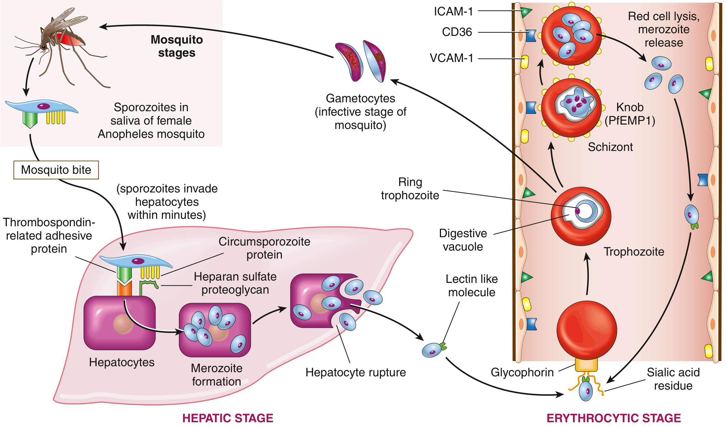
Why P. falciparum is the Deadliest
- Cytoadherence - PfEMP1 causes infected RBCs to stick to vessel walls → capillary occlusion → organ failure
- Cerebral malaria - congestion of brain capillaries → seizures, coma, death (especially in children)
- Infects ALL ages of RBCs - so the parasitemia can be very high (>5% of RBCs infected)
- No dormant liver stage - no relapses, but the acute infection is overwhelming
- Severe anemia - massive RBC destruction
- Metabolic acidosis, hypoglycemia, ARDS, acute kidney injury - multi-organ failure in severe cases
Complications
- Cerebral malaria (most feared)
- Severe anemia
- Acute kidney injury (blackwater fever - hemoglobinuria from hemolysis)
- Acute Respiratory Distress Syndrome (ARDS)
- Hypoglycemia (parasite consumes glucose; quinine treatment also lowers glucose)
- Splenic rupture
- Death if untreated
Treatment
- Artemisinin-based Combination Therapy (ACT) - Artemether-Lumefantrine (Coartem) - first-line for uncomplicated falciparum malaria
- IV Artesunate - severe/cerebral malaria (hospital setting)
- Supportive care - IV fluids, anti-seizure drugs, blood transfusion
This covers every single question in the KMTC General Medicine paper with full pathophysiology, clinical reasoning, and detail relevant to your level of study. Let me know which topic you'd like to explore even further!
This is a shared conversation. Sign in to Orris to start your own chat.