
Shoulder dislocation in details according to orthopaedics text books
shoulder dislocation anterior glenohumeral X-ray diagram
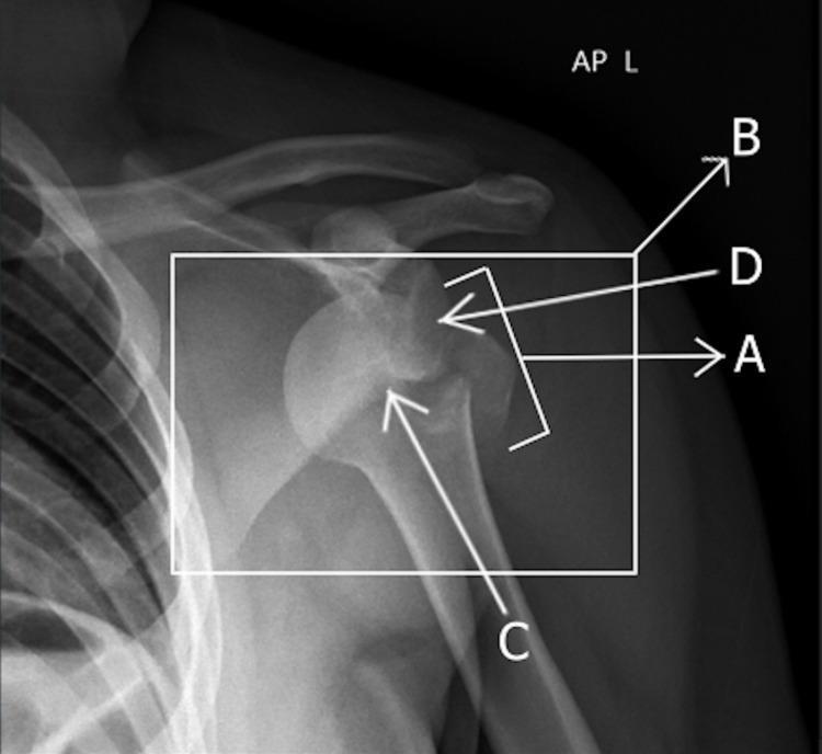
Anterior-posterior (AP) X-ray of the left shoulder demonstrating an anterior glenohumeral joint dislocation accompanied by an associated fracture. The humeral head (labeled C) is displaced anteriorly and inferiorly relative to the glenoid fossa (labeled D), appearing medially beneath the coracoid process. An avulsion fracture of the greater tuberosity is visible (labeled A and B), appearing as a cortical fragment separated from its original anatomical position on the proximal humerus. The diagnostic image highlights key orthopedic pathology including joint space widening, loss of normal articulation, and traumatic osseous injury. This visual is significant for medical education regarding shoulder trauma, dislocation classifications, and the identification of associated fractures in surgical and emergency medicine contexts.
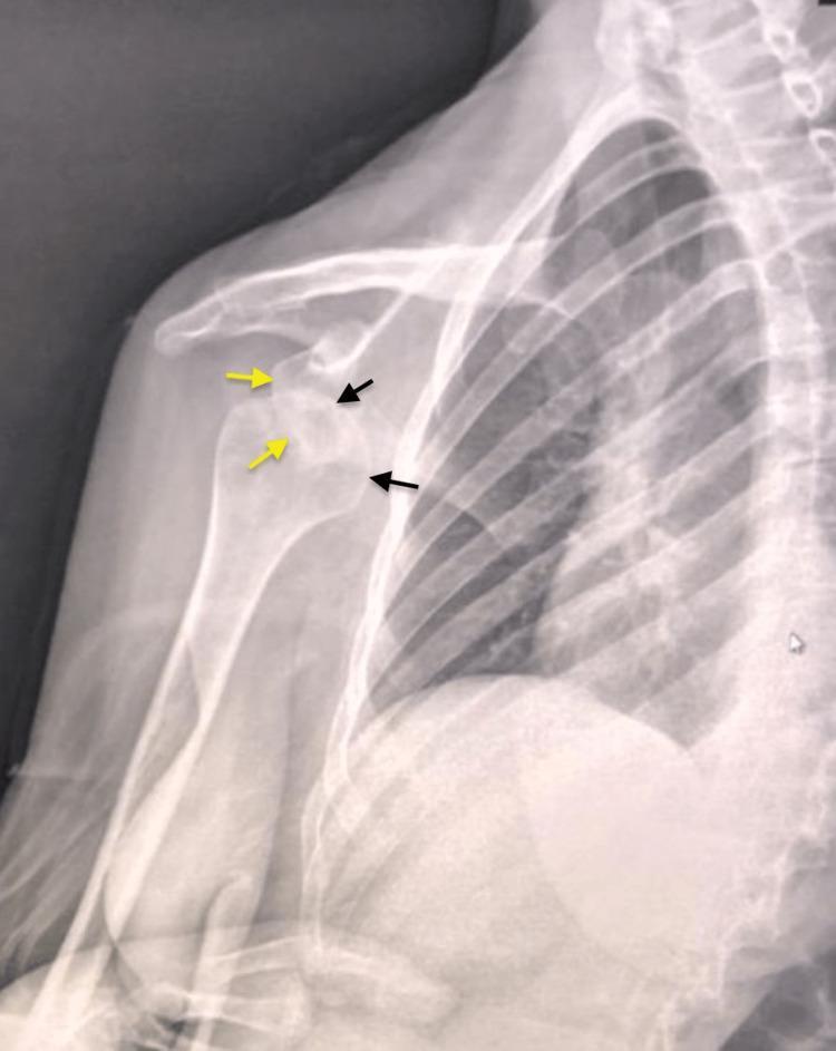
This diagnostic image is a preoperative anteroposterior (AP) X-ray of the right shoulder in a 30-year-old patient. The radiograph demonstrates a significant anterior and medial dislocation of the glenohumeral joint. Key anatomical landmarks are highlighted: yellow arrows point to the empty glenoid cavity of the scapula, while black arrows indicate the displaced humeral head. The humeral head is positioned medially and slightly inferiorly to the glenoid fossa, resting in a subcoracoid position. There is a complete loss of articular congruence between the humeral head and the glenoid. This chronic presentation (three months post-injury) highlights the typical radiological appearance of a neglected shoulder dislocation. The image serves as an educational example for orthopedic trauma and radiology, illustrating joint disruption, displacement patterns in anterior shoulder instability, and the visual assessment of glenohumeral alignment.
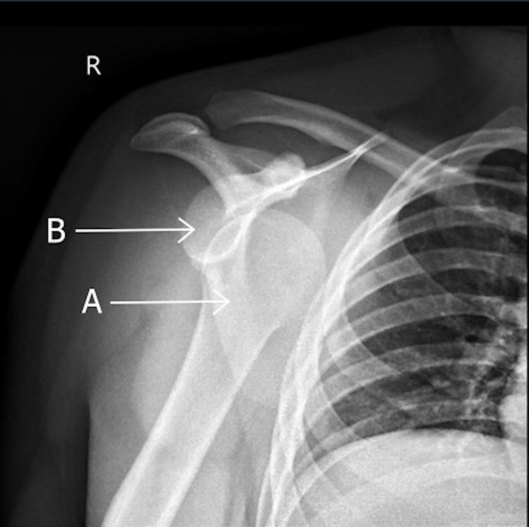
This anterior-posterior (AP) X-ray radiography shows an anterior dislocation of the right glenohumeral joint. The image demonstrates a loss of normal anatomical congruity between the proximal humerus and the scapula. Specifically, the humeral head (labeled A) is displaced inferiorly and medially relative to the glenoid fossa (labeled B), characteristic of an anterior subcoracoid dislocation. Key anatomical structures visible include the right clavicle, the acromion process, the coracoid process, and the upper rib cage. No acute cortical fractures or bony defects are clearly demarcated in this view. This diagnostic image serves as a clinical example of common shoulder trauma and illustrates the spatial relationship disruption between the glenoid and the humerus used to confirm joint instability. It is an essential resource for medical education regarding musculoskeletal radiology and orthopedic emergency management.
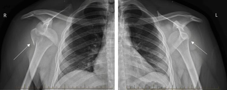
Anteroposterior (AP) X-ray radiographs of the right (R) and left (L) shoulders demonstrating bilateral anterior glenohumeral dislocations. In both views, the humeral heads are displaced anteriorly and inferiorly relative to the glenoid fossa. On the right shoulder, an arrow highlights a concomitant fracture of the greater tuberosity, characterized by a cortical disruption and slight displacement of the bone fragment. On the left shoulder, an arrow indicates the malpositioned humeral head beneath the coracoid process, consistent with subcoracoid dislocation. This clinical presentation is frequently associated with high-energy trauma or generalized tonic-clonic seizures. The images serve as an educational example of complex orthopedic injuries and the importance of checking for associated fractures in the setting of shoulder dislocations.
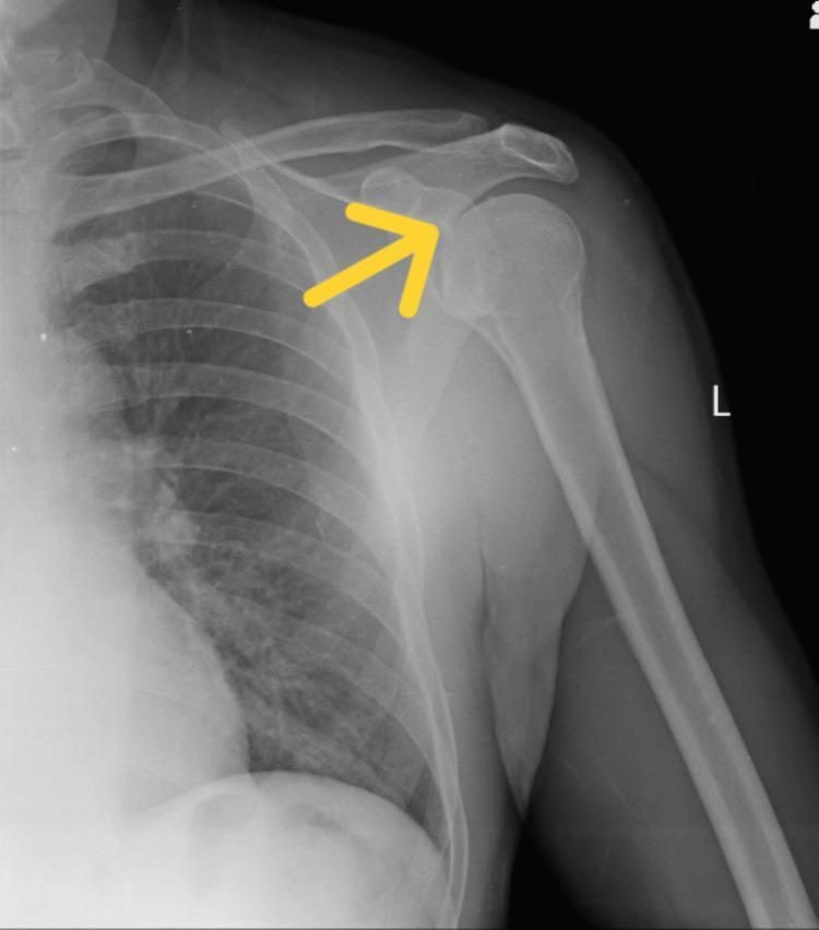
This diagnostic image is an anteroposterior (AP) X-ray of the left shoulder girdle. It illustrates the primary anatomical structures of the glenohumeral joint, including the humerus, scapula (acromion process and glenoid fossa), and clavicle, with the adjacent rib cage and lung field visible in the medial aspect. The radiograph demonstrates a classic anterior shoulder dislocation, characterized by the displacement of the humeral head anteriorly and inferiorly relative to the glenoid fossa. A yellow annotation arrow is positioned at the vacated glenoid rim, highlighting the misalignment where the humeral head has lost its normal anatomical articulation. The bone density appears preserved, with no immediate evidence of associated fractures such as a Hill-Sachs or Bankart lesion visible in this view. This image serves as a clinical teaching tool for medical students and clinicians to identify acute traumatic joint instability and the radiographic presentation of glenohumeral misalignment.
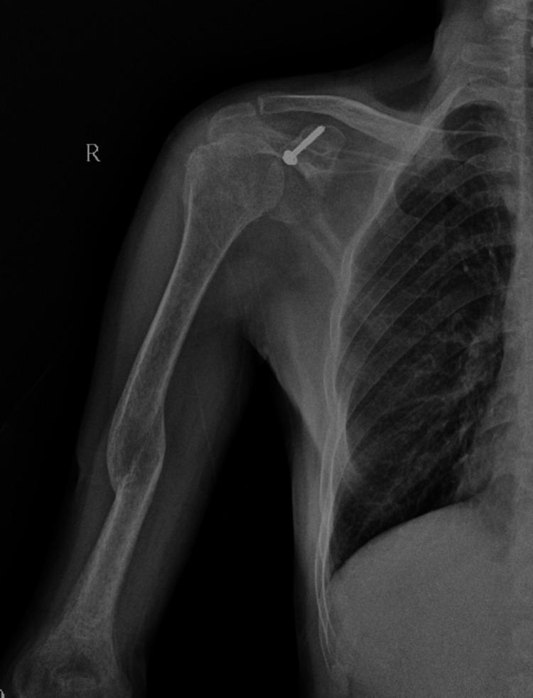
Anterior-posterior (AP) X-ray of the right shoulder and proximal humerus showing postoperative changes following a Latarjet procedure. The glenohumeral joint is well-reduced with anatomic alignment of the humeral head and glenoid fossa. A single metallic cannulated screw with a washer is visible, fixating the coracoid process graft to the anteroinferior glenoid rim to address anterior shoulder instability. The clavicle and acromioclavicular joint appear intact. Along the mid-diaphysis of the humerus, there is a localized area of cortical thickening and increased bone density, possibly representing a healed fracture or chronic stress response. The adjacent rib cage and apical lung field are unremarkable. This image demonstrates the surgical management of chronic anterior shoulder dislocation through coracoid transfer.
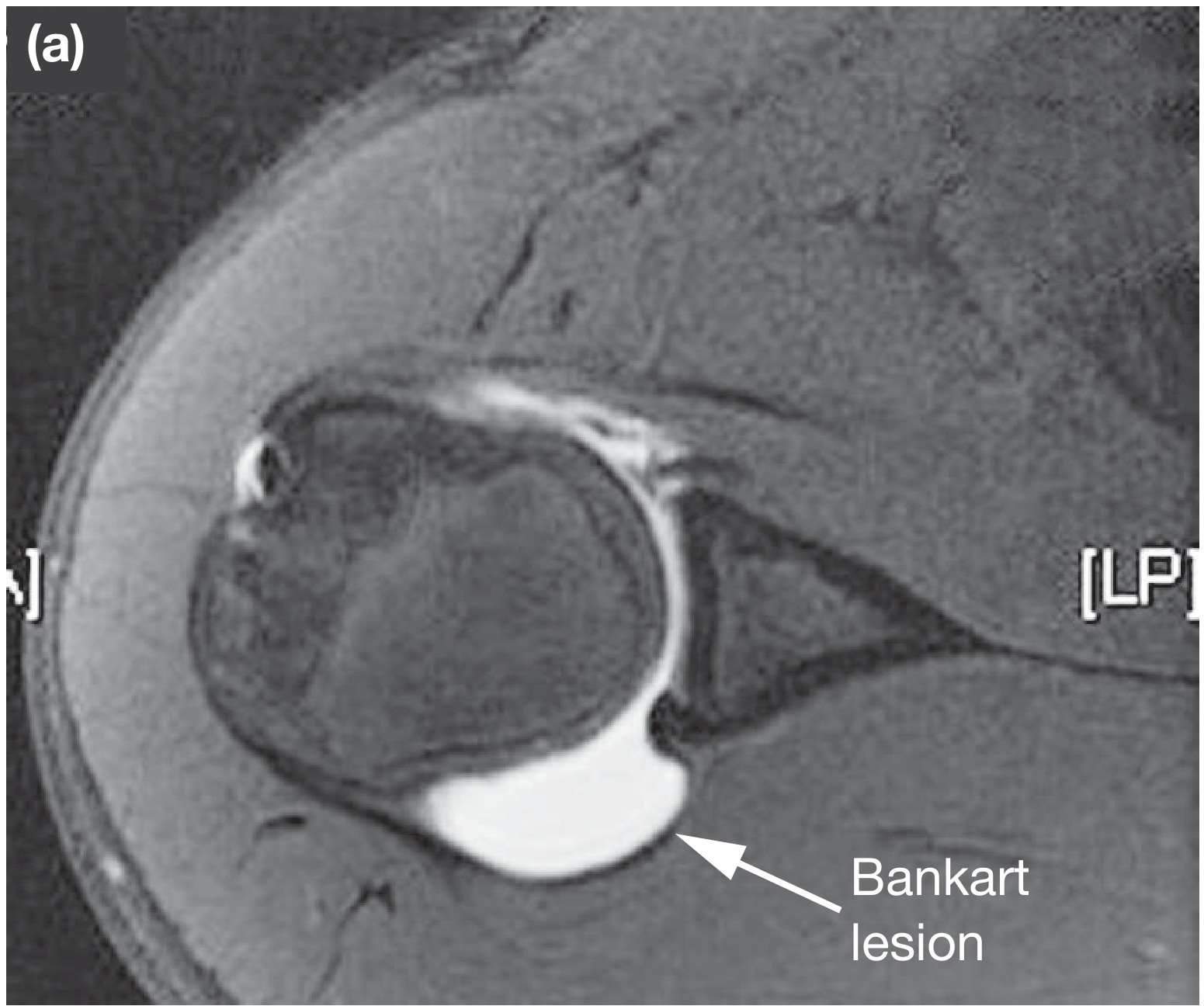
Hill-Sachs lesion Bankart lesion shoulder instability MRI
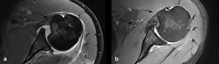
Two side-by-side axial T2-weighted or proton density fat-saturated MRI scans of the shoulder demonstrating the progression of a reverse Hill-Sachs lesion (RHSL) and posterior glenohumeral instability. Image (a) shows the immediate posttraumatic state, characterized by an acute impaction fracture of the anterior humeral head (reverse Hill-Sachs defect) and a concomitant posterior bony Bankart lesion. Image (b) shows the same joint after 5 years of conservative treatment. Comparison reveals morphological changes: the reverse Hill-Sachs defect demonstrates a decrease in depth and filling, likely through fracture hematoma consolidation and cortical remodeling. The posterior bony Bankart lesion shows signs of consolidation. However, both images demonstrate persistent static posterior glenohumeral decentering, where the humeral head remains abnormally positioned posteriorly relative to the glenoid fossa. This visual progression is clinically significant for evaluating the outcomes of conservative management in posterior shoulder instability and the risk of developing posterior decentering osteoarthritis.
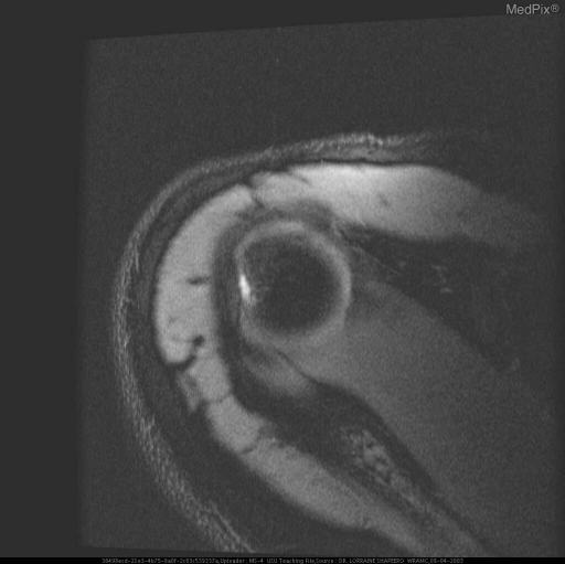
**Imaging Modality:** Magnetic Resonance Imaging (MRI) **Anatomical Region:** Right shoulder (glenohumeral joint) **View:** Axial section **Observed Pathology:** The image demonstrates signs of chronic anterior shoulder instability. There is evidence of a **Bankart lesion**, characterized by a disruption or fracture of the anterior-inferior glenoid labrum/rim. This is accompanied by a **Hill-Sachs deformity**, visible as a cortical depression or flattening of the posterolateral aspect of the humeral head. **Characteristic Visual Features:** - **Bankart Lesion:** Loss of the normal triangular low-signal intensity of the anterior labrum, with associated irregularity of the glenoid margin. - **Hill-Sachs Lesion:** A wedge-shaped defect in the humeral head, resulting from forceful impaction against the glenoid during an anterior dislocation event. - **Joint Space:** The relationship between the humeral head and the glenoid fossa is visualized, showing post-traumatic structural changes. **Clinical Context:** These findings are pathognomonic for recurrent anterior glenohumeral dislocations. The combination of the bony/labral Bankart and the Hill-Sachs lesion represents the "bipolar" nature of many instability-related injuries. **Key Diagnostic Features:** - Anterior-inferior glenoid rim disruption. - Posterolateral humeral head impaction fracture. - Presence of these dual osseous/cartilaginous defects in a patient with a history of shoulder instability.
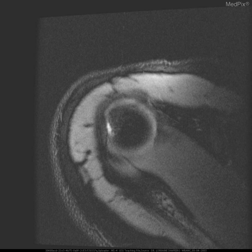
**Imaging Modality:** Magnetic Resonance Imaging (MRI) **Anatomical Region:** Right shoulder girdle, specifically the glenohumeral joint. **Observed Pathology and Visual Features:** This axial T2-weighted or proton density (PD) MRI sequence demonstrates characteristic findings of post-traumatic glenohumeral instability: * **Hill-Sachs Deformity:** A distinct cortical depression and bone marrow signal abnormality are visible on the posterolateral aspect of the humeral head, representing a compression fracture. * **Bankart Lesion:** There is evidence of disruption or tearing of the anteroinferior glenoid labrum, accompanied by irregularity of the adjacent glenoid rim. * **Joint Space:** Mild joint effusion or intra-articular fluid is present, outlining the glenohumeral structures. * **Soft Tissues:** The surrounding deltoid musculature and subcutaneous tissues appear intact, though minor perivascular or interstitial edema may be present. **Clinical Context:** The combination of a Hill-Sachs lesion and a Bankart lesion is highly indicative of prior anterior glenohumeral dislocation. **Diagnostic Significance:** These findings are critical for indexing cases of recurrent shoulder instability and preoperative planning for capsulolabral repair (e.g., Bankart repair) and assessing the risk of future subluxation events.
Shoulder Dislocation - Detailed Orthopaedic Review
1. Anatomy and Why the Shoulder Dislocates
- The glenoid fossa is shallow - its articular surface is 3-4 times smaller than the humeral head
- The glenoid labrum (a fibrocartilaginous rim ~5 mm wide at its base) deepens the socket but provides limited stability
- The joint capsule and ligaments are inherently weak
- Primary stability depends on the rotator cuff tendons (SITS muscles) rather than bony congruency
2. Classification of Glenohumeral Instability
| Type | Features | Surgery |
|---|---|---|
| Traumatic | Unidirectional; involuntary | Usually successful |
| Atraumatic | Multidirectional; painful; involuntary | Responds to surgery |
| Habitual | Voluntary; ligament laxity; painless | Usually contraindicated |
3. Types of Dislocation by Direction
Anterior Dislocation (>95% of all cases)
- Subcoracoid - most common; head rests below the coracoid process
- Subglenoid - head rests below the glenoid fossa
- Subclavicular - head lies medial to the coracoid, below the clavicle
- Intrathoracic - very rare; head penetrates between the ribs into the thoracic cavity
Posterior Dislocation (<5%)
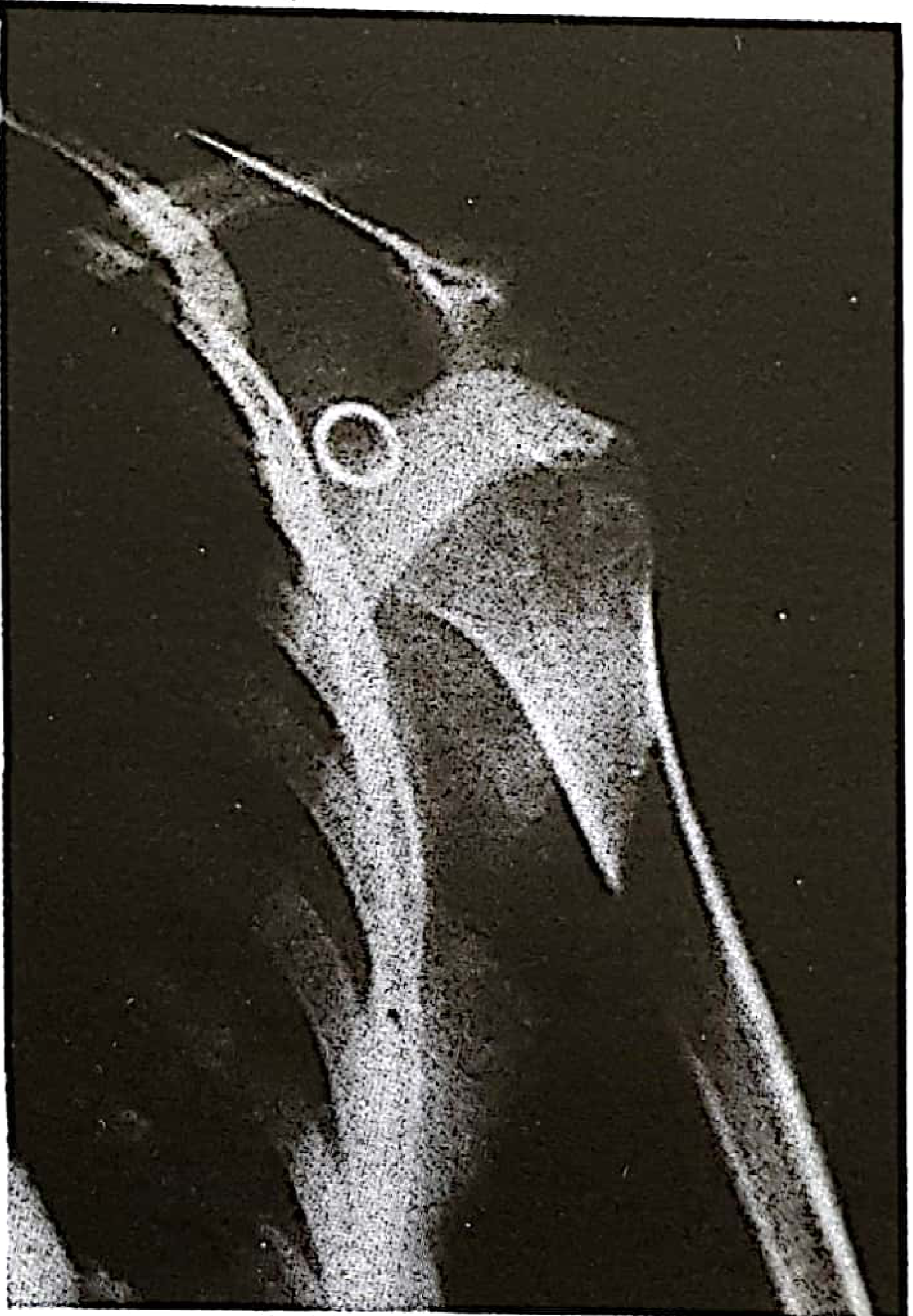
- On a standard AP radiograph, the shoulder may look deceptively normal - the "rim sign" or "light bulb sign" may be the only clue
- High index of suspicion from the history is essential
- May result in a "locked" posterior dislocation where the posterior glenoid rim becomes embedded in the humeral head - requiring open reduction
Inferior Dislocation (Luxatio Erecta) - Rare
- The humeral head displaces straight inferiorly
- Arm is locked in full elevation above the head - a characteristic and dramatic appearance
- High rate of neurovascular complications
4. Mechanism of Injury
5. Clinical Features
Anterior Dislocation
| Finding | Description |
|---|---|
| Arm position | Slight abduction and external rotation; patient supports with opposite arm |
| Shoulder contour | "Squared off" - normal rounded contour lost; shoulder drops in a straight line from acromion tip |
| Deltoid fullness | Flat/empty - "flattening of the deltoid" |
| Humeral head | Palpable anteriorly beneath deltoid |
| Restricted movement | Patient resists adduction and internal rotation; cannot touch contralateral shoulder |
Posterior Dislocation
- Patient may be post-ictal or recovering from electric shock, making examination difficult
- Often presents with pain and limited external rotation
- Standard AP radiograph can appear deceptively normal
6. Investigations
Radiography
- Minimum two views are required (AP + axillary or Velpeau view)
- AP view: shows loss of articular congruence, humeral head displaced from glenoid
- Axillary view: essential for confirming posterior dislocations
CT Scan
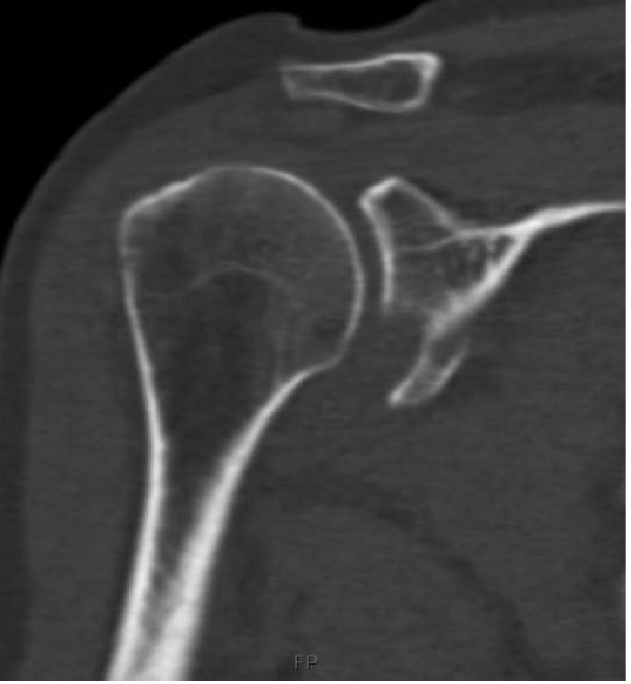
- Defines bony defects (Hill-Sachs lesion, bony Bankart lesion, glenoid rim fractures)
- Essential preoperatively
MRI / MR Arthrography
- Best for soft tissue assessment - labral tears (Bankart lesion), rotator cuff tears
- CT arthrography also useful when MRI unavailable
7. Pathological Lesions
Bankart Lesion

- Soft Bankart: labral tear only
- Bony Bankart: labral detachment + fracture of the anteroinferior glenoid rim
Hill-Sachs Lesion
8. Reduction Techniques
1. Stimson (Gravity/Prone) Technique
- Patient prone, dislocated arm hanging over the edge of the stretcher
- A 10-lb weight attached to the wrist
- Intra-articular lidocaine injected
- Complete muscle relaxation required
- Reduction occurs in 20-30 minutes by gravity
- Safe, effective, easy to learn; main drawback is time required
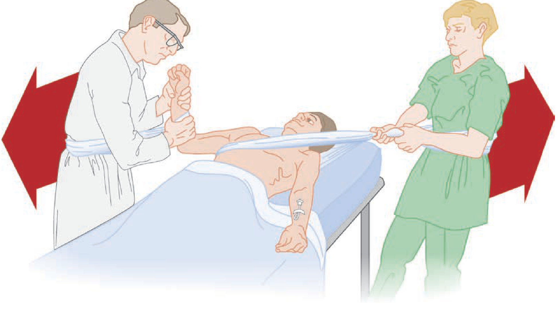
2. Traction-Countertraction (Modified Hippocratic)
- Patient supine, arm abducted, elbow flexed 90°
- A sheet across the thorax provides countertraction via an assistant
- Another sheet around the forearm provides traction by the physician
- Gradual traction + gentle internal/external rotation aids reduction
- The original Hippocratic method: operator applies foot to the axilla while pulling the arm - now superseded by the modified sheet method
3. Scapular Manipulation
- Patient prone or sitting, gentle forward traction on the arm
- The inferior scapular tip is rotated medially while the superior angle is pushed laterally
- This repositions the glenoid to meet the humeral head
- Effective and requires minimal force
4. External Rotation (Hennepin) Technique
- Patient supine, arm adducted, elbow flexed to 90°
- Physician slowly externally rotates the arm in a slow, controlled arc
- Reduction typically occurs before 90° of external rotation is reached
- Minimal force; can often be done without sedation
5. FARES Method (Fast, Reliable, Safe)
- Supine patient; physician holds wrist, traction applied
- Arm oscillated vertically while gradually abducting to 90°, then externally rotating
- Increasingly favored for requiring less analgesia
6. Arlt Method
- Patient sits in a chair, affected arm hanging over an upholstered armrest
- Reposition achieved through longitudinal traction with the armrest as a fulcrum
- Simple and effective (THIEME Atlas, p. 283)
9. Post-Reduction Management
- Apply shoulder immobilizer or sling maintaining adduction and internal rotation
- Orthopedic follow-up within 1 week for uncomplicated cases
- Follow-up within 1-2 days if complicated by bony/soft tissue injury
- Young patients (<25): longer immobilization (3-6 weeks) to reduce recurrence
- Older patients (>40): early mobilization to prevent stiffness is prioritized
10. Complications
Neurovascular Injuries
| Structure | Frequency | Notes |
|---|---|---|
| Axillary nerve | Most common (10-25% of cases) | Traction neurapraxia; test sensation over deltoid insertion ("regimental badge" area); usually resolves spontaneously |
| Brachial plexus | Less common | High-energy injuries |
| Axillary artery | Rare; elderly patients | Presents with absent radial pulse, axillary hematoma, axillary bruit; vascular emergency |
| Radial, ulnar, median nerves | Rare | --- |
Bony Injuries
- Hill-Sachs lesion (humeral head compression fracture)
- Bony Bankart lesion (glenoid rim fracture)
- Greater tuberosity fracture - common; often visible on post-reduction films
Soft Tissue Injuries
- Soft Bankart lesion (labral tear) - predisposes to recurrence
- Rotator cuff tear - especially in patients >40 years; rotator cuff weakens with age. Suspect if weakness of external rotation persists post-reduction
Recurrent Dislocation
- Most common long-term complication
- Children and young adults: recurrence rate >90%
- With each subsequent dislocation, progressively less force is required
- Eventually the shoulder may redislocate with minor movements (e.g., arm rotation during sleep)
11. Recurrent Shoulder Dislocation
- Follows one or more traumatic dislocations; some patients have a developmental predisposition
- After several episodes, the shoulder dislocates with minimal trauma or simply from external rotation
- Episodes are painful but the patient may sometimes self-reduce
- The shoulder must be seen and confirmed dislocated before deciding on surgery
- Confirm whether the instability is anterior or posterior - the operative procedure for one can worsen the other
Investigations for Recurrence
- CT or MR arthrography: detaches anteroinferior labrum (Bankart's lesion); humeral head defect (Hill-Sachs lesion); bony glenoid defects
- Apprehension test (Figure 38.23 from Bailey & Love): forced abduction + external rotation elicits apprehension (sense of impending dislocation) - positive in anterior instability
Surgical Options
| Procedure | Mechanism | Notes |
|---|---|---|
| Bankart repair (arthroscopic or open) | Reattaches the labrum to the glenoid rim | Gold standard for soft tissue Bankart lesion |
| Putti-Platt procedure | Tightens and overlaps subscapularis + anterior capsule | Older technique; restricts external rotation |
| Latarjet procedure | Transfers the coracoid process to the anteroinferior glenoid | Used when significant bony Bankart defect (>25% glenoid loss) |
| Posterior capsule repair | Tightens posterior capsule + labral repair | For recurrent posterior instability |
12. Fracture-Dislocation of the Shoulder
- Closed reduction may be attempted but is often impossible
- Open reduction + internal fixation is usually required
- In elderly patients with comminuted fracture-dislocations, hemiarthroplasty or reverse total shoulder arthroplasty may be the best option
Summary Table
| Feature | Anterior | Posterior | Inferior (Luxatio Erecta) |
|---|---|---|---|
| Frequency | >95% | <5% | Rare |
| Mechanism | Abduction + ER + extension | Forced IR (seizure, electrocution) | Hyperabduction/axial load |
| Arm position | Slight abduction + ER | Adduction + IR | Fixed elevation (arm above head) |
| Bony lesions | Hill-Sachs + Bankart | Reverse Hill-Sachs + posterior Bankart | --- |
| Nerve at risk | Axillary nerve | Axillary nerve | Axillary nerve + brachial plexus |
| Diagnosis pitfall | Usually obvious | Easy to miss on AP X-ray | Usually obvious |
| Recurrence | Very common (>90% in young) | Less common | Low |
- Bailey & Love's Short Practice of Surgery, 28th ed. pp. 556-558
- Tintinalli's Emergency Medicine, 9th ed., pp. 1867-1870
- Pye's Surgical Handicraft, 22nd ed., pp. 184-186
- THIEME Atlas of Anatomy - General Anatomy and Musculoskeletal System, pp. 283-284
Recurrent shoulder dislocation in details according to orthopaedics text books
recurrent shoulder dislocation Bankart repair Latarjet surgery
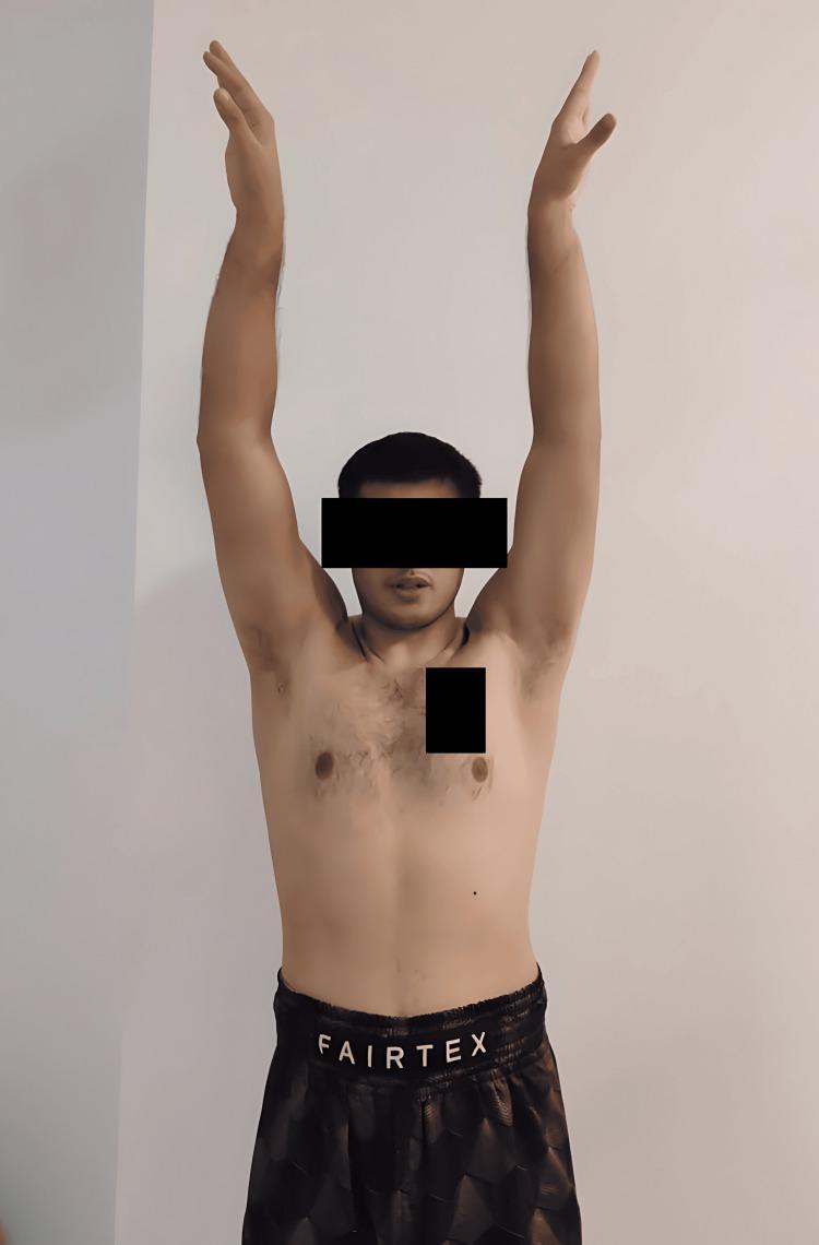
This clinical photograph shows the anterior view of a standing male patient one year following a bilateral open Latarjet procedure for recurrent shoulder dislocation. The patient is demonstrating active shoulder abduction, achieving a full range of motion (approximately 180 degrees) with both arms extended vertically overhead. The image illustrates a successful postoperative functional outcome following surgical stabilization for anterior shoulder instability, Bankart lesions, and Hill-Sachs defects. The patient exhibits a mesomorphic physique with well-healed surgical sites in the deltopectoral regions. This visual serves as a clinical assessment of joint mobility and muscle function in the context of orthopedic rehabilitation and sports medicine. Key educational concepts include the evaluation of postoperative glenohumeral stability and the restoration of athletic function in high-demand patients.
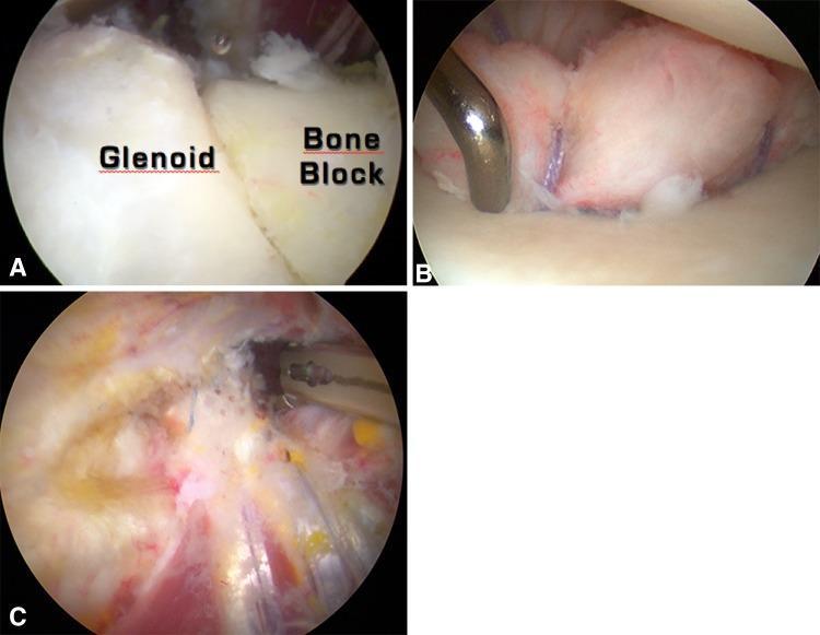
Arthroscopic views of a combined Bristow-Latarjet-Bankart procedure for recurrent anterior shoulder instability. Image A (intra-articular view) shows a coracoid bone graft positioned in a 'standing' orientation, fixed flush with the glenoid surface to restore bone loss; the graft is secured with a cortical screw. Image B demonstrates the associated capsulolabral repair (Bankart repair) where suture anchors are used to reattach the labrum over the bone block, moving the graft to an extra-articular position and enhancing glenoid concavity (bumper effect). Image C (extra-articular view) visualizes the transferred conjoined tendon passing through a split in the subscapularis muscle to provide a dynamic 'sling' or 'belt' effect, reinforcing the anteroinferior capsule. This '2B3' triple-locking technique combines osseous restoration, dynamic soft tissue reinforcement, and labral repair to stabilize the glenohumeral joint.
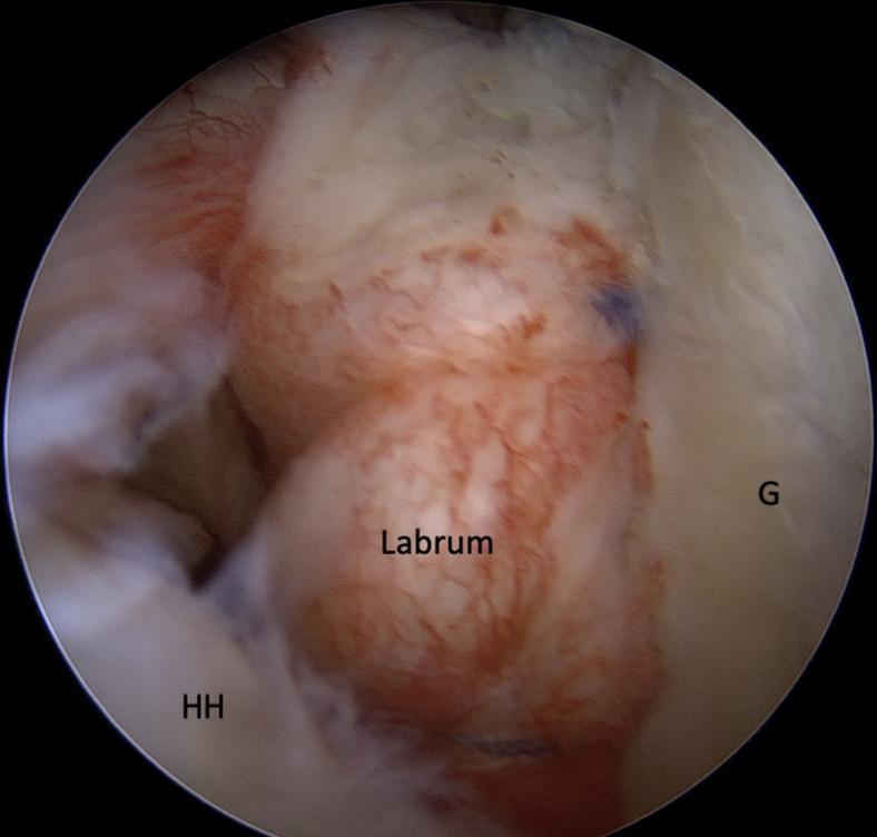
This diagnostic image is an intra-articular arthroscopic photograph of a left shoulder joint, captured from the posterior viewing portal. The image demonstrates the final result of a standard Bankart repair, an orthopedic procedure used to treat anterior shoulder instability. The primary anatomical structures are labeled: the glenoid (G) on the right and the humeral head (HH) in the lower-left. The labrum, specifically the fibrocartilaginous rim, is visible and labeled in the center, appearing re-attached and tensioned against the glenoid rim. Three suture anchors are strategically placed along the anterior-inferior glenoid rim at approximately the 7, 8, and 9 o'clock positions. The educational focus is the visual confirmation of the 'labral bumper effect' and the tensioning of the capsuloligamentous structures. This clinical imaging illustrates surgical management for recurrent shoulder dislocation and labral tears within the field of sports medicine and orthopedic surgery.
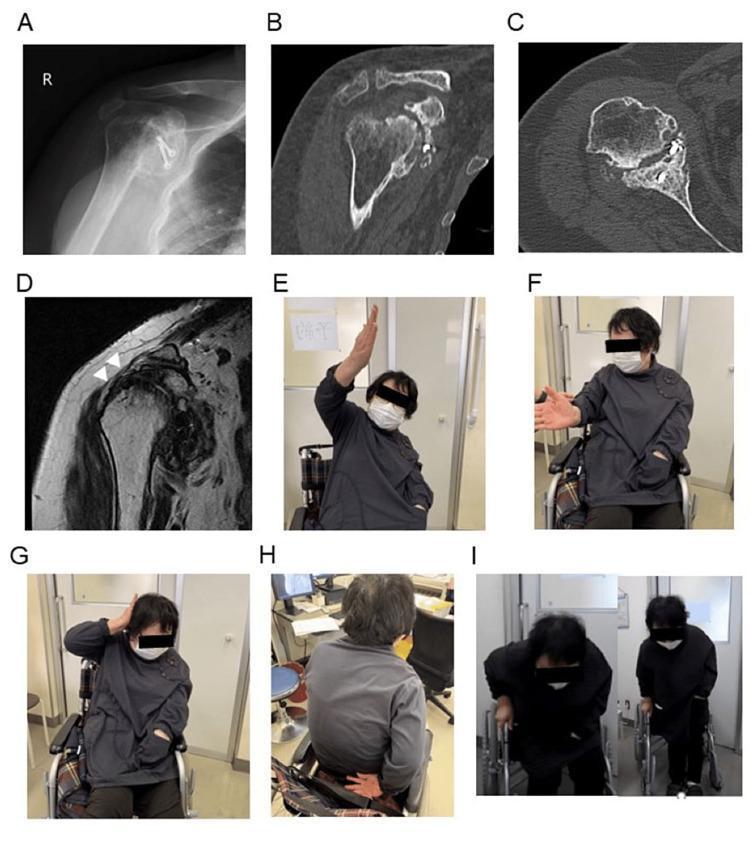
This composite figure illustrates the 50-month post-operative outcomes of a patient following a Latarjet procedure for recurrent shoulder dislocation. Panels A-D display multi-modal diagnostic imaging of the right shoulder: (A) An anteroposterior radiograph showing glenohumeral osteoarthritis and surgical hardware; (B-C) Sagittal and axial CT scans demonstrating successful coracoid bone graft transfer and integration into the anterior glenoid rim; (D) A coronal T2-weighted MRI where white arrowheads indicate the structural integrity of the repaired supraspinatus tendon. Panels E-I are clinical photographs assessing functional recovery and active range of motion: (E) Shoulder flexion reaching 120 degrees; (F) External rotation to 30 degrees; (G) Functional ability to reach the hand behind the head with the elbow back; (H) Internal rotation to the level of the fifth lumbar spine; and (I) The patient demonstrating weight-bearing capability by standing independently from a wheelchair. The collection serves as an educational example of managing complex shoulder instability through bone block reconstruction and rotator cuff repair in a patient with hemiplegia.
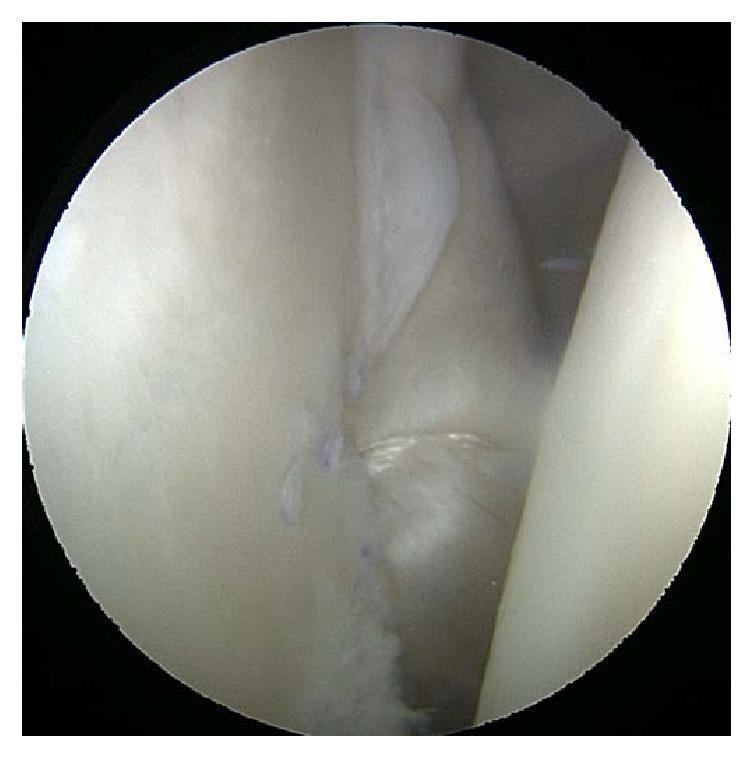
Intraoperative arthroscopic clinical photograph demonstrating the final appearance of a shoulder joint following an anterior and posterior capsular shift with Bankart repair. The image showcases the glenohumeral joint space where light-colored, creamy-white soft tissue, primarily the joint capsule and labrum, appears smooth and glistening. Key surgical features include blue synthetic sutures embedded within the tissue margins, indicating the successful plication and stabilization of the redundant capsule. The fold of tissue in the upper right quadrant reflects the tightened capsular volume intended to address multidirectional instability. This visual is significant for illustrating orthopedic surgical techniques for recurrent shoulder dislocation and instability, specifically highlighting tissue approximation and the structural outcomes of a capsulolabral reconstruction.
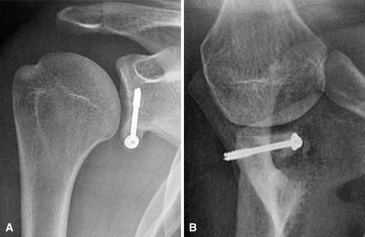
Two-panel postoperative radiographic study of the right shoulder demonstrating successful Bristow-Latarjet-Bankart procedure for recurrent anterior shoulder instability. Panel A is an anteroposterior (AP) view showing a coracoid bone graft fixated to the anteroinferior glenoid rim with a single radiopaque orthopedic screw. The graft is positioned optimally below the glenoid equator and flush with the articular surface. Panel B is a lateral (Bernageau) or scapular Y-view, illustrating the horizontal trajectory of the fixation screw through the bone block into the glenoid neck. There is visible evidence of osseous integration and bone healing between the transferred coracoid process and the native glenoid. The humeral head is centered within the glenohumeral joint, indicating restored stability and the successful recreation of the glenoid's concave architecture. This visual serves as an educational example of ideal bone block positioning to address severe glenoid bone deficiency.
Hill-Sachs lesion humeral head compression fracture X-ray CT
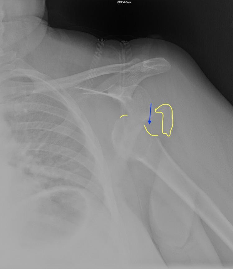
This diagnostic radiograph (X-ray) of the left shoulder in an anteroposterior (AP) view demonstrates an anterior-inferior glenohumeral dislocation. The humeral head is displaced from the glenoid fossa and is positioned medially and inferiorly. A key educational feature is the Hill-Sachs lesion, a compression fracture of the posterolateral humeral head, which is highlighted with a blue arrow and a yellow outline indicating the cortical depression. This lesion typically occurs when the humeral head impacts the anterior glenoid rim during dislocation. Additionally, a small radio-opaque fragment is outlined in yellow near the medial aspect of the joint, suggestive of a possible bony Bankart lesion or a loose fracture fragment. The image serves as a clinical example of trauma-induced shoulder instability and associated secondary fractures, which are critical for surgical planning and risk assessment for recurrent dislocations in orthopedics.
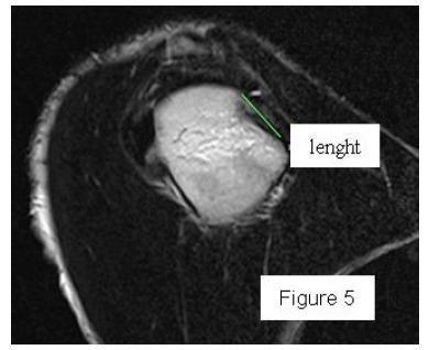
This diagnostic image is a CT scan in a sagittal view focused on the humeral head of the shoulder. It demonstrates a Hill-Sachs lesion, which is a posterolateral compression fracture of the humeral head typically resulting from anterior shoulder dislocation. The image shows a cortical depression or defect on the posterior-superior aspect of the bone. A green solid line spans the edge of the indentation to measure the length of the lesion, which is an important clinical parameter in determining shoulder instability and surgical planning (on-track versus off-track lesions). The high-contrast bone appears bright against the surrounding darker soft tissues, highlighting the altered morphology of the articular surface. This visual aid is intended for intermediate to advanced orthopedic or radiological education regarding the quantification of glenohumeral joint trauma.
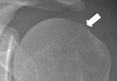
This diagnostic image is a conventional plain film radiograph (X-ray) of the human shoulder, specifically focusing on the humeral head. The image demonstrates a close-up view of the proximal humerus and its articulation with the glenoid. A prominent white arrow points to a localized bony protuberance on the postero-superior aspect of the humeral head. This finding represents a variation of a Hill-Sachs lesion, which is typically a compression fracture or depression following an anterior shoulder dislocation. In this specific case, the lesion appears paradoxically as an 'osteophyte-like' outward projection rather than a cortical indentation. This visual phenomenon occurs when the X-ray beam is tangent to the elevated, hypertrophic margin of the bony defect. The image illustrates a critical diagnostic pitfall in musculoskeletal radiology, where the morphology of a Hill-Sachs lesion can be misinterpreted based on the radiographic projection. The internal trabecular pattern and cortical density of the humeral head are well-preserved, highlighting the focal nature of the contour abnormality at the postero-superior head-neck junction.
Recurrent Shoulder Dislocation - Detailed Orthopaedic Review
1. Definition and Epidemiology
- Traumatic shoulder dislocation is the commonest of all dislocations, usually first presenting in patients under 25 years
- In young adults (<25 years), the recurrence rate after first-time anterior dislocation exceeds 90%
- Subsequent dislocations require progressively less force than the initial event
- The shoulder usually dislocates anteroinferiorly
2. Pathological Anatomy (Why Recurrence Happens)
A. Bankart Lesion (Primary Lesion)
- Soft Bankart: labral detachment only, no bony involvement
- Bony Bankart: labral detachment + avulsion fracture of the anteroinferior glenoid rim - causes significant glenoid bone loss
B. Hill-Sachs Lesion
- Visible on AP X-ray in external rotation as a notch on the posterolateral humeral head
- Classified as "on-track" or "off-track" based on the Glenoid Track concept (see below)
C. Capsular Laxity and Stretching
D. HAGL Lesion (Humeral Avulsion of Glenohumeral Ligament)
E. Glenoid Bone Loss
3. Classification of Shoulder Instability
| Class | Mechanism | Direction | Volition | Surgery |
|---|---|---|---|---|
| Traumatic (TUBS) | Significant force required for first dislocation | Unidirectional (usually anterior) | Involuntary | Usually successful |
| Atraumatic (AMBRI) | No significant trauma | Multidirectional | Involuntary; painful | Responds to surgery |
| Habitual | Voluntary | Variable | Voluntary | Usually contraindicated |
4. Clinical Features and Examination
History
- First dislocation required significant trauma (fall on outstretched arm in abduction + external rotation)
- Subsequent episodes occur with progressively less force
- Patient may describe shoulder "slipping out" during sleep, overhead activities, or swimming
- May have episodes of subluxation (shoulder slips then spontaneously reduces) - these can be as disabling as complete dislocation
- Some patients learn to self-reduce their shoulder
Examination
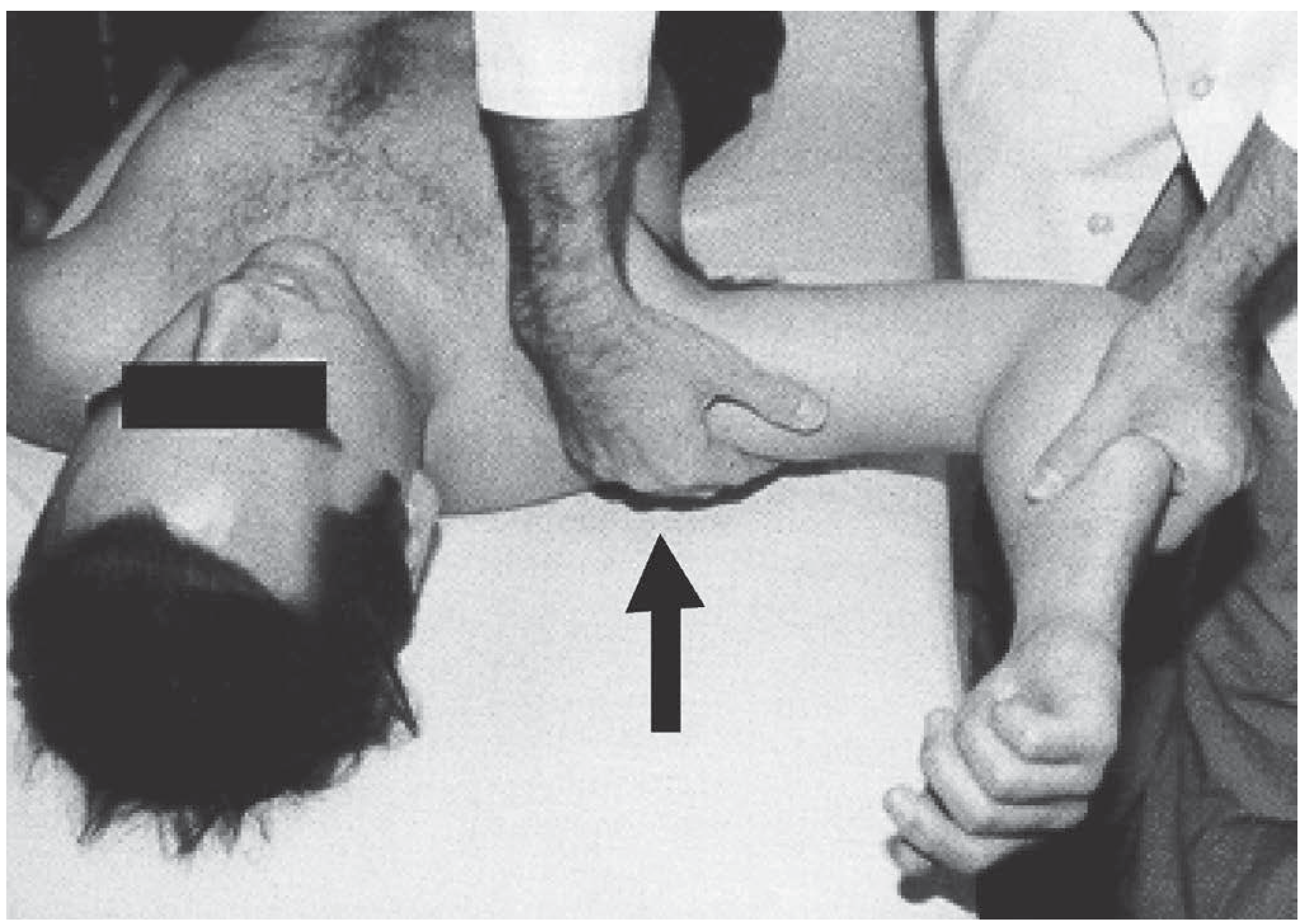
1. Apprehension Test (Crank Test)
- Patient supine, arm abducted to 90° and progressively externally rotated
- Positive when patient experiences apprehension - "a sense of impending doom as the shoulder feels about to dislocate"
- Patient resists further movement; may grab the examiner's hand
- Most reliable test for anterior instability
2. Relocation Test (Jobe's Test)
- Following a positive apprehension test, posterior pressure is applied to the humeral head
- If apprehension resolves with posterior pressure → confirms anterior instability
- Helps differentiate true instability from impingement
3. Anterior Drawer / Load-and-Shift Test
- Patient seated or supine
- Humeral head loaded into glenoid then translated anteriorly and posteriorly
- Graded by amount of translation: Grade I (to rim), Grade II (over rim, spontaneous reduction), Grade III (over rim, no spontaneous reduction)
4. Sulcus Sign
- Arm hangs at the side; downward traction applied
- A gap (sulcus) forming below the acromion indicates inferior laxity
- Suggests multidirectional instability if bilateral
5. Investigations
Plain Radiography
- AP view: May show Hill-Sachs notch on the posterolateral humeral head (visible in external rotation), glenoid erosion, or loose bodies
- Axillary lateral view: Essential - shows glenoid rim erosion and Hill-Sachs defect in profile
- West Point view: Tangential view of anteroinferior glenoid - best for bony Bankart
- Stryker Notch view: Best for Hill-Sachs lesion
CT Scan
- Gold standard for quantifying bone loss - both glenoid and humeral side
- Essential for surgical planning
- Glenoid bone loss calculated using the "bare spot" method or best-fit circle method
MRI / MR Arthrography
- Best modality for labral pathology - detects soft Bankart lesion, posterior labral injury, HAGL lesion, rotator cuff tears
- MR arthrography (with intra-articular contrast) is superior to standard MRI for labral tears
6. Instability Severity Index Score (ISIS)
| Prognostic Factor | Points |
|---|---|
| Age at surgery <20 years | 2 |
| Age at surgery >20 years | 0 |
| Competitive sport (preoperative) | 2 |
| Recreational or no sport | 0 |
| Contact or forced overhead sport | 1 |
| Other sport | 0 |
| Shoulder hyperlaxity (anterior or inferior) | 1 |
| Hill-Sachs lesion visible on AP X-ray in external rotation | 2 |
| Glenoid loss of contour on AP X-ray | 2 |
| Total | 10 |
- Score ≤3: Low risk of recurrence - arthroscopic repair suitable
- Score ≥4: Higher risk - consider open Bankart or Latarjet procedure
- Score ≥6: Bone block surgery (Latarjet) strongly preferred
7. Surgical Treatment
- Low rates of recurrence, complication, and reoperation
- Does no harm (avoids arthritis)
- Maintains motion
- Applicable in most cases
- Corrects the specific pathologic condition
Procedure Selection Based on Pathology
| Pathology | Preferred Procedure |
|---|---|
| Traumatic Bankart (soft tissue) | Jobe capsulolabral reconstruction (Modified Bankart) |
| Acute bony Bankart | Screw or anchor fixation |
| Bankart + hyperlaxity | Bankart + rotator interval closure |
| HAGL lesion | Suture anchor repair (humeral side) |
| Multidirectional instability | Capsular shift (Neer or glenoid-side) |
| Glenoid bone loss >25% | Latarjet procedure |
| Glenoid bone loss >40% | Eden-Hybinette procedure |
| Humeral head defect 20-25% (6mm deep) | Remplissage + Bankart repair |
| Humeral head defect >40% | Allograft reconstruction |
| Anterior humeral head loss >30% | McLaughlin procedure |
| Capsular deficiency | Achilles allograft reinforcement |
A. Bankart Repair (Capsulolabral Reconstruction)
- Arthroscopic: now the dominant approach (~90-95% of cases per Campbell's); uses suture anchors; lower morbidity
- Open (Modified Bankart / Jobe Capsulolabral Reconstruction): gold standard for complex cases
- Deltopectoral interval approach; incision 2 cm distal and lateral to coracoid
- Retract deltoid + cephalic vein laterally; pectoralis major medially; conjoined tendon intact, retracted medially
- Split subscapularis tendon transversely at junction of upper 2/3 and lower 1/3; dissect from underlying anterior capsule
- Horizontal anterior capsulotomy from humeral insertion to anterior glenoid neck
- Insert humeral head retractor; elevate capsule on anterior neck subperiosteally
- Decorticate anterior scapular neck to bleeding bone (maximizes healing potential)
- Drill bone tunnels or place suture anchors at the articular margin of the glenoid
- Restore glenoid concavity by reattaching labrum at the edge of the articular surface
- Capsular advancement and imbrication - superior capsular flap is advanced superiorly and inferiorly, recreating physiological capsular tension
- Repair subscapularis; close deltopectoral interval
- Abrade the scapular neck to maximize healing potential
- Restore glenoid concavity
- Secure anatomic capsular fixation at the edge of the glenoid articular surface
- Recreate physiologic capsular tension by superior and inferior capsular advancement
- Supervised goal-oriented rehabilitation
B. Putti-Platt Operation
- One of the most widely used older procedures (alongside Bankart)
- Usually successful at controlling instability but non-anatomic
C. Latarjet Procedure (Coracoid Transfer)
- Bone block effect: Enlarges the glenoid articular arc, reducing bone deficiency
- Sling effect (dynamic): The conjoined tendon acts as a dynamic anterior sling - in abduction/external rotation, the tendon tightens across the inferior subscapularis and prevents anterior translation
- Capsulodesis effect: The transferred structures reinforce the anterior capsule
D. Remplissage ("Filling In")
- Combined with arthroscopic Bankart repair
- In contact/collision athletes - any significant Hill-Sachs lesion is treated with remplissage
- In throwing athletes - larger Hill-Sachs defects may undergo lateralized remplissage (anchors in lateral aspect of defect) to preserve external rotation; smaller defects may be left alone (Campbell's, p. 5050)
E. Eden-Hybinette Procedure
F. Capsular Shift (Neer Inferior Capsular Shift)
- For anteroinferior-predominant MDI: humeral-side shift (Neer)
- For posterior-predominant MDI: glenoid-side shift
8. Conservative Management
- Physiotherapy and rotator cuff strengthening - primary treatment for atraumatic/MDI
- Proprioceptive retraining and neuromuscular control exercises
- Activity modification (avoidance of provocative positions)
- At least 6 months of structured rehabilitation before considering surgery for atraumatic MDI
9. Summary of Surgical Procedures
| Procedure | Approach | Indication | Key Feature | Drawback |
|---|---|---|---|---|
| Bankart repair (arthroscopic) | Arthroscopic | Soft Bankart, bone loss <25% | Anatomic; preserves motion | Higher recurrence in high-risk patients |
| Open Bankart (Jobe) | Open deltopectoral | Complex Bankart; failed arthroscopic | Best long-term results (2% recurrence at 17 yr) | Open surgery morbidity |
| Putti-Platt | Open | Older technique; anterior instability | Simple; reliable | Restricts external rotation |
| Latarjet | Open or arthroscopic-assisted | Bone loss >25%; off-track Hill-Sachs; ISIS ≥6 | Triple mechanism (bone + sling + capsule) | Potential for hardware failure, neurovascular risk |
| Remplissage | Arthroscopic | Hill-Sachs 20-25%; off-track | Fills humeral defect; combined with Bankart | May reduce external rotation |
| Eden-Hybinette | Open | Bone loss >40%; failed Latarjet | Free bone graft | Donor site morbidity; complex |
| Capsular shift | Open or arthroscopic | MDI | Reduces capsular volume | Technical complexity |
10. Complications of Recurrent Dislocation and its Surgery
Complications of the Recurrence Itself
- Progressive glenoid bone erosion (bony Bankart) with each episode
- Enlarging Hill-Sachs lesion on the humeral head
- Rotator cuff damage (especially in patients >40)
- Axillary nerve injury (rare with each recurrence but cumulative risk)
- Glenohumeral arthritis - long-term consequence of repeated instability
Complications of Surgery
- Recurrent instability (main failure mode)
- Loss of external rotation - especially Putti-Platt and overtightened Bankart repairs
- Hardware failure - screw backout in Latarjet
- Neurological injury - axillary or musculocutaneous nerve
- Glenohumeral arthritis - particularly after non-anatomic procedures (Putti-Platt); the Latarjet also has a rate of late arthritis (coracoid arthroplasty or "Latarjet arthropathy")
- Subscapularis failure - failure of repair after open surgery
11. Summary Box (Bailey & Love, Summary Box 38.8)
- An appreciable force leads to the first dislocation or subluxation
- Subsequent dislocations/subluxations require less force
- The commonest direction of dislocation is anteroinferior
- There is a positive apprehension sign on examination
- Surgical treatment repairs the labral lesion (Bankart repair) and reverses traumatic laxity of the capsule
- Up to 90-95% success rate with Bankart repair (arthroscopic or open)
- Bony defects of the glenoid (and occasionally large Hill-Sachs lesions) may require bone grafting
- Campbell's Operative Orthopaedics, 15th ed. (2026), pp. 2897-2900
- Bailey & Love's Short Practice of Surgery, 28th ed., pp. 556-558
- Pye's Surgical Handicraft, 22nd ed., pp. 185-186
- Balg F, Boileau P. The instability severity index score. J Bone Joint Surg 2007;89B:1470-1477