
A patient reports sudden onset of blindness in the right eye. Examination reveals loss of direct light reflex in the right eye, but consensual light reflex in right eye is intact. Which structure is most likely damaged? • Left optic tract O Right lateral geniculate body O Right oculomotor nerve • Right optic nerve O Right pretectal nucleus
- Afferent limb: Retina → optic nerve → optic chiasm → pretectal nucleus (bilaterally)
- Efferent limb: Pretectal nucleus → Edinger-Westphal nucleus (both sides) → oculomotor nerve (CN III) → ciliary ganglion → sphincter pupillae
- Loss of direct light reflex in the right eye = shining light in the right eye produces no constriction in the right pupil
- Intact consensual reflex in the right eye = shining light in the LEFT eye causes the right pupil to constrict normally
- The efferent pathway to the right eye is intact (right CN III and right sphincter work fine - proven by the intact consensual response)
- The afferent signal from the right eye is absent - light entering the right eye cannot reach the pretectal nuclei
| Option | Why incorrect |
|---|---|
| Left optic tract | Affects the visual field (homonymous hemianopia) but not the pupillary reflex in this pattern |
| Right lateral geniculate body | Geniculate fibers project to visual cortex, not to pretectal nuclei; no pupillary defect |
| Right oculomotor nerve (CN III) | Would cause loss of BOTH direct AND consensual response in the right eye, plus ptosis and lateral deviation |
| Right pretectal nucleus | Would cause bilateral pupillary dysfunction, not a unilateral afferent defect |
- No direct reflex (afferent signal from right eye is lost)
- Intact consensual reflex (the efferent pathway via CN III is intact, driven by the left eye's input reaching both pretectal nuclei)
Create a visual diagram of the pupillary reflex arc
A detailed anatomical diagram of the pupillary light reflex arc showing both eyes. The diagram includes: - Two eyes (left and right) at the top with pupils - Afferent pathway: light entering right eye → right retina → right optic nerve → optic chiasm (showing partial decussation) → both pretectal nuclei (left and right) in the midbrain - Efferent pathway: pretectal nuclei → bilateral Edinger-Westphal nuclei → oculomotor nerve (CN III) on both sides → ciliary ganglion → sphincter pupillae muscle in both eyes - Arrows showing direction of signal flow - Color coding: afferent pathway in blue, efferent pathway in red - Labels for: Retina, Optic Nerve, Optic Chiasm, Optic Tract, Pretectal Nucleus, Edinger-Westphal Nucleus, CN III (Oculomotor Nerve), Ciliary Ganglion, Sphincter Pupillae - Direct reflex (same side) and consensual reflex (opposite side) clearly labeled - Clean white background, medical illustration style, educational diagram
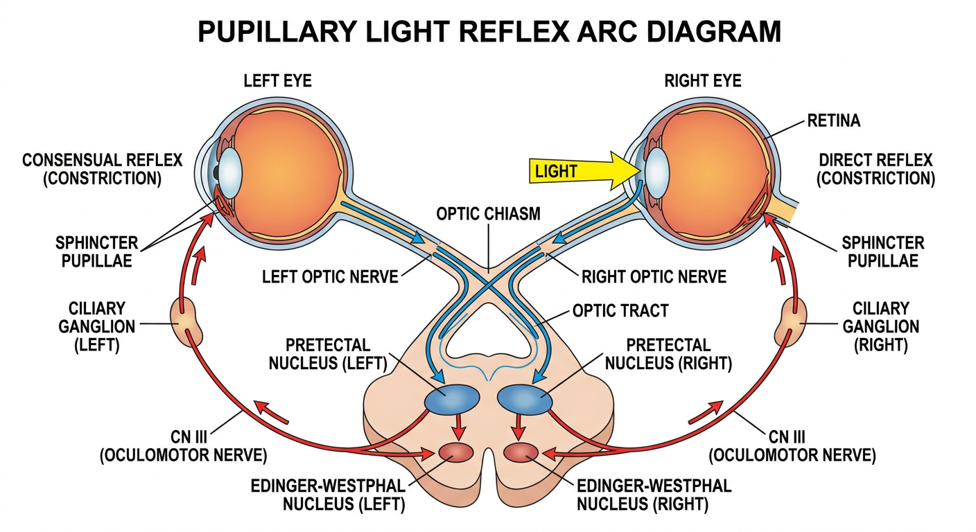
- Light enters the eye → stimulates retinal photoreceptors
- Signal travels via the optic nerve (CN II)
- Fibers partially cross at the optic chiasm (nasal fibers cross, temporal fibers stay ipsilateral)
- Signals reach both pretectal nuclei in the dorsal midbrain via the optic tracts
- One pretectal nucleus connects to BOTH EW nuclei
- This is why shining light in ONE eye causes BOTH pupils to constrict:
- Direct reflex = same eye as light source constricts
- Consensual reflex = opposite eye also constricts
Is pretactal nuclei connect both the ender watsfall nucles
Light in RIGHT eye
        ↓
Right Pretectal Nucleus
        ↓           ↓
   (ipsilateral)  (crosses via posterior commissure)
        ↓           ↓
Right EW Nucleus   Left EW Nucleus
        ↓                ↓
Right CN III         Left CN III
        ↓                ↓
Right pupil          Left pupil
constricts           constricts
(DIRECT reflex)    (CONSENSUAL reflex)
| Lesion site | Direct reflex | Consensual reflex |
|---|---|---|
| Right optic nerve (afferent lost) | Absent right | Absent LEFT (when light in right eye) |
| Right CN III (efferent lost) | Absent right | Absent right |
| Right pretectal nucleus | Reduced both | Reduced both |

Give me a clear image of these structures present abow the mid brain just like in the picture
A detailed hand-drawn style whiteboard anatomical diagram showing structures above and at the midbrain level, viewed from a posterior-superior angle, similar to a neuroscience lecture diagram. Include: TOP STRUCTURES (Epithalamus/Diencephalon): - Habenular nucleus - small paired rounded structures dorsomedially at posterior diencephalon, labeled clearly - Pineal body (P.B.) - midline, posterior to habenular commissure, labeled - Stria medullaris thalami - fiber pathway running along dorsomedial thalamus to habenular nucleus - Thalamus - large paired oval masses on both sides of third ventricle, labeled MIDDLE STRUCTURES (Pretectal area / Midbrain roof): - Pretectal nucleus - paired, just anterior to superior colliculus, labeled with arrows - Superior colliculus - paired rounded bumps on dorsal midbrain tectum - Posterior commissure - transverse fiber bundle crossing midline just dorsal to cerebral aqueduct, labeled - Lateral geniculate body and Medial geniculate body - shown as bumps on posterolateral thalamus, labeled LOWER STRUCTURES (Midbrain tegmentum): - Edinger-Westphal nucleus (EW) - paired, labeled with "E-W" near oculomotor nucleus - Oculomotor nucleus labeled "3" - Optic tract fibers curving toward pretectal nucleus and lateral geniculate body shown in orange/red arrows CONNECTIONS shown with colored arrows: - Blue arrows: pretectal nucleus connecting bilaterally to BOTH Edinger-Westphal nuclei via posterior commissure - Orange: optic tract input to pretectal nucleus - Labels: Tuber cinereum, Median eminence at bottom Style: clean whiteboard lecture diagram with colored marker lines (orange, blue, teal), cursive handwritten labels, educational neuroanatomy illustration, clear and well-spaced
400, message: Got more than 4194304 bytes when reading: b'data: {"candidates": [{"content": {"role": "model","parts": [{"inlineData": {"mimeType": "image/png"...'.
We encountered an error while processing your request. Please try again or reach out to us for help.
??
Neuroanatomy whiteboard lecture diagram showing structures above the midbrain related to the pupillary reflex arc. Posterior-superior view. Include these labeled structures: Habenular nucleus (paired, dorsomedial), Pineal body (midline), Thalamus (large paired ovals), Pretectal nucleus (paired, anterior to superior colliculus), Superior colliculus (paired bumps on midbrain tectum), Posterior commissure (crossing fibers connecting both pretectal nuclei), Lateral geniculate body, Medial geniculate body, Edinger-Westphal nucleus (EW, paired in midbrain tegmentum), Oculomotor nucleus (CN III), Optic tract. Show colored arrows: orange for optic tract input, blue for pretectal nucleus connecting bilaterally to both Edinger-Westphal nuclei via posterior commissure. Clean educational diagram with handwritten-style labels, white background.
400, message: Got more than 4194304 bytes when reading: b'data: {"candidates": [{"content": {"role": "model","parts": [{"inlineData": {"mimeType": "image/png"...'.
We encountered an error while processing your request. Please try again or reach out to us for help.