
Development of pancreas
Development of the Pancreas
Overview
1. Pancreatic Buds - Origin and Timing
| Bud | Origin | Timing | What it Forms |
|---|---|---|---|
| Dorsal pancreatic bud | Arises directly from the duodenum, slightly cranial to the ventral bud | Appears first | Head (part), body, tail |
| Ventral pancreatic bud | Arises near the entry of the bile duct into the duodenum; grows within the ventral mesentery | Appears slightly later | Uncinate process + part of the head |
2. Rotation and Fusion (Weeks 5-8)
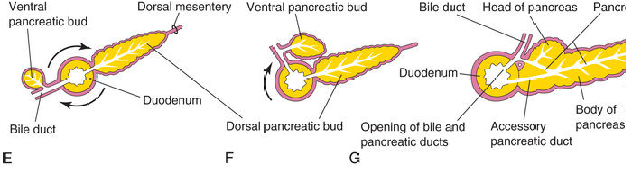
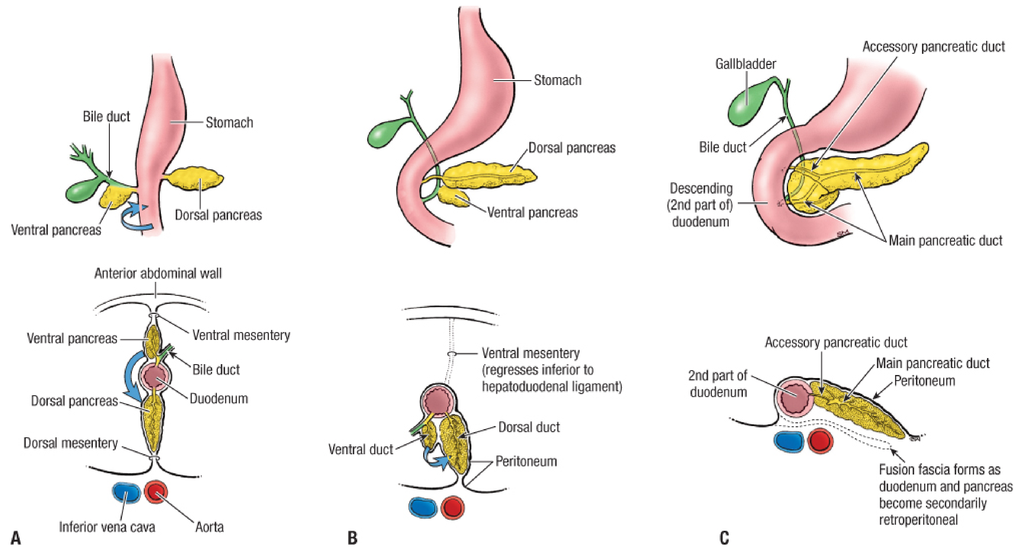
- As the duodenum rotates to the right and becomes C-shaped, the ventral pancreatic bud is carried dorsally along with the bile duct.
- The ventral bud swings behind the dorsal bud and comes to lie posterior to it.
- The two buds fuse to form a single organ.
3. Duct Formation
- The main pancreatic duct (of Wirsung) = duct of the ventral bud + distal part of the duct of the dorsal bud.
- The proximal part of the dorsal duct often persists as the accessory pancreatic duct (of Santorini), which opens at the minor duodenal papilla (~2 cm cranial to the main duct).
- In ~9% of people, the ducts fail to fuse, resulting in pancreas divisum (two separate ducts).
4. Retroperitoneal Position
- The pancreas is carried to lie along the dorsal abdominal wall.
- It becomes secondarily retroperitoneal - the peritoneum on the right side of the duodenum and mesoduodenum fuses with the dorsal parietal peritoneum and degenerates, forming a fusion (Toldt's) fascia.
5. Histogenesis (Cellular Differentiation)
Exocrine Component
- Endodermal cells form a network of tubules (primordial pancreatic ducts).
- Pancreatic acini develop from cell clusters around the ends of these tubules early in the fetal period.
- Chemokine SDF-1 (stromal cell-derived factor-1), expressed in the mesenchyme, controls tubule formation and branching.
Endocrine Component (Islets of Langerhans)
- Develop in the 3rd month from groups of cells that separate from the tubules and scatter among the acini.
- Neurogenin-3 transcription factor is required for endocrine cell differentiation.
- Insulin secretion begins at ~10 weeks (early fetal period).
- Alpha cells (glucagon) and delta cells (somatostatin) differentiate before beta cells.
- Glucagon is detectable in fetal plasma at 15 weeks.
Connective Tissue
- Derived from the surrounding splanchnic (visceral) mesoderm - forms the capsule and interlobular septa.
6. Molecular Regulation
| Signal | Source | Role |
|---|---|---|
| FGF-2 | Notochord + developing heart | Represses Shh in foregut endoderm; induces dorsal pancreatic bud |
| Activin (TGF-β family) | Notochord | Represses Shh in endoderm destined to form dorsal bud |
| PDX1 (Pdx1 homeobox gene) | Expressed in ventral duodenal region | Upregulated after Shh repression; master regulator of pancreas development |
| PAX4 + PAX6 co-expression | Endocrine progenitors | Specifies β, δ, and γ (PP) cells |
| PAX6 alone | Endocrine progenitors | Specifies α (glucagon) cells |
| Neurogenin-3 | Endocrine progenitors | Required for islet cell differentiation |
| Myte | Endocrine progenitors of dorsal bud | Transcription factor in endocrine progenitor cells |
7. Congenital Anomalies
Annular Pancreas
- The ventral bud is bifid (splits into two parts); one part migrates normally, the other migrates in the opposite direction, surrounding the duodenum with pancreatic tissue.
- Can cause duodenal stenosis or obstruction in neonates.
- Associated with Down syndrome, intestinal malrotation, and cardiac defects.
- More common in females.
Ectopic Pancreas (Heterotopic Pancreas)
- Pancreatic tissue found separately from the main pancreas.
- Common locations: mucosa of the stomach, proximal duodenum, jejunum, pyloric antrum, Meckel's diverticulum.
- Usually asymptomatic (found incidentally on CT); may rarely cause GI symptoms, obstruction, bleeding, or malignant transformation.
Pancreas Divisum
- Failure of the two pancreatic ducts to fuse (~9% of people).
- Results in two separate drainage systems.
Hypertrophy of Beta Cells (in maternal diabetes)
- When the mother has diabetes mellitus, the fetal beta cells are chronically exposed to high glucose.
- Beta cells undergo hypertrophy, increasing insulin secretion - leads to fetal macrosomia.
Summary Timeline
| Week | Event |
|---|---|
| Week 4-5 | Dorsal pancreatic bud appears from caudal foregut |
| Week 5 | Ventral pancreatic bud appears near bile duct |
| Week 5-8 | Duodenum rotates right (C-shape); ventral bud rotates dorsally |
| Week 7-8 | Two buds fuse; ducts anastomose |
| Week 8+ | Pancreas becomes secondarily retroperitoneal |
| Month 3 | Islets of Langerhans develop |
| Week 10 | Insulin secretion begins |
| Week 15 | Glucagon detectable in fetal plasma |
- The Developing Human: Clinically Oriented Embryology (Moore et al.), pp. 625-628
- Langman's Medical Embryology, pp. 263-264
- Fischer's Mastery of Surgery, 8th ed., p. 8181
Development of Kidney & associated Congenital Anomalies
Development of the Kidney & Associated Congenital Anomalies
Overview
| Kidney | Week | Region | Fate |
|---|---|---|---|
| Pronephros | Week 3-4 | Cervical/Thoracic | Vestigial; completely degenerates by week 5 |
| Mesonephros | Week 4-8 | Thoracolumbar | Transient; partially regresses; duct persists |
| Metanephros | Week 5 onward | Sacral/Pelvic | Permanent kidney |
1. Pronephros (Week 3-5)
- First evidence appears in the late 3rd week in the cervical/upper thoracic nephrogenic cord.
- Tubules form in a craniocaudal wave, immediately followed by craniocaudal degeneration.
- Non-functional in humans (analogous to the kidney of primitive fish).
- Key legacy: generates the pronephric duct, which grows caudally and, as pronephric tubules degenerate, becomes renamed the mesonephric (Wolffian) duct.
2. Mesonephros (Week 4-8)
- Develops in the thoracolumbar region; more developed than the pronephros.
- Its tubules acquire glomeruli and may function temporarily as an excretory organ while the metanephros is forming.
- Regresses mostly by week 8, but the mesonephric (Wolffian) duct is retained and plays critical roles:
- In males: becomes epididymis, vas deferens, seminal vesicles, ejaculatory duct.
- In females: largely degenerates (vestigial remnants = epoophoron, paroophoron).
- The ureteric bud sprouts from the mesonephric duct near the cloaca - the key initiating event for permanent kidney development.
3. Metanephros - The Permanent Kidney (Week 5 onward)
A. Ureteric Bud (Metanephric Diverticulum)
- An outgrowth of the mesonephric duct, close to its entrance to the cloaca, at ~week 5.
- Penetrates the metanephric mesenchyme (blastema).
B. Metanephric Mesenchyme (Metanephric Blastema)
- Derived from the caudal portion of the nephrogenic cord.
- Caps the ureteric bud distally.
- Provides signals that drive ureteric bud branching.
4. Collecting System Development (Ureteric Bud Derivatives)
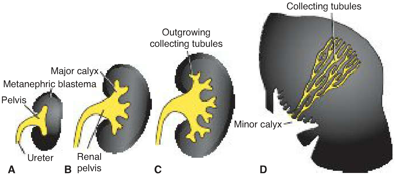
- The bud dilates to form the primitive renal pelvis, then splits into cranial and caudal portions = major calyces.
- Each calyx forms two new buds that further subdivide into minor calyces.
- Continued branching to 12 or more generations forms the collecting tubules (1-3 million total).
- Tubules of the 2nd generation enlarge and absorb those of the 3rd and 4th generations = minor calyces.
- 5th generation and beyond elongate and converge = renal pyramids.
- Ureter
- Renal pelvis
- Major calyces
- Minor calyces
- ~1-3 million collecting tubules
5. Nephron Formation (Metanephric Mesenchyme Derivatives)
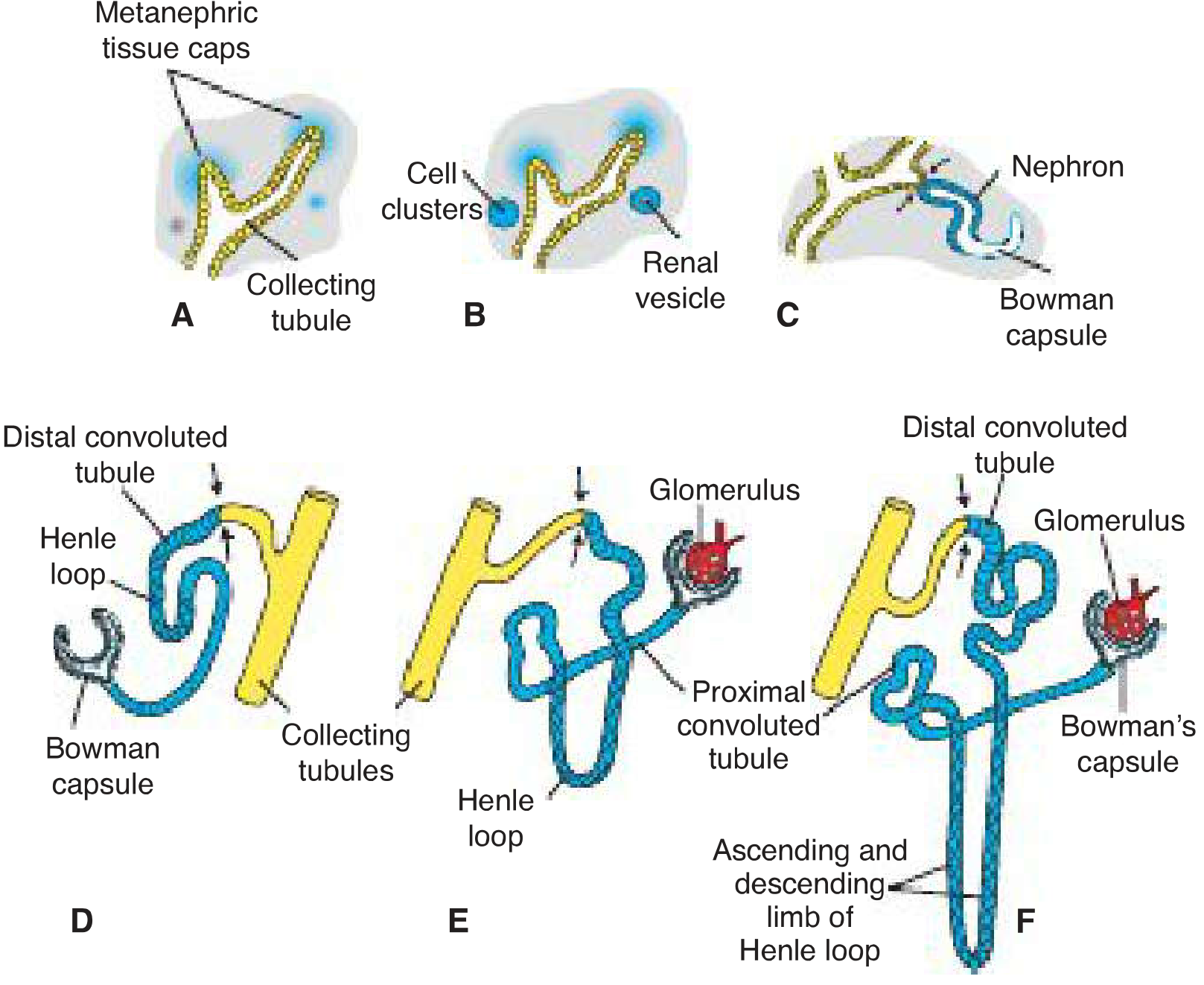
- Metanephric tissue cap condenses around each collecting tubule tip.
- Cap cells form renal vesicles.
- Vesicles elongate into S-shaped tubules.
- Capillaries invade one end of the S-shape → differentiate into glomeruli.
- The proximal end forms Bowman's capsule (deeply indented by the glomerulus).
- Continued elongation forms:
- Proximal convoluted tubule (PCT)
- Loop of Henle
- Distal convoluted tubule (DCT)
- The distal end opens into a collecting tubule, completing the nephron.
- Nephron formation continues until birth (~1 million nephrons per kidney at birth).
- Urine production begins at week 12 (after glomerular capillary differentiation beginning at week 10).
- At birth, kidneys appear lobulated - lobulation disappears in infancy as nephrons grow (but number does not increase after birth).
6. Positional Changes - Ascent of Kidney
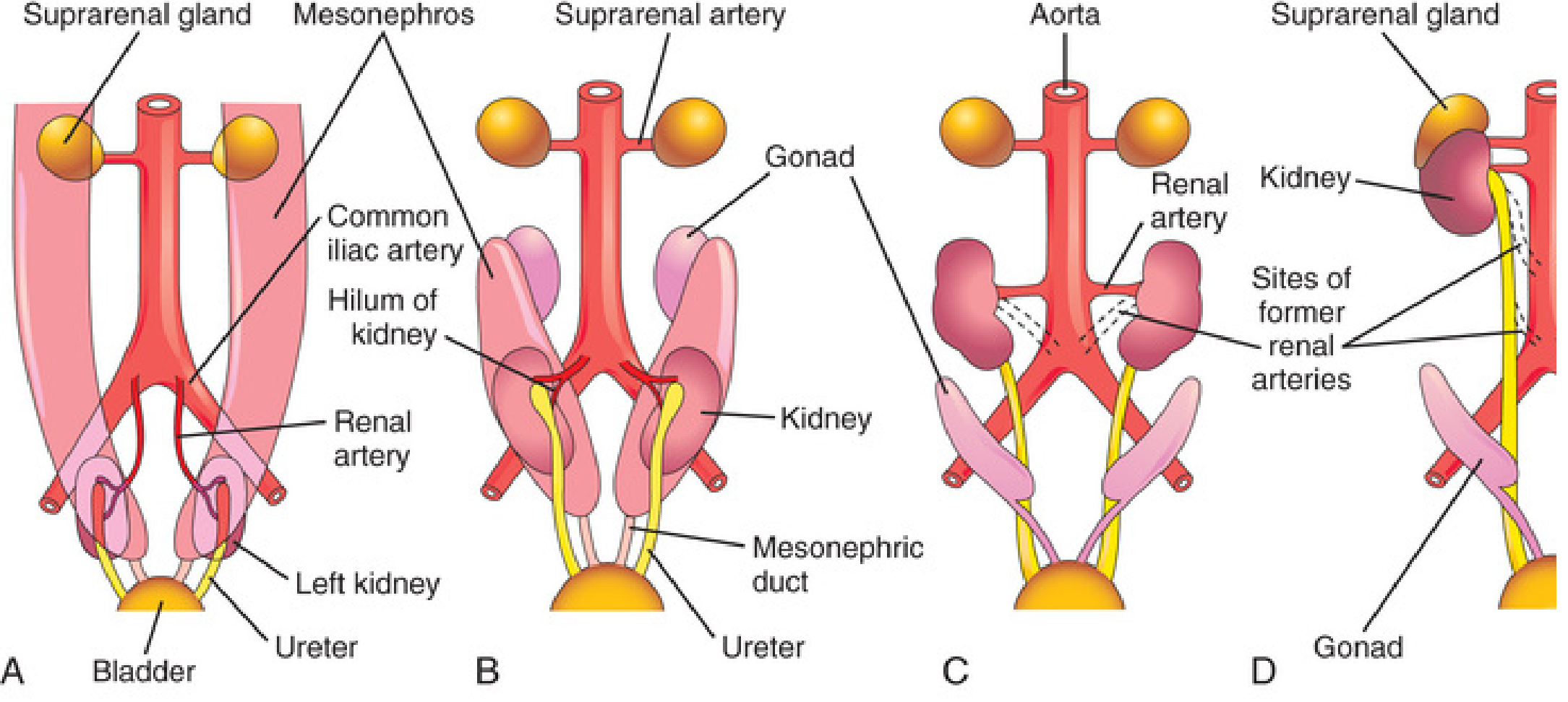
- Not true active migration; the caudal part of the embryo grows away from the kidneys, so they appear to "ascend."
- Kidneys reach their adult position (flanking the vertebral column at T12-L3) by the beginning of the fetal period.
- Initially the hilum faces ventrally.
- As the kidney ascends, it rotates medially ~90 degrees.
- By week 9, the hila face anteromedially.
- Kidneys become retroperitoneal and come in contact with the suprarenal glands (which fixes their position).
- Initially supplied by common iliac arteries.
- As they ascend, they receive new branches from the distal aorta, then the abdominal aorta at higher levels.
- Caudal branches involute and disappear normally.
7. Molecular Regulation
| Molecule | Source | Role |
|---|---|---|
| GDNF (glial-derived neurotrophic factor) | Metanephric mesenchyme | Activates RET receptor on ureteric bud; drives bud outgrowth and branching |
| RET (tyrosine kinase receptor) | Ureteric bud epithelium | Receptor for GDNF |
| HGF (hepatocyte growth factor) | Mesenchyme | Stimulates branching via MET receptor on ureteric bud |
| WT1 | Metanephric mesenchyme | Transcription factor; makes mesenchyme competent to respond to ureteric bud; also regulates GDNF and HGF |
| FGF2, BMP7 | Ureteric bud | Signals back to mesenchyme; block apoptosis, stimulate proliferation |
| LIM1, PAX2 | Intermediate mesoderm | Early transcription factors; LIM1 essential for nephric duct formation |
| Neurogenin-3 | Endocrine progenitors (note: shared with pancreas) | Islet differentiation |
| SIX2 | Nephron progenitors | Promotes progenitor self-renewal |
| WNT9b | Ureteric bud | Drives mesenchyme induction; promotes differentiation |
- Fibronectin, collagen I, III (mesenchymal matrix) are replaced by laminin and type IV collagen (epithelial basal lamina).
- Cell adhesion molecules syndecan and E-cadherin are expressed.
8. Congenital Anomalies of the Kidney & Ureter
A. Renal Agenesis
- Incidence: ~1 in 1000 neonates.
- Males > Females; left kidney more often absent.
- Usually asymptomatic (contralateral kidney undergoes compensatory hypertrophy).
- Suspect in infants with a single umbilical artery.
- Mechanism: ureteric bud fails to develop OR fails to penetrate the metanephric blastema.
- Incidence: ~1 in 3000 births; incompatible with postnatal life.
- Causes oligohydramnios (little/no urine excreted into amniotic fluid).
- Results in Potter sequence (syndrome):
- Potter facies: widely spaced eyes, epicanthic folds, low-set ears, broad flat nose, receding chin.
- Compressed uterine cavity → clubfeet.
- Oligohydramnios → pulmonary hypoplasia → respiratory insufficiency (main cause of death).
- ~85% of cases have associated defects (absence of vagina/uterus, vas deferens; cardiac, tracheal, duodenal anomalies).
B. Renal Dysplasia & Multicystic Dysplastic Kidney
- Spectrum of severe malformations - primary reason for dialysis/transplant in early life.
- Numerous ducts surrounded by undifferentiated cells; nephrons fail to develop; collecting ducts never form (ureteric bud fails to branch properly).
- Can involute completely → renal agenesis.
C. Polycystic Kidney Disease
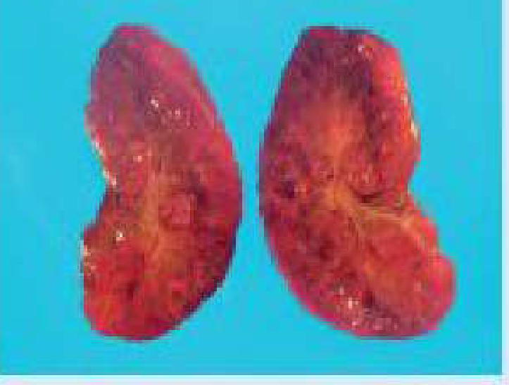
| Feature | ARPKD | ADPKD |
|---|---|---|
| Inheritance | Autosomal recessive | Autosomal dominant |
| Incidence | 1/5,000 | 1/500-1,000 |
| Cyst origin | Collecting ducts | All nephron segments |
| Onset | Infancy/childhood | Usually adulthood |
| Progression | Rapidly progressive | Slower |
| Gene | PKHD1 | PKD1, PKD2 |
- Bardet-Biedl syndrome: renal cysts + obesity + intellectual disability + limb defects.
- Meckel-Gruber syndrome: renal cysts + hydrocephalus + microphthalmia + cleft palate + polydactyly.
D. Malrotated Kidney
- If the kidney fails to rotate: hilum faces anteriorly (retained fetal position).
- If rotated too far: hilum faces posteriorly.
- If lateral rotation: hilum faces laterally.
- Often associated with ectopic kidneys.
E. Ectopic Kidneys
- One or both kidneys remain in an abnormal position due to failure of ascent.
- Most ectopic kidneys are in the pelvis (pelvic kidney).
- Pelvic kidneys are close together and may fuse to form a discoid/pancake kidney.
- Supplied by multiple vessels from nearby structures (internal/external iliac arteries).
- Crossed renal ectopia: one kidney crosses to the opposite side; 90% are fused.
F. Horseshoe Kidney
- Most common renal fusion defect; occurs in 0.2% of the population.
- Usually the inferior poles fuse (rarely the superior poles).
- The U-shaped kidney typically lies anterior to L3-L5 vertebrae.
- Mechanism: the two kidneys fuse across the midline during pelvic ascent, and the inferior mesenteric artery blocks further ascent; horseshoe kidney therefore lies below the inferior mesenteric artery in 60% of cases.
- Usually asymptomatic (collecting system develops normally, ureters enter bladder).
- Can cause urinary obstruction (stones, hydronephrosis) or infection.
- Associated with Turner syndrome (~7% of patients).
G. Accessory (Supernumerary) Renal Arteries
- Occur in ~25% of adult kidneys (2-4 renal arteries).
- Arise from persistence of embryonic renal vessels that normally involute during ascent.
- Accessory artery to inferior pole may cross anterior to the ureter and obstruct it → hydronephrosis.
- Accessory arteries are end arteries - damage causes ischemia of the supplied segment.
H. Ureteric Duplication (Duplex Collecting System)
- Results from early splitting or formation of two ureteric buds.
- Incomplete division: bifid ureter (two ureters joining before entering bladder).
- Complete division: double kidney with bifid ureter or completely separate ureters.
- One of the duplicated ureters may be ectopic, entering the vagina, urethra, or vestibule in females.
I. Ectopic Ureter
- Does not enter the bladder normally; results from failure of incorporation into the trigone.
- Males: opens into neck of bladder, prostatic urethra, ductus deferens, seminal vesicles.
- Females: opens into urethra, vagina, or vestibule → continuous urine dribbling (incontinence despite normal voiding).
J. Renal Tumors (Developmental Basis)
- Wilms Tumor (Nephroblastoma): mutation in WT1 gene (chromosome 11p13); usually in children under 5.
- WAGR syndrome: Wilms tumor + Aniridia + Gonadoblastoma + mental Retardation - microdeletion of chromosome 11 involving both PAX6 and WT1 genes.
- Denys-Drash syndrome: renal failure + ambiguous genitalia + Wilms tumor.
Summary Timeline
| Week | Event |
|---|---|
| Week 3-4 | Pronephros appears and degenerates; pronephric duct forms |
| Week 4 | Mesonephros develops; mesonephric duct grows caudally to cloaca |
| Week 5 | Ureteric bud sprouts from mesonephric duct; penetrates metanephric mesenchyme |
| Week 5-8 | Ureteric bud branches repeatedly; collecting system forms |
| Week 6-9 | Kidneys ascend from pelvis; medial rotation of hilum ~90 degrees |
| Week 9 | Kidneys fixed in adult position by contact with suprarenal glands |
| Week 10 | Glomerular capillary differentiation begins |
| Week 12 | Urine production begins |
| Until birth | Nephron formation continues (~1 million per kidney at birth) |
- The Developing Human: Clinically Oriented Embryology (Moore et al.), pp. 692-698
- Langman's Medical Embryology, pp. 274-282
- Campbell-Walsh-Wein Urology, 3-Volume Set, pp. 1949-2088
Why rbc count is more in newborns
Why RBC Count is Higher in Newborns
Normal Values at a Glance
| Parameter | Newborn | Adult (male) |
|---|---|---|
| RBC count | ~5.5-6.5 million/µL | ~4.5-5.5 million/µL |
| Hemoglobin | 160-240 g/L (16-24 g/dL) | 130-170 g/L |
| Hematocrit | 55-68% | 40-52% |
The Core Reason: Fetal Hypoxia
Mechanism 1: Increased RBC Production (Erythropoiesis)
- The kidney detects tissue hypoxemia and responds by secreting erythropoietin (EPO).
- EPO drives the bone marrow (and before that, the liver and spleen in fetal life) to produce more red blood cells.
- This results in physiological polycythemia of the newborn - more RBCs in circulation to carry as much oxygen as possible from the placenta to fetal tissues.
- Hyperglycemia (e.g., in infants of diabetic mothers) is an additional powerful stimulus for fetal EPO production, probably mediated by further decreases in fetal oxygen tension - leading to even higher RBC counts in those neonates.
Mechanism 2: Fetal Hemoglobin (HbF) with Higher Oxygen Affinity
- Newborn RBCs contain primarily fetal hemoglobin (HbF), which is structurally different from adult hemoglobin (HbA).
- HbF has higher oxygen affinity than HbA - its oxygen-dissociation curve is shifted to the left.
- This means HbF binds oxygen more readily at the low oxygen levels present in the placental vascular bed, allowing efficient loading of O₂ from maternal blood.
- The downside: HbF releases oxygen less efficiently at the tissue level.
- Working together, the higher RBC count (more carriers) + HbF (better at capturing available O₂) ensures adequate oxygen delivery to fetal tissues.
What Happens After Birth?
- EPO secretion falls sharply from the kidneys (no longer driven by hypoxia).
- New RBC production drops dramatically.
- The large number of excess RBCs are rapidly destroyed (hemolyzed), especially the older, shorter-lived fetal RBCs.
- HbF is progressively replaced by adult HbA over the first 6 months of postnatal life.
- Hemoglobin falls rapidly in the first 3 months of life.
Summary Flow
Fetal life → Low PO₂ (placental gas exchange)
     ↓
Kidney senses hypoxia → ↑ Erythropoietin secretion
     ↓
↑ RBC production in marrow/liver/spleen (fetal hematopoiesis)
     ↓
High RBC count at birth (~5.5-6.5 million/µL) + HbF predominance
     ↓
Birth → Lungs take over → PO₂ rises → EPO drops
     ↓
RBC production stops → Excess RBCs destroyed
     ↓
↓ RBC count over weeks (physiological anemia of infancy by ~8-12 weeks)
↓ Bilirubin load → Physiological neonatal jaundice (day 2-3)
Clinical Significance
| Condition | Link to High Neonatal RBC Count |
|---|---|
| Physiological jaundice (day 2-3) | Bilirubin from excess RBC breakdown overwhelms immature liver |
| Pathological polycythemia (Hb >20 g/dL) | Vascular sludging, ischemia, kernicterus risk |
| Physiological anaemia of infancy (~8-12 weeks) | Post-birth EPO suppression + rapid growth diluting RBC mass |
| Infants of diabetic mothers | Higher EPO from hyperglycemia-induced hypoxia → higher polycythemia risk |
- Miller's Anesthesia, 10e, p. 10557 - direct textbook quotation: "The fetus uses two compensatory mechanisms to assure adequate oxygen delivery in the relatively hypoxemic in utero environment. One of them is the increased red blood cell production resulting from increased fetal renal erythropoietin secretion in response to hypoxemia."
- Barash, Cullen & Stoelting's Clinical Anesthesia, 9e, p. 3613
- Creasy & Resnik's Maternal-Fetal Medicine, p. 1440
- Grainger & Allison's Diagnostic Radiology, p. 2950
Can u give development of kidney in flowchart
A detailed medical educational flowchart titled "Development of the Kidney" showing the following sequential and branching information in a clean, professional style with colored boxes and arrows: TOP LEVEL: "INTERMEDIATE MESODERM" (wide blue box) Arrow down to: "NEPHROGENIC CORD → UROGENITAL RIDGE" Then splits into THREE parallel columns labeled: COLUMN 1 - "1. PRONEPHROS (Week 3-4)" [light red box] - Cervical/Thoracic region - Non-functional (vestigial) - Degenerates by Week 5 - Arrow: "Duct retained → becomes Mesonephric (Wolffian) Duct" COLUMN 2 - "2. MESONEPHROS (Week 4-8)" [light orange box] - Thoracolumbar region - Temporary excretory function - Partially regresses - Arrow down: "URETERIC BUD sprouts from Mesonephric Duct (Week 5)" COLUMN 3 - "3. METANEPHROS (Week 5+)" [green box] - PERMANENT KIDNEY - Sacral/Pelvic region From URETERIC BUD arrow to: "Penetrates METANEPHRIC MESENCHYME (Blastema)" [yellow box] Then TWO branches side by side: LEFT BRANCH - "URETERIC BUD DERIVATIVES (Collecting System)" [blue box]: Ureteric Bud → Dilates → Primitive Renal Pelvis → splits → Major Calyces → Minor Calyces → 12+ generations of branching → Collecting Tubules (1-3 million) → Renal Pyramids Also forms: Ureter RIGHT BRANCH - "METANEPHRIC MESENCHYME DERIVATIVES (Nephrons)" [purple box]: Metanephric cap → Renal Vesicles → S-shaped tubules → Glomerulus (capillaries invade) + Bowman's Capsule → PCT → Loop of Henle → DCT → connects to Collecting Tubule ~1 million nephrons at birth; nephron formation complete at birth Both branches merge at: "COMPLETE NEPHRON UNIT" [dark green box] BELOW that, a section titled "POSITIONAL CHANGES": Week 5-9 box: Kidneys initially in PELVIS → Ascend to abdomen (embryo grows caudally) → Hilum rotates medially 90° → Hila face anteromedially → Kidneys become RETROPERITONEAL TIMELINE at the bottom as a horizontal bar: Week 3-4: Pronephros | Week 4: Mesonephros | Week 5: Ureteric bud | Week 9: Adult position fixed | Week 10: Glomeruli form | Week 12: Urine production begins | Birth: ~1 million nephrons Use medical textbook style, clear readable font, color-coded columns, arrows between all steps
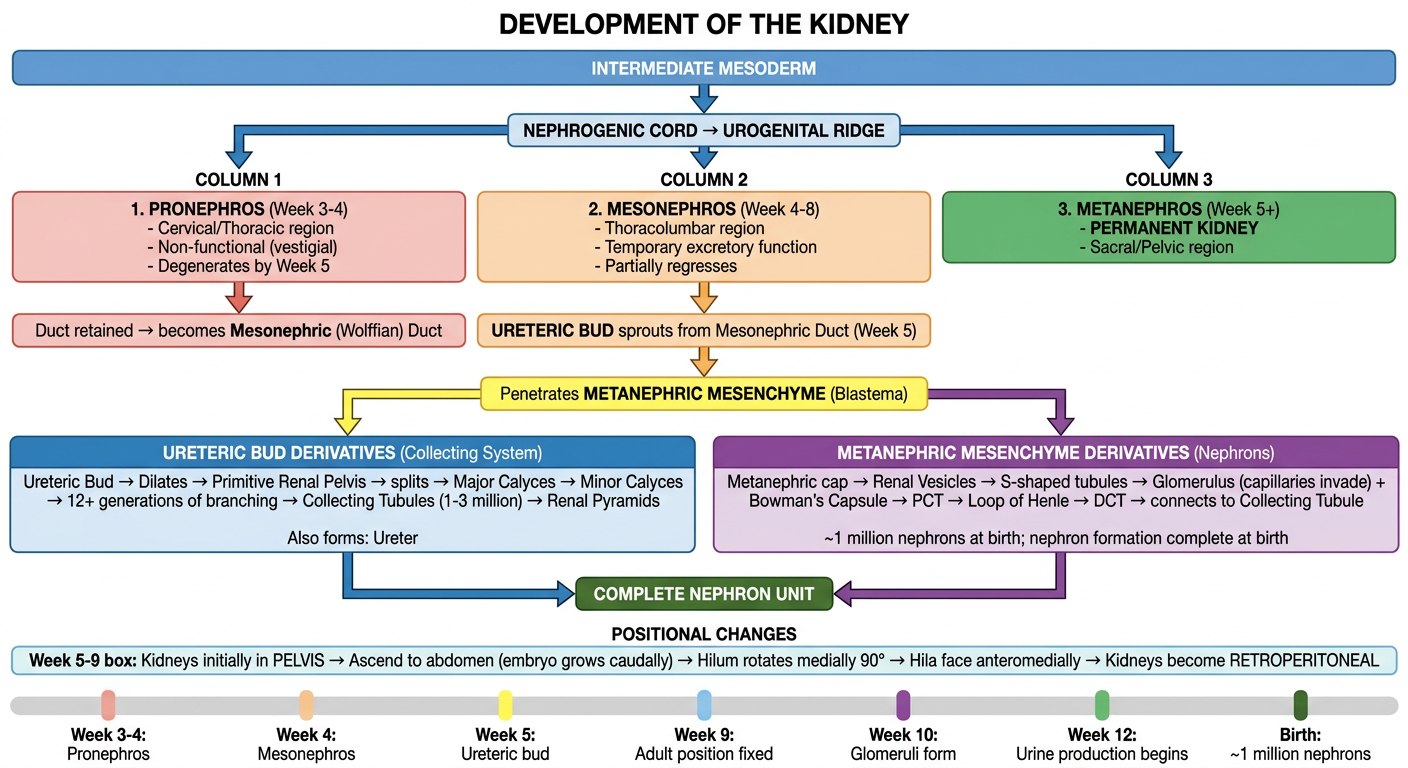
- Top: Everything originates from the intermediate mesoderm → nephrogenic cord
- Three columns show the three kidney systems in order (pronephros → mesonephros → metanephros)
- Two branches from the metanephros show what each tissue component contributes:
- Ureteric bud → entire collecting system (pelvis, calyces, collecting tubules, ureter)
- Metanephric mesenchyme → all nephrons (glomerulus to DCT)
- Positional changes section shows the ascent and medial rotation
- Timeline bar at the bottom gives week-by-week landmarks
1
- More flowcharts — e.g., development of pancreas flowchart, RBC count flowchart?
- A specific question — did you mean to type something and it got cut off?
- Option 1 from a previous list?
1
What did you mean by '1'?
A detailed medical educational flowchart titled "Development of the Pancreas" in a clean professional style with color-coded boxes and arrows, suitable for medical students. TOP BOX (wide dark blue): "CAUDAL FOREGUT ENDODERM (Week 4-5)" Arrow down splits into TWO parallel branches: LEFT BRANCH - labeled "DORSAL PANCREATIC BUD" (green box): - Appears FIRST - Cranial to ventral bud - Grows between layers of dorsal mesentery - Larger bud Arrow down: "Forms → Body, Tail, and part of Head" RIGHT BRANCH - labeled "VENTRAL PANCREATIC BUD" (orange box): - Near entry of bile duct into duodenum - Grows in ventral mesentery - Smaller bud Arrow down: "Forms → Uncinate process + part of Head" Between the two branches, a bold arrow labeled "DUODENUM ROTATES RIGHT (C-shape) Week 5-8" pointing right From ventral bud: "Ventral bud carried DORSALLY with Bile Duct → comes to lie POSTERIOR to dorsal bud" Both branches merge with a large arrow labeled "FUSION of both buds (Week 7-8)" into: CENTER BOX (dark green): "SINGLE PANCREATIC ORGAN" Arrow down splits into TWO sub-branches: LEFT SUB-BRANCH "DUCT FORMATION" (blue box): - Ducts of both buds anastomose - Main Pancreatic Duct (Wirsung) = Ventral duct + Distal part of dorsal duct - Accessory Pancreatic Duct (Santorini) = Proximal part of dorsal duct → opens at Minor Duodenal Papilla (~2 cm cranial) - 9% of people: ducts fail to fuse → PANCREAS DIVISUM RIGHT SUB-BRANCH "HISTOGENESIS" (purple box): - Endoderm forms network of TUBULES - Acini develop from cell clusters at tubule ends (Exocrine) - Islets of Langerhans separate from tubules (Month 3) (Endocrine) - Insulin secretion begins: Week 10 - Alpha cells (glucagon) + Delta cells (somatostatin) develop BEFORE beta cells - Glucagon detectable in plasma: Week 15 - Connective tissue from splanchnic mesoderm BELOW both sub-branches: "RETROPERITONEAL POSITION" box (grey): As stomach + duodenum + ventral mesentery rotate → Pancreas lies along dorsal abdominal wall → Becomes secondarily retroperitoneal CONGENITAL ANOMALIES section at bottom (red-bordered box) with three side-by-side boxes: Box 1 "Annular Pancreas": Bifid ventral bud wraps around duodenum → Duodenal stenosis/obstruction. Associated: Down syndrome Box 2 "Ectopic Pancreas": Pancreatic tissue in stomach/duodenum/jejunum/Meckel's diverticulum. Usually asymptomatic Box 3 "Pancreas Divisum": Failure of duct fusion (~9%). Two separate drainage systems TIMELINE bar at very bottom: Week 4-5: Dorsal bud appears | Week 5: Ventral bud appears | Week 5-8: Duodenum rotates | Week 7-8: Buds fuse, ducts anastomose | Month 3: Islets form | Week 10: Insulin secretion | Week 15: Glucagon detectable Use medical textbook style with clean arrows, readable font, color-coded sections
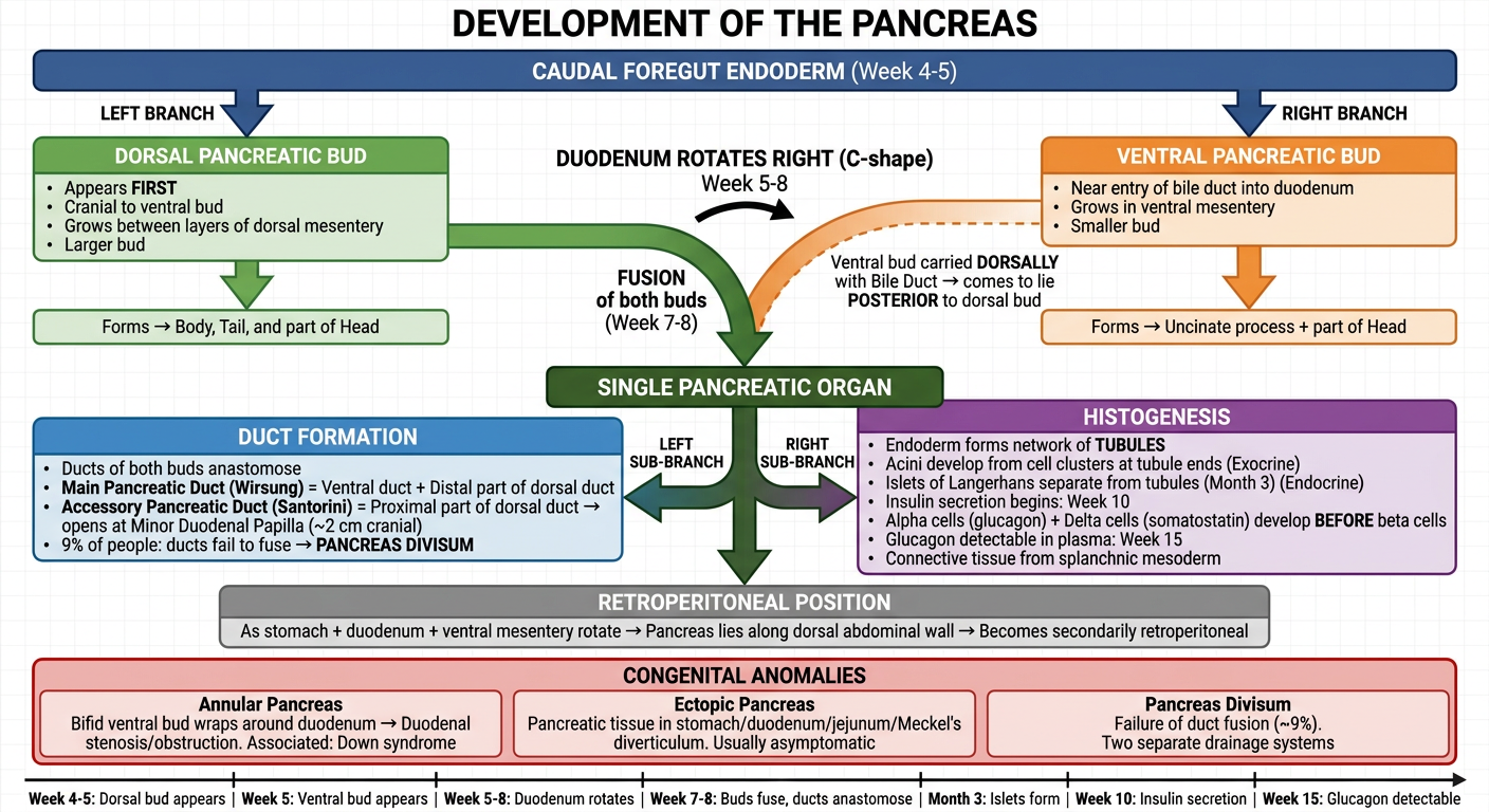
- Two buds arise from the caudal foregut simultaneously
- Duodenal rotation is the key mechanical event that swings the ventral bud behind the dorsal bud
- Fusion creates the single organ
- After fusion, two things happen in parallel - duct formation (left) and histogenesis/cell differentiation (right)
- Congenital anomalies at the bottom all trace back to errors in the rotation/fusion step
- Ventral bud → uncinate process + part of head
- Main duct = ventral duct + distal dorsal duct
- Accessory duct = proximal dorsal duct (Santorini)
- Annular pancreas = bifid ventral bud wrapping duodenum
- Pancreas divisum = 9% of people, ducts never fuse