
Pouch of Douglas nd it’s clinical features
Reading File
Now let me search the medical library for Pouch of Douglas content in parallel.
Finding Sources
Finding Sources
Excellent results. Let me now read the key sections in parallel.
Reading File
Reading File
Reading File
Reading File
Now let me fetch the anatomy image and search for more clinical features content simultaneously.
Loading Image
Finding Sources
Let me read the culdocentesis anatomy section and the gynecology section for fuller clinical context.
Reading File
Reading File
Finding Sources
I now have comprehensive information from multiple authoritative textbooks. Let me compile the full answer.
Pouch of Douglas (Rectouterine Pouch)
Definition and Anatomy
The Pouch of Douglas (also called the rectouterine pouch or cul-de-sac of Douglas) is a peritoneal recess formed by the reflection of peritoneum between the rectum (posteriorly) and the uterus/upper vagina (anteriorly) in females. It is the deepest point of the peritoneal cavity in the upright and supine positions.
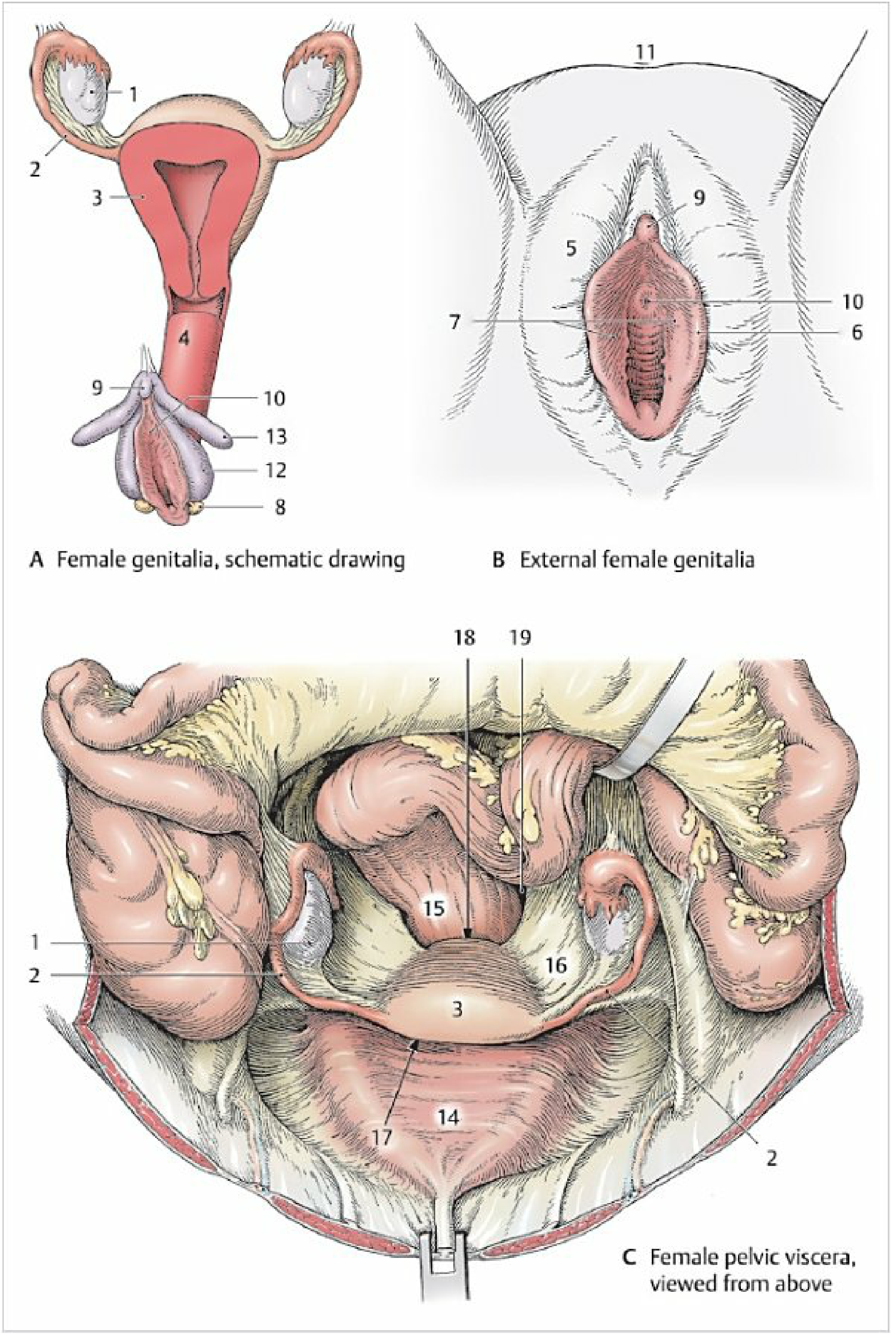
Boundaries:
- Anteriorly: Posterior surface of the uterus and upper vagina
- Posteriorly: Anterior surface of the rectum
- Laterally: Rectouterine folds (sacrouterine/uterosacral ligaments) containing subserous fibrous connective tissue and the inferior hypogastric nerve plexus
In males, the equivalent space is the rectovesical pouch (between rectum and bladder), which is shallower. The posterior vaginal wall is adjacent to the pouch only at its uppermost ~2 cm, with the vaginal wall being less than 5 mm thick in this region.
Contents (normally): Small intestine and a small amount of physiologic peritoneal fluid.
Clinical Significance
1. Collection of Free Fluid (Most Important Clinical Feature)
Because it is the most dependent part of the peritoneal cavity, free fluid from any source gravitates here. This is exploited diagnostically in:
- Ectopic pregnancy (hemoperitoneum): Free fluid or an adnexal mass seen on ultrasound in the pouch of Douglas strongly suggests ectopic pregnancy. The Bailey & Love's Short Practice of Surgery (28th ed.) notes: "An ectopic pregnancy is more likely if free fluid is seen in the pouch of Douglas or an adnexal mass is identified on ultrasound scan."
- Ruptured corpus luteum cyst, ruptured viscus, liver/spleen injury - blood pools here
- FAST exam (Focused Assessment with Sonography for Trauma): The pouch of Douglas is one of four standard windows assessed for free fluid - Harriet Lane Handbook, 23rd ed.
2. Pelvic Abscess
A pelvic abscess in the pouch of Douglas can complicate any abdominal disease or operation (appendicitis, diverticulitis, PID, post-operative). Clinical features:
- Swinging (spiking) pyrexia developing after a few days
- Mucous discharge per rectum (due to rectal wall irritation)
- Urinary frequency (irritation of the bladder base)
- Rectal examination: Tender, boggy/fluctuant mass palpable anteriorly
- Treatment: Abscess may discharge spontaneously into the rectum; if not, drainage via proctoscope (sinus forceps plunged through the bulging anterior rectal wall) - Pye's Surgical Handicraft, 22nd ed.
3. Endometriosis
The pouch of Douglas is one of the most common sites for endometriotic deposits (along with ovaries, uterine ligaments, fallopian tubes, and rectovaginal septum). Advanced endometriosis can:
- Obliterate the pouch of Douglas entirely
- Cause fibrotic plaques involving the rectosigmoid
- Form "chocolate cysts" (endometriomas) with brown fluid from old hemorrhage
- Present with deep dyspareunia, pelvic pain, and dysmenorrhea - Robbins Basic Pathology
4. Malignant Deposits ("Rectal Shelf" / Blumer's Shelf)
Malignant cells from intra-abdominal carcinomas (e.g., stomach, ovary, colon) drop to this lowest point. On rectal examination, hard, irregular nodular deposits can be felt anteriorly in the pouch of Douglas - this is called Blumer's shelf or "rectal shelf sign." This is felt on rectal exam as pus, blood, or malignant deposit.
- S Das Manual on Clinical Surgery (13th ed.) specifically notes: "Presence of pus, blood, malignant deposit (e.g., from carcinoma of the stomach) or tumour of the sigmoid colon may be felt through the pouch."
5. Fistulae
Disease in the pouch of Douglas (Crohn disease, diverticular disease, carcinoma, radiation injury) can cause fistulae through the posterior fornix of the vagina, resulting in rectovaginal fistulae. These present with passage of flatus or feces per vagina - Fischer's Mastery of Surgery, 8th ed.
6. Herniation (Enterocele / Douglas Hernia)
A Pouch of Douglas hernia originates in the cul-de-sac and protrudes via the anterior rectal wall, potentially presenting as rectal prolapse through the anus.
Clinical Examination
On rectal examination (index finger fully inserted reaches ~1 inch above the floor of the pouch in females, ~half that in males):
- Normally: softness, no tenderness
- Pathological: tender mass (abscess), hard nodules (malignant drop metastases), free fluid
On vaginal examination: the pouch can be palpated through the posterior vaginal fornix.
Culdocentesis
The shallow vaginal wall overlying the pouch (< 5 mm thick) allows culdocentesis - needle aspiration through the posterior fornix. This is used to:
- Confirm hemoperitoneum (non-clotting blood) in suspected ectopic pregnancy
- Diagnose PID/pelvic infection (turbid or purulent fluid)
- Differentiate inflammatory from hemorrhagic pelvic pathology
- Most useful when ultrasound is unavailable or the patient is hemodynamically unstable - Roberts & Hedges' Clinical Procedures in Emergency Medicine
Summary Table
| Clinical Condition | Feature in Pouch of Douglas |
|---|---|
| Ectopic pregnancy / hemoperitoneum | Free blood (non-clotting) on culdocentesis; echogenic fluid on USS |
| Pelvic abscess | Tender boggy mass per rectum; swinging fever; mucous PR discharge |
| Endometriosis | Obliteration of pouch; deep dyspareunia; pelvic pain |
| Malignant deposits | Hard nodular shelf (Blumer's shelf) on PR exam |
| Rectovaginal fistula | From disease eroding through the pouch |
| Peritonitis (any cause) | Fluid accumulation detectable by USS or culdocentesis |
This is a shared conversation. Sign in to Orris to start your own chat.