
HISTORY A 47 year old man with recently diagnosed type 2 Diabetes mellitus presented with high grade fever for 20 days which was intermittent more during the night , associated with chills and rigors. He subsequently developed a persistent dry cough and right upper abdominal pain for 3 days , fatigue and reduced oral intake . There was no history of vomiting , diarrhea , blood or mucus in stool, dysuria , jaundice , breathlessness , significant weight loss EXAMINATION FINDINGS The patient was febrile and requiring 2 L/min oxygen, with RR 22/min. Abdomen was soft with right hypochondrial and epigastric tenderness, and liver was palpable 2 cm below the right costal margin. Respiratory examination revealed right basal bronchial breath sounds. Give me a proper ddx for this
liver abscess differential diagnosis fever hepatomegaly pleuropulmonary complications diabetic
pyogenic liver abscess CT scan rim enhancement
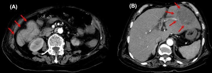
Two-panel axial contrast-enhanced CT scan of the abdomen demonstrating primary colonic malignancy and a secondary hepatic complication. Image (A) shows a transverse colon tumor in the right abdominal region, indicated by red arrows. The mass displays circumferential wall thickening, heterogeneous contrast enhancement, and irregular borders, suggestive of adenocarcinoma. Image (B) shows a large, complex liver abscess within the right hepatic lobe, highlighted by red arrows. The abscess presents as a multiloculated, low-attenuation fluid collection with peripheral rim enhancement and internal septations, characteristic of a pyogenic liver abscess. The surrounding hepatic parenchyma exhibits subtle inflammatory changes. These diagnostic images illustrate the clinical association between obstructive or perforated colorectal cancer and the development of pyogenic liver abscesses, often caused by portal venous seeding of enteric flora like Klebsiella pneumoniae. This visual material is suitable for teaching diagnostic radiology of the gastrointestinal system and hepatobiliary complications of malignancy.
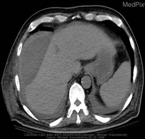
**Imaging Modality:** Computed Tomography (CT) scan of the upper abdomen, axial section.
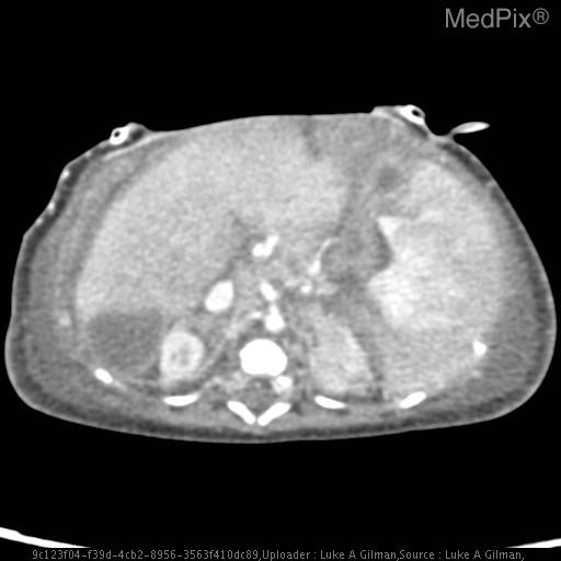
**Imaging Modality:** Axial contrast-enhanced Computed Tomography (CT) scan of the upper abdomen.
Differential Diagnosis
Case Summary
Working Diagnosis (Most Likely)
1. Pyogenic Liver Abscess (PLA) with Right Pleuropulmonary Complication
- Diabetes mellitus is one of the strongest independent risk factors for PLA, including Klebsiella pneumoniae liver abscess, which is disproportionately associated with T2DM. As noted by Bailey & Love: "There is an increased incidence in the elderly, those with diabetes and the immunosuppressed."
- 20 days of fever with chills/rigors is the classic presentation - PLA often causes a prolonged "fever of unknown origin" pattern before localizing symptoms emerge
- The dry cough and right basal bronchial breath sounds are explained by the abscess exerting upward pressure on the right hemidiaphragm, causing a reactive right pleural effusion and basal consolidation (bronchial breath sounds = consolidation/effusion). StatPearls confirms pleuropulmonary fistula and subphrenic extension as recognized complications
- RUQ pain + hepatomegaly develop as the abscess enlarges and stretches the capsule
- Causative organisms: Klebsiella pneumoniae (especially in diabetics - can cause a hypervirulent metastatic syndrome), E. coli, Streptococcus milleri, anaerobes
Close Second - Must Exclude
2. Amoebic Liver Abscess (ALA)
- Young-middle aged male
- 2-4 week subacute onset
- Fever, cough, RUQ pain, hepatomegaly
- No jaundice (ALA is characteristically non-icteric)
- No diarrhea (concurrent colitis is absent in most ALA cases)
- Newly diagnosed diabetes mellitus - per Maingot's: "amebic liver abscesses are more likely to occur in males younger than 50 years who have immigrated or traveled to an endemic country... The patient will also not be jaundiced or have biliary disease or diabetes mellitus" - diabetes is specifically cited as a feature favouring pyogenic over amoebic
- No history of travel to endemic region or dysentery
- Age 47 is slightly above the typical peak for ALA (20s-30s)
Important Differentials to Keep on the Radar
3. Subphrenic Abscess
4. Hydatid Cyst of Liver (Echinococcal Disease)
5. Complicated Cholecystitis / Ascending Cholangitis
6. Right-Sided Community-Acquired Pneumonia / Parapneumonic Empyema
7. Hepatocellular Carcinoma with Secondary Infection / Necrosis
8. Visceral Leishmaniasis (Kala-Azar)
9. Infective Endocarditis with Septic Hepatic Emboli
Summary Table
| Diagnosis | Key Supporting Features | Key Against |
|---|---|---|
| Pyogenic Liver Abscess (top pick) | Diabetes, prolonged fever + chills, RUQ pain, hepatomegaly, pleuropulmonary signs | Need imaging confirmation |
| Amoebic Liver Abscess | Subacute fever, RUQ pain, cough, hepatomegaly, no diarrhea/jaundice | Diabetes, age, no travel history |
| Subphrenic Abscess | Right pleural signs, fever, RUQ pain | No preceding abdominal surgery/perforation history |
| Hydatid Cyst (infected) | Long subacute course, hepatomegaly | No livestock/dog exposure, no eosinophilia |
| Cholangitis / Cholecystitis | Biliary source most common for PLA, diabetes | No jaundice |
| Right CAP / Empyema | Fever, cough, bronchial sounds, O2 need | Hepatomegaly + prolonged RUQ symptoms primary |
| HCC ± infection | New T2DM, hepatomegaly, constitutional symptoms | No chronicity or risk factors for cirrhosis mentioned |
| Visceral Leishmaniasis | Prolonged nocturnal fever, hepatomegaly | Requires endemic area exposure |
Recommended Initial Workup (Diagnostic Hierarchy)
- Bloods: CBC (leukocytosis), CMP (ALP, bilirubin, transaminases), blood cultures x2-3 before antibiotics, CRP/ESR, LDH, AFP
- Serology: Anti-amoebic IgG (ELISA), Echinococcus serology
- Imaging: Abdominal ultrasound (first line - fast, available), followed by contrast-enhanced CT abdomen+chest for characterization, source identification, and complications (fistula, subphrenic extension)
- CXR: Right pleural effusion, elevated right hemidiaphragm, basal consolidation
- Aspirate: Ultrasound-guided aspiration - "anchovy paste" pus = amoebic; turbid/purulent = pyogenic; send for culture, Gram stain, cytology
Would sarcoidosis be one of the differential
Can Sarcoidosis Explain This Case? - A Critical Appraisal
What Sarcoidosis CAN Produce (That Overlaps Here)
- Hepatomegaly (most common clinical finding)
- Low-grade fever and fatigue
- RUQ abdominal pain and reduced appetite
- Elevated ALP (infiltrative pattern)
- Pulmonary involvement - cough, bilateral hilar adenopathy, interstitial infiltrates
Why Sarcoidosis Does NOT Fit This Case
| Feature in This Patient | Why It Argues Against Sarcoidosis |
|---|---|
| High-grade fever with chills and rigors | Sarcoidosis produces low-grade, indolent fever. High-grade fever with rigors is a hallmark of infection, not granulomatous inflammation |
| 20-day course with acute progression | Sarcoidosis is chronic and insidious - subacute to chronic over months/years |
| Right basal bronchial breath sounds requiring O2 | Sarcoidosis causes bilateral upper/mid-zone interstitial changes, hilar adenopathy, and diffuse infiltrates - NOT unilateral right-basal consolidation. Unilateral basal bronchial sounds + O2 requirement strongly point to consolidation or pleural effusion, not sarcoid |
| Neutrophilic clinical picture | Rigors with swinging fever = bacteraemia or abscess until proven otherwise |
| Newly diagnosed T2DM | This is a risk factor for infection, not sarcoidosis (though sarcoidosis can paradoxically cause hypercalcaemia-driven renal complications, it does not drive new-onset T2DM) |
| Unilateral RUQ-epicentred pain | Sarcoidosis rarely causes acute, localised hepatic pain like this |
Where Sarcoidosis WOULD Be Relevant (Hypothetically)
- Chronic dry cough + bilateral hilar lymphadenopathy on CXR
- Low-grade, prolonged fever without rigors
- Hepatomegaly with elevated ALP but normal bilirubin
- Uveitis, skin lesions (erythema nodosum), or peripheral lymphadenopathy
- Hypercalcaemia / elevated ACE