
Featured if healthy gingiva
healthy gingiva normal appearance
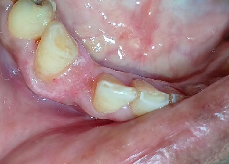
This intraoral clinical photograph shows a postoperative view of the mandibular right anterior region, specifically focusing on the gingiva surrounding the lateral incisor and canine. The image demonstrates a three-month follow-up after the surgical excision of a pyogenic granuloma. The gingival tissue appears healthy, exhibiting a normal pale pink color and firm texture with no signs of edema, erythema, or residual swelling. The interdental papillae are intact, and the surgical site shows complete secondary intention healing without visible scar tissue or recurrence of the pedunculated lesion. The adjacent teeth show yellowish physiologic staining. This image serves as an educational example of successful periodontal soft tissue healing and the clinical appearance of healthy gingiva following electrosurgical intervention.
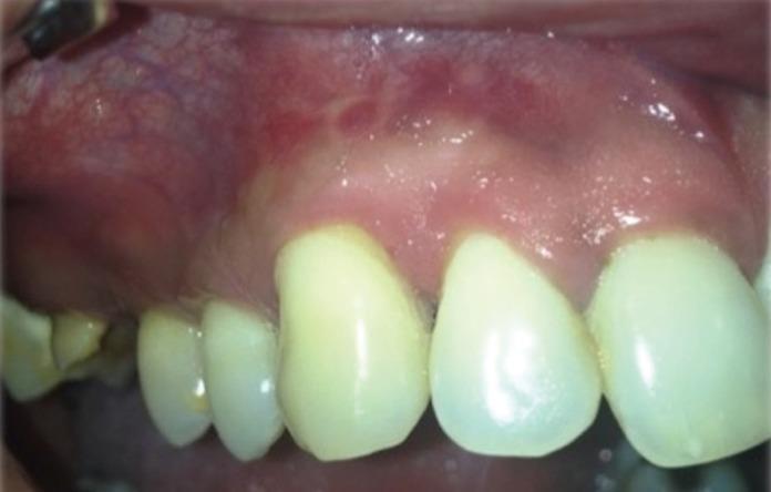
This clinical photograph displays an intraoral view of the maxillary gingiva and oral mucosa following chemotherapy for plasmablastic lymphoma. The image serves as a post-treatment control, demonstrating the resolution of previously documented violaceous, ulcerated masses. The alveolar mucosa appears healthy and pink with normal vascularity and a smooth texture, showing no evidence of residual tumor or recurrent lesions. The attached gingiva maintains a firm appearance with a well-defined gingival margin, though slight recession is visible in the interproximal spaces. The visible maxillary teeth show uniform yellowish-white coloration and mild physiological wear on the incisal edges. This visual is significant for medical education in oncology and oral medicine, illustrating the appearance of clinical remission in the oral cavity after systemic treatment for aggressive hematologic malignancies.
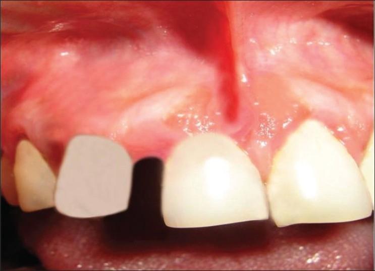
This intraoral clinical photograph provides a postoperative view of the maxillary anterior gingiva and teeth three months following the excision of a peripheral cemento-ossifying fibroma. The image focuses on the right maxillary quadrant, showing the labial gingiva, alveolar mucosa, and the labial frenum. The gingiva appears generally healthy and coral pink, though localized areas of mild erythema and slight edema are visible, particularly at the interdental papilla and the surgical site margins. The site of previous pathology shows successful tissue healing with no evidence of immediate recurrence. There is a notable edentulous space (diastema) between the existing anterior teeth, consistent with previous pathological migration or tooth loss. The visible teeth show varying shades, with one tooth presenting a flatter, opaque appearance suggestive of a provisional restoration or prosthetic. The surrounding alveolar mucosa shows normal vascularity. This image is used to demonstrate long-term follow-up and clinical healing after the surgical management of reactive gingival lesions.
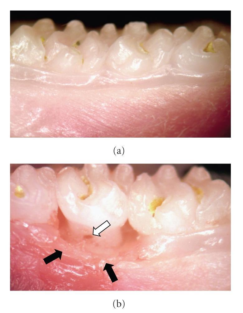
This intraoral clinical photograph, taken at 15X magnification, provides a side-by-side comparison of healthy and periodontal-diseased tissue in a dental model. Panel (a) illustrates healthy gingiva, characterized by a smooth, light pink appearance, firm texture, and intact structural integrity around the teeth with no signs of inflammation. In contrast, panel (b) demonstrates the macroscopic manifestations of progressive periodontal disease. Key diagnostic features highlighted by black arrows include significant gingival inflammation, marked by intense redness (erythema), swelling (edema), and the destruction of normal gingivomucosal structures. A white arrow specifically points to a furcation lesion, indicating advanced tissue and bone loss between the roots of the affected molar. This image serves as a visual educational tool to identify the clinical differences between healthy periodontal support and the inflammatory-destructive processes characteristic of periodontitis.
Features of Healthy Gingiva
- Coral pink / pale pink, due to vascularity of the tissue modified by epithelial thickness and degree of keratinization
- May show physiologic pigmentation (melanin) in darker-skinned individuals - appears as diffuse brown/black patches, which is normal
- No redness (erythema), which would indicate inflammation
- Follows a scalloped outline around each tooth
- Marginal gingiva is knife-edge (thin) and hugs the tooth like a collar
- Interdental papillae are pointed/pyramidal and completely fill the interproximal space up to the contact point, with no blunting or cratering
- Firm and resilient, tightly bound to the underlying periosteum and tooth (except the free/marginal gingiva)
- Does not bleed easily on gentle probing
- Attached gingiva shows stippling (an orange-peel like texture) in many individuals, caused by connective tissue papillae projecting into the epithelium - considered a sign of health, though its absence doesn't always mean disease
- Free gingiva is smooth
- Gingival margin sits at (or just coronal to) the cementoenamel junction
- Normal sulcus depth is about 1-3 mm on probing
- Size corresponds to the sum of the bulk of cellular and intercellular elements and their vascular supply - no edematous enlargement
- No spontaneous bleeding or bleeding on probing
- No suppuration (pus)
- Absence of plaque/calculus accumulation at the gingival margin
- Attached gingiva of adequate width, firmly bound to underlying bone