
MENSTRUATION,PRIMARY AMENORRHOEA FUNDAMENTALSOF REPRODUCTION,PLACENTA AND MEMBRANES,FETUS DEVELOPMENT OF GENITAL ORGANS AND GONADS,CONGENITAL MALFORMATIONS OF GENITAL ORGANS MATERNAL PHYSIOLOGICAL CHANGES IN PREGNANCY
I. MENSTRUATION & PRIMARY AMENORRHOEA
Normal Menstruation
- The hypothalamus secretes GnRH in a pulsatile fashion
- GnRH stimulates the pituitary to release FSH and LH
- FSH/LH promote ovarian follicular development and ovulation
- The follicle secretes estrogen pre-ovulation
- Post-ovulation, the corpus luteum secretes progesterone + estrogen - stimulating endometrial maturation
- If no pregnancy occurs, estrogen/progesterone drop, triggering withdrawal bleeding
- Predominantly arterial (75%); 25% venous
- Contains tissue debris, prostaglandins, and fibrinolysin (which lyses clots - so no clots normally)
- Duration: 3-5 days (range 1-8 days is normal)
- Volume: average 30 mL; up to 80 mL is normal; >80 mL is abnormal (menorrhagia)
- Estrogen makes cervical mucus thinner, more alkaline - promotes sperm survival (fern pattern at ovulation, spinnbarkeit 8-12 cm)
- Progesterone makes mucus thick, cellular, no fern pattern
- No corpus luteum forms, so no progesterone effect
- Estrogen alone causes endometrial growth - eventually sloughs (variable timing, often <28 days)
- Common in first 12-18 months after menarche and premenopausally
Primary Amenorrhoea
Definition
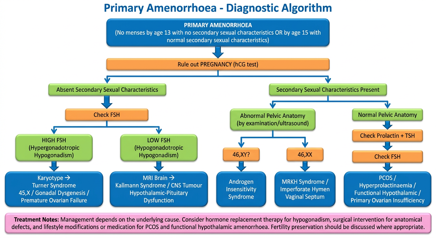
- Primary amenorrhoea = absence of menses by age 13 when there is no visible development of secondary sexual characteristics, OR by age 15 in the presence of normal secondary sexual characteristics
- Failure to begin breast development by age 13 always warrants investigation
- These ages represent 2 standard deviations above the mean age of developing secondary sexual characteristics and menses
Diagnostic Key: Presence of Secondary Sexual Characteristics?
-
Hypergonadotropic Hypogonadism (high FSH, low estrogen - primary ovarian failure)
- Turner syndrome (45,X) - most common cause; streak gonads
- Other chromosomal/gonadal dysgenesis
- When gonadal failure occurs with primary amenorrhoea, there is a high incidence of abnormal karyotype
-
Hypogonadotropic Hypogonadism (low FSH/LH, low estrogen - hypothalamic/pituitary cause)
- Genetic disorders (e.g., Kallmann syndrome - anosmia + GnRH deficiency)
- Other hypothalamic/pituitary dysfunction
- CNS tumors, cranial irradiation
-
Outflow/Müllerian anomalies (normal karyotype 46,XX but absent uterus/vagina)
- Mayer-Rokitansky-Küster-Hauser (MRKH) syndrome - absent uterus + vagina, normal ovaries
- Imperforate hymen or transverse vaginal septum (cryptomenorrhoea - blood accumulates)
-
Androgen Insensitivity Syndrome (AIS) - 46,XY; testes present but end-organ insensitivity to androgens; normal female external appearance; absent/scant pubic/axillary hair; absent uterus
-
Ovotesticular Disorder of Sexual Development
- Polycystic Ovarian Syndrome (PCOS)
- Hyperprolactinaemia
- Primary Ovarian Insufficiency (POF)
- Hypothalamic/pituitary lesions
- Functional hypothalamic amenorrhoea: eating disorders, weight loss, exercise, stress, obesity
- Thyroid disorders
Diagnostic Workup
- Physical exam for secondary sexual characteristics + pelvic anatomy
- Pregnancy test (hCG) - always first
- Serum prolactin + TSH
- FSH + estradiol - distinguish hypergonadotropic vs hypogonadotropic
- Anti-Müllerian hormone (AMH) - useful
- Karyotype if gonadal failure suspected
Treatment Principles
- Specific therapy targeting cause (medical or surgical)
- Hormone therapy to initiate/maintain secondary sexual characteristics
- Maximize bone mass (estrogen + calcium + vitamin D in low-estrogen states)
- Ovulation induction for those desiring pregnancy
II. FUNDAMENTALS OF REPRODUCTION, PLACENTA & MEMBRANES, FETUS
The Placenta
- Fetal part = chorionic sac (outermost fetal membrane - villous chorion)
- Maternal part = decidua basalis (derived from endometrium)
The Decidua (endometrium in pregnancy)
| Region | Location |
|---|---|
| Decidua basalis | Deep to the conceptus - forms maternal part of placenta |
| Decidua capsularis | Superficial, overlying the conceptus |
| Decidua parietalis | Remaining decidua lining the rest of uterus |
Placental Structure
- Cotyledons: The septa from decidua basalis project toward the chorionic plate and divide the fetal placenta into irregular convex areas called cotyledons
- Each cotyledon = 2+ main stem villi + many branch villi
- By end of 4th month, decidua basalis is almost entirely replaced by cotyledons
- Separates maternal blood (in intervillous space) from fetal blood (in villous capillaries)
- Early: thick (4 layers: syncytiotrophoblast, cytotrophoblast, connective tissue, endothelium)
- At term: very thin (mainly syncytiotrophoblast + endothelium)
- Hofbauer cells (fetal macrophages) are present in chorionic villi from early pregnancy
Placental Functions
- Respiration - O2 in, CO2 out
- Nutrition - glucose, amino acids, fatty acids in
- Excretion - urea, creatinine, bilirubin out
- Hormone production - hCG, hPL, estrogen, progesterone
- Protection - partial barrier to microbes (not all viruses)
- Immunologic - maternal antibodies (IgG) transferred to fetus
Umbilical Cord
- Contains 2 umbilical arteries (carry deoxygenated blood from fetus to placenta) and 1 umbilical vein (carries oxygenated blood from placenta to fetus)
- Embedded in Wharton's jelly (mucoid connective tissue)
Fetal Membranes
- Amnion - inner membrane; produces and contains amniotic fluid
- Chorion - outer membrane; fuses with amnion later in gestation
- By week 22, amnion and chorion fuse with each other and the decidua parietalis, obliterating the uterine cavity
Amniotic Fluid
- Produced by: fetal urine (major source after 12 weeks), fetal lung secretions, amniotic epithelium
- Absorbed by: fetal swallowing, membranous absorption
- Volume: ~30 mL at 10 weeks; ~350 mL at 20 weeks; ~700-1000 mL at term
- Functions: cushions fetus, allows movement, maintains temperature, prevents adhesions, lung maturation
III. DEVELOPMENT OF GENITAL ORGANS AND GONADS; CONGENITAL MALFORMATIONS
Sexual Determination
- Chromosomal sex determined at fertilization by sperm (X or Y)
- Male/female morphologic characteristics do NOT begin to develop until the 7th week
- Before week 7: indifferent (bipotential) stage - genital systems are identical
Development of Gonads
- Mesothelium (mesodermal epithelium of posterior abdominal wall)
- Underlying mesenchyme (embryonic connective tissue)
- Primordial germ cells (earliest undifferentiated sex cells)
- Week 5: Thickened mesothelium on medial side of mesonephros forms the gonadal ridge; epithelial cords (gonadal cords) grow into underlying mesenchyme
- Week 7: Sex differentiation begins
- Genes required for bipotential gonad development: FOG2, WT1, NR5A1
- First recognizable at 24 days post-fertilization in endodermal cells of the yolk sac near allantois origin
- Migrate along dorsal mesentery of hindgut to reach gonadal ridges
- If PGCs fail to reach the gonadal ridges, gonads do not develop (gonadal dysgenesis)
Testicular Development (XY)
- The SRY gene (on Y chromosome) codes for testis-determining factor (TDF)
- TDF causes the medulla of the indifferent gonad to differentiate into a testis; cortex regresses
- Sertoli cells (from surface epithelium) form seminiferous tubules; produce AMH (Müllerian-inhibiting factor) which causes regression of paramesonephric ducts
- Leydig cells (from mesenchyme) produce testosterone - causes mesonephric (Wolffian) ducts to develop into male genital ducts (epididymis, vas deferens, seminal vesicles)
- DHT (dihydrotestosterone) causes external genitalia to masculinize
Ovarian Development (XX)
- Without SRY, the cortex of the indifferent gonad differentiates into an ovary; medulla regresses
- Ovary not histologically identifiable until ~10th week
- Cortical cords from surface epithelium break up into primordial follicles at ~16 weeks
- Each primordial follicle = primary oocyte surrounded by follicular cells
- Without AMH, paramesonephric (Müllerian) ducts develop
- Without testosterone, mesonephric (Wolffian) ducts regress
Development of Female Genital Tract
Paramesonephric (Müllerian) Duct Derivatives
| Structure | Derivative |
|---|---|
| Cranial unfused parts | Fallopian tubes |
| Caudal fused portion (uterovaginal primordium) | Uterus + superior vagina |
| Fusion also forms | Broad ligament, rectouterine + vesicouterine pouches |
- Endometrial stroma and myometrium derived from splanchnic mesenchyme
- Uterine development regulated by HOXA10 homeobox gene
- The vagina: upper 1/3 from Müllerian ducts; lower 2/3 from urogenital sinus
Development of Male Genital Tract
| Structure | Derivative |
|---|---|
| Mesonephric tubules | Efferent ductules |
| Mesonephric duct | Epididymis, vas deferens, ejaculatory duct, seminal vesicle |
| Urogenital sinus contribution | Prostate, bulbourethral glands |
Congenital Malformations of Genital Organs
| Condition | Defect |
|---|---|
| Uterus didelphys | Complete failure of fusion - double uterus, double cervix, double vagina |
| Bicornuate uterus | Partial fusion failure - 2 uterine horns, 1 cervix |
| Septate uterus | Failure of median septum to resorb - most common Müllerian anomaly |
| Unicornuate uterus | One Müllerian duct fails to develop |
| MRKH Syndrome | Complete Müllerian aplasia - absent uterus + upper vagina; normal ovaries; 46,XX |
| Imperforate hymen | Failure of canalization of hymen - cryptomenorrhoea, haematocolpos |
| Vaginal atresia | Failure of vaginal plate to canalize |
| Condition | Defect |
|---|---|
| Androgen Insensitivity (AIS) | 46,XY; absent androgen receptor; female phenotype; no uterus; testes in labia/inguinal canal |
| Congenital Adrenal Hyperplasia (CAH) | 46,XX; excess androgens (21-hydroxylase deficiency); virilised female external genitalia |
| 5-alpha reductase deficiency | 46,XY; inability to convert testosterone to DHT; female/ambiguous external genitalia; virilises at puberty |
IV. MATERNAL PHYSIOLOGICAL CHANGES IN PREGNANCY
Pituitary Changes
- Anterior pituitary can double or triple in size (hyperplasia/hypertrophy of lactotrophs)
- Pituitary volume increases: 40% (2nd trimester), 75% (3rd trimester), 120% (immediate postpartum)
- Prolactin: ~10-fold increase throughout gestation (driven by placental estrogens)
- GH: Pituitary GH only in 1st trimester; placental GH variant takes over later - stimulates IGF-1
- LH and FSH: Concentrations decline (despite rising GnRH from placenta)
- ACTH: 2-4 fold increase (partly from placental production); diurnal variation is retained but blunted
Adrenocortical Changes
- CRH: Rises progressively in 2nd and 3rd trimester - largely from placenta
- Cortisol: Corticosteroid-binding globulin triples → total plasma cortisol increases
- Free (unbound) cortisol also increases - urinary free cortisol rises 2-3 fold in last 2 trimesters
- 11-β-hydroxysteroid dehydrogenase 2 in the placenta inactivates cortisol to cortisone, protecting the fetus; but 25% of fetal cortisol still derives from maternal cortisol
Renin-Angiotensin-Aldosterone System (RAAS)
- Renin (juxtaglomerular apparatus) converts angiotensinogen → Angiotensin I → Angiotensin II
- Aldosterone rises significantly in pregnancy
- Angiotensin II: pressor action + stimulates aldosterone secretion
- These changes lead to sodium and water retention (plasma volume expansion)
Cardiovascular Changes
- Plasma volume increases by 40-50% starting from 6-8 weeks
- Red blood cell mass increases by ~20-30% → physiologic dilutional anaemia
- Cardiac output increases by 30-50% (increased stroke volume + heart rate)
- Systemic vascular resistance decreases (progesterone effect)
- Blood pressure: falls slightly in 1st/2nd trimester (vasodilation), returns to near normal in 3rd
Renal Changes
- GFR increases by 40-50% early in pregnancy
- Creatinine and BUN are lower than non-pregnant values
- Glucosuria may occur (tubular maximum overwhelmed by high GFR)
- Physiological dilatation of ureters and renal pelvis (progesterone effect + mechanical)
Respiratory Changes
- Tidal volume increases by ~40%; respiratory rate unchanged
- Minute ventilation increases → mild respiratory alkalosis
- Functional residual capacity decreases (diaphragm elevated by gravid uterus)
Haematological Changes
- Leukocytosis (WBC rises to 9-12 × 10⁹/L; up to 25 in labour)
- Hypercoagulable state: Fibrinogen increases, factors VII, VIII, X, XII increase; protein S decreases → DVT risk
- ESR increases (not clinically useful in pregnancy)
- Platelets may decrease slightly
Endocrine - Thyroid
- TBG rises (estrogen-stimulated) → total T3 and T4 rise; free T3 and T4 remain normal
- First trimester: hCG has mild TSH-like activity → can transiently suppress TSH
- Iodine requirements increase
Musculoskeletal / Other
- Progesterone relaxes smooth muscle (GI: constipation, GORD; uterus: prevents premature contraction)
- Relaxin + progesterone: relax ligaments → pelvic girdle pain, lumbar lordosis
- Insulin resistance increases in 2nd/3rd trimester (hPL, cortisol, progesterone)
- Skin: hyperpigmentation (MSH rises), linea nigra, chloasma (mask of pregnancy), striae gravidarum
- Berek & Novak's Gynecology (Amenorrhoea chapter)
- Ganong's Review of Medical Physiology, 26th Ed. (Normal Menstruation)
- The Developing Human: Clinically Oriented Embryology - Moore & Persaud (Placenta, Genital development)
- Creasy & Resnik's Maternal-Fetal Medicine: Principles and Practice (Physiological changes in pregnancy)
U can flow charts and diagrams
Medical flowchart titled "Primary Amenorrhoea - Diagnostic Algorithm". Clean white background, professional medical style with colored boxes and arrows. Flow starts at top: "PRIMARY AMENORRHOEA (No menses by age 13 with no secondary sexual characteristics OR by age 15 with normal secondary sexual characteristics)". First step: "Rule out PREGNANCY (hCG test)". Then splits into two branches: LEFT branch "Absent Secondary Sexual Characteristics" and RIGHT branch "Secondary Sexual Characteristics Present". LEFT branch: check FSH → splits into "HIGH FSH (Hypergonadotropic Hypogonadism)" leading to box "Karyotype → Turner Syndrome 45,X / Gonadal Dysgenesis / Premature Ovarian Failure" and "LOW FSH (Hypogonadotropic Hypogonadism)" leading to box "MRI Brain → Kallmann Syndrome / CNS Tumour / Hypothalamic-Pituitary Dysfunction". RIGHT branch splits into: "Abnormal Pelvic Anatomy (by examination/ultrasound)" → "46,XY? → Androgen Insensitivity Syndrome" and "46,XX → MRKH Syndrome / Imperforate Hymen / Vaginal Septum". And "Normal Pelvic Anatomy" → check Prolactin + TSH → then FSH → boxes for "PCOS / Hyperprolactinaemia / Functional Hypothalamic / Primary Ovarian Insufficiency". Color code: blue boxes for diagnoses, orange for tests, green for decisions, pink for treatment notes at bottom. Arrow style: clean with labels.
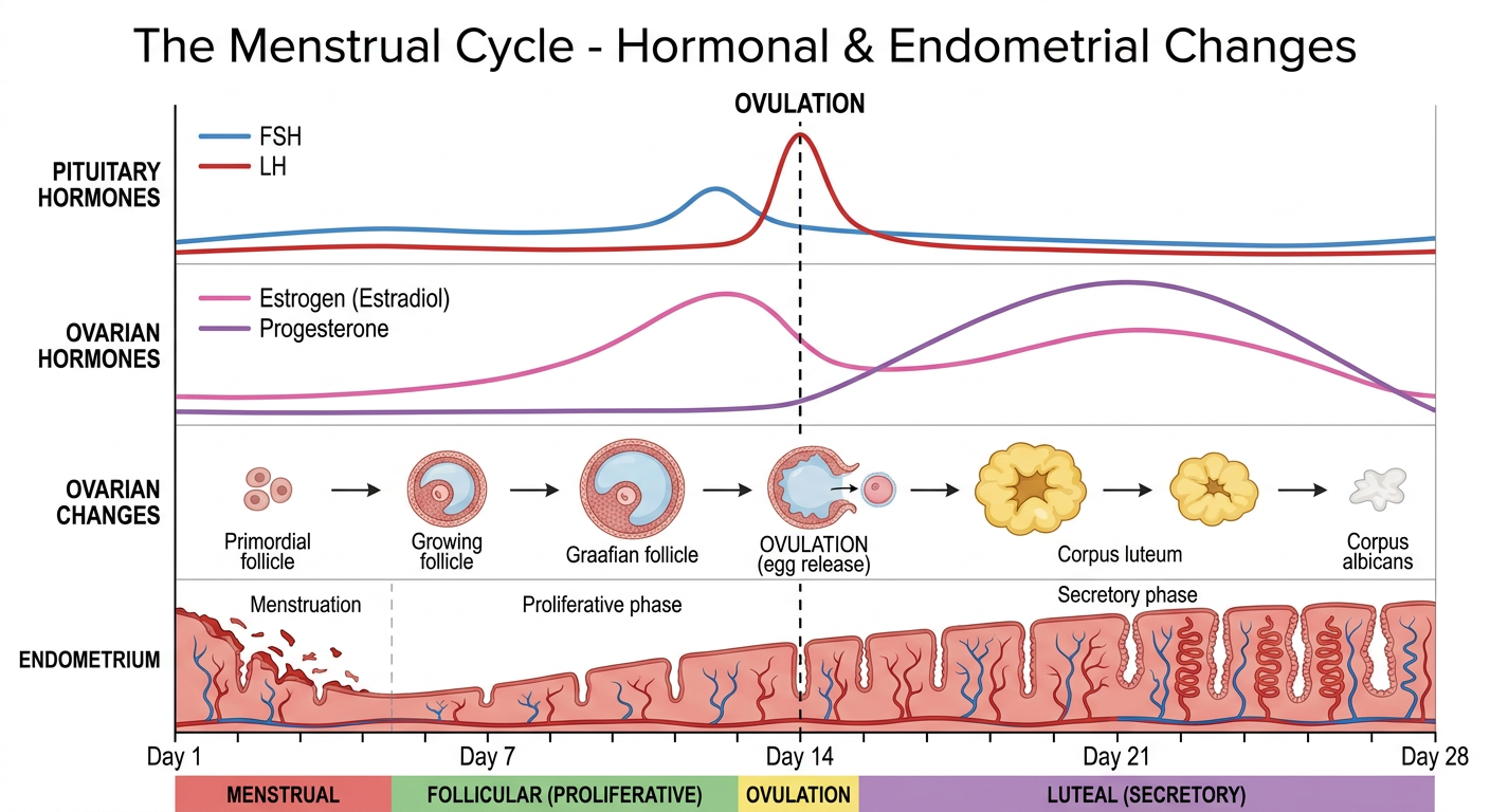
Medical educational diagram titled "The Menstrual Cycle - Hormonal & Endometrial Changes". Clean white background, professional textbook style. The diagram has multiple horizontal panels stacked vertically all aligned to the same 28-day horizontal time axis (Day 1 to Day 28), with a vertical dashed line at Day 14 labeled "OVULATION". Panels from top to bottom: 1) "PITUITARY HORMONES" - FSH curve peaks around day 12 (smaller peak), LH large sharp spike at day 13-14 (LH surge), both labeled, in blue and red curves. 2) "OVARIAN HORMONES" - Estrogen (estradiol) rises in follicular phase with a peak just before ovulation then a second smaller rise in luteal phase; Progesterone flat in follicular phase then rises steeply after ovulation peaking day 21 then falling - drawn in pink and purple. 3) "OVARIAN CHANGES" - drawn as small labeled diagrams: primordial follicle → growing follicle → Graafian follicle → OVULATION (egg release) → corpus luteum → corpus albicans. 4) "ENDOMETRIUM" - drawn as thickness bar: menstruation (days 1-5, thin, shedding shown), proliferative phase (days 6-14, thickening), secretory phase (days 15-28, thick, glandular). 5) "PHASE" labels at bottom: MENSTRUAL | FOLLICULAR (PROLIFERATIVE) | OVULATION | LUTEAL (SECRETORY). Color coded and clearly labeled, educational style.
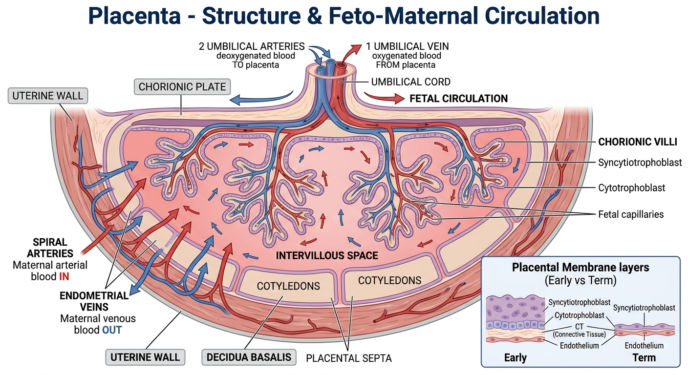
Medical educational diagram titled "Placenta - Structure & Feto-Maternal Circulation". Clean white background, professional anatomy textbook style. Large cross-sectional diagram of the placenta showing: UTERINE WALL at the outer edge with SPIRAL ARTERIES (red arrows pointing inward, labeled "Maternal arterial blood IN") and ENDOMETRIAL VEINS (blue arrows pointing outward, labeled "Maternal venous blood OUT"). INTERVILLOUS SPACE in the center filled with maternal blood (shown in pink). CHORIONIC VILLI hanging down into the intervillous space, labeled with: Syncytiotrophoblast (outer layer), Cytotrophoblast, Fetal capillaries inside villi. DECIDUA BASALIS at bottom separating uterine wall from intervillous space, with PLACENTAL SEPTA dividing it into COTYLEDONS (label 2-3 cotyledons). At top: CHORIONIC PLATE with UMBILICAL CORD inserting, showing 2 UMBILICAL ARTERIES (blue, deoxygenated blood TO placenta) and 1 UMBILICAL VEIN (red, oxygenated blood FROM placenta). Fetal side labeled "FETAL CIRCULATION" with arrows. Inset box in corner showing "Placental Membrane layers (Early vs Term)": Early = 4 layers (Syncytiotrophoblast / Cytotrophoblast / CT / Endothelium), Term = 2 layers (Syncytiotrophoblast / Endothelium). Clear labels, color coded red=oxygenated, blue=deoxygenated.
Medical educational flowchart titled "Development of Gonads and Genital Organs". Clean white background, professional embryology textbook style. Top center: large box "INDIFFERENT (BIPOTENTIAL) GONAD (Week 5-6)" containing: "Gonadal ridge from mesothelium + mesenchyme + Primordial germ cells migrate from yolk sac". Below it: central box "WEEK 7: SEX DETERMINATION". Two large arrows branch LEFT and RIGHT. LEFT branch labeled "46,XY (SRY gene → Testis Determining Factor)": Box "TESTIS develops" → "Medulla differentiates into testis / Cortex regresses" → Two sub-boxes: "Sertoli cells → secrete AMH → Paramesonephric (Mullerian) ducts REGRESS" and "Leydig cells → secrete Testosterone → Wolffian (Mesonephric) ducts DEVELOP". Wolffian duct derivatives in blue box: "Epididymis, Vas deferens, Seminal vesicles, Ejaculatory duct". DHT box: "Testosterone → DHT → External genitalia masculinize (penis, scrotum, prostate)". RIGHT branch labeled "46,XX (No SRY)": Box "OVARY develops" → "Cortex differentiates / Medulla regresses" → "No AMH → Paramesonephric (Mullerian) ducts DEVELOP" → "No Testosterone → Wolffian ducts REGRESS". Mullerian duct derivatives in pink box: "Fallopian tubes (cranial unfused) / Uterus + upper vagina (caudal fused - uterovaginal primordium) / Broad ligament". Bottom box: "Lower vagina from Urogenital Sinus". Color: blue for male pathway, pink for female, orange for indifferent stage, green boxes for key hormones.
Error calling model 'gemini-3-pro-image' (RESOURCE_EXHAUSTED): 429 RESOURCE_EXHAUSTED. {'error': {'code': 429, 'message': 'Resource has been exhausted (e.g. check quota).', 'status': 'RESOURCE_EXHAUSTED'}}
We encountered an error while processing your request. Please try again or reach out to us for help.
Can u ?
1. Primary Amenorrhoea - Diagnostic Flowchart
2. The Menstrual Cycle - Hormonal & Endometrial Changes
3. Placenta - Structure & Feto-Maternal Circulation
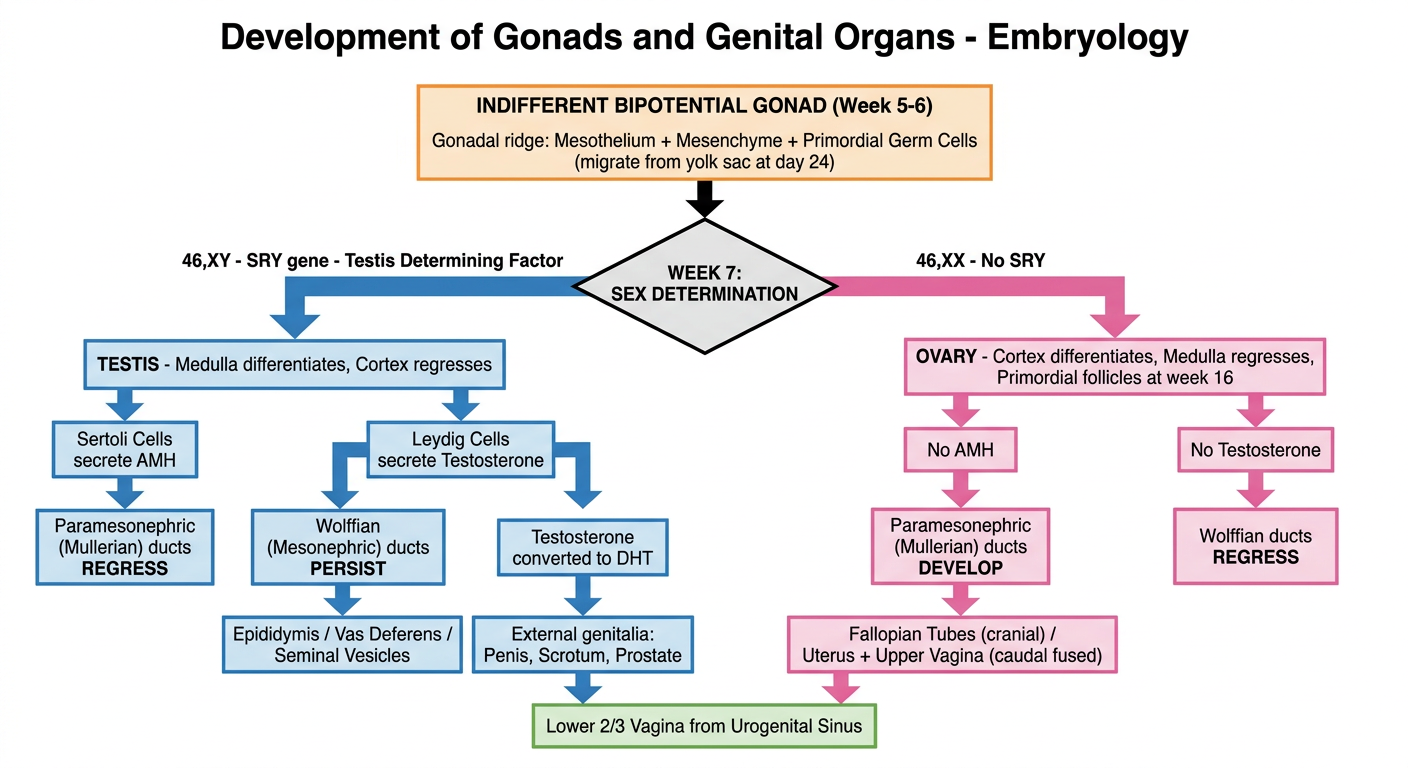
Medical educational flowchart titled "Development of Gonads and Genital Organs - Embryology". Clean white background, textbook style. Top center box in orange: "INDIFFERENT BIPOTENTIAL GONAD (Week 5-6) - Gonadal ridge: Mesothelium + Mesenchyme + Primordial Germ Cells (migrate from yolk sac at day 24)". Arrow down to central diamond: "WEEK 7: SEX DETERMINATION". Two large arrows branch LEFT (blue) and RIGHT (pink). LEFT blue branch "46,XY - SRY gene - Testis Determining Factor": Box "TESTIS - Medulla differentiates, Cortex regresses". Two arrows down: Left sub-arrow to blue box "Sertoli Cells secrete AMH" leading to "Paramesonephric (Mullerian) ducts REGRESS". Right sub-arrow to blue box "Leydig Cells secrete Testosterone" leading to "Wolffian (Mesonephric) ducts PERSIST" then to labeled box "Epididymis / Vas Deferens / Seminal Vesicles". Below: "Testosterone converted to DHT" arrow to "External genitalia: Penis, Scrotum, Prostate". RIGHT pink branch "46,XX - No SRY": Box "OVARY - Cortex differentiates, Medulla regresses, Primordial follicles at week 16". Two arrows: Left to pink box "No AMH" leading to "Paramesonephric (Mullerian) ducts DEVELOP" then labeled box "Fallopian Tubes (cranial) / Uterus + Upper Vagina (caudal fused)". Right to pink box "No Testosterone" leading to "Wolffian ducts REGRESS". Bottom shared box green: "Lower 2/3 Vagina from Urogenital Sinus". All boxes clearly labeled with arrows and colors.
Medical educational infographic titled "Maternal Physiological Changes in Pregnancy". Clean white background, professional style with organ system sections arranged in a grid. Each section has an icon and colored header. Sections: 1) CARDIOVASCULAR (red heart icon): "Plasma volume +40-50% / Cardiac output +30-50% / Heart rate +15-20 bpm / SVR decreases / BP falls in 1st-2nd trimester". 2) HAEMATOLOGICAL (blood drop icon): "Dilutional anaemia (RBC +20%, plasma +50%) / WBC rises to 9-12 x10^9 / Hypercoagulable: Fibrinogen up, Factors VII VIII X XII up, Protein S down / ESR raised". 3) RENAL (kidney icon): "GFR +40-50% / Creatinine and BUN lower / Physiological glucosuria / Ureteric dilatation (progesterone + mechanical)". 4) RESPIRATORY (lung icon): "Tidal volume +40% / Minute ventilation up / FRC decreases (diaphragm elevated) / Mild respiratory alkalosis / pCO2 falls to ~30 mmHg". 5) ENDOCRINE (gland icon): "Pituitary doubles/triples in size / Prolactin x10 increase / Cortisol rises (CBG triples) / Aldosterone rises (RAAS activated) / TBG rises - total T3/T4 up, FREE T3/T4 normal / Insulin resistance increases in 2nd-3rd trimester". 6) PITUITARY (brain icon): "LH and FSH fall / ACTH x2-4 rise / GH: placental variant takes over / CRH rises from placenta". 7) GI/OTHER (body icon): "Progesterone relaxes smooth muscle: constipation, GORD / Relaxin loosens ligaments / Hyperpigmentation, linea nigra, chloasma". Color each section differently, clean boxes, organized grid layout.
Error calling model 'gemini-3-pro-image' (RESOURCE_EXHAUSTED): 429 RESOURCE_EXHAUSTED. {'error': {'code': 429, 'message': 'Resource has been exhausted (e.g. check quota).', 'status': 'RESOURCE_EXHAUSTED'}}
We encountered an error while processing your request. Please try again or reach out to us for help.
?
1. Primary Amenorrhoea - Diagnostic Flowchart
2. Menstrual Cycle - Hormonal & Endometrial Changes
3. Placenta - Structure & Feto-Maternal Circulation
4. Gonad & Genital Organ Development - Embryology Flowchart
5. Maternal Physiological Changes in Pregnancy
| System | Key Changes |
|---|---|
| Cardiovascular | Plasma volume +40-50% |
| Haematological | Dilutional anaemia (plasma +50%, RBC +20%) |
| Renal | GFR +40-50% |
| Respiratory | Tidal volume +40% |
| Pituitary | Size doubles/triples |
| Adrenal/Cortisol | CBG triples → ↑Total cortisol |
| Thyroid | ↑TBG → ↑Total T3/T4 |
| Metabolic | ↑Insulin resistance (2nd/3rd trimester) |
| GI/MSK/Skin | Progesterone → ↓GI motility (constipation, GORD) |