
Treatment of infected sebaceous cyst
sebaceous cyst infected treatment incision drainage excision
infected sebaceous cyst incision drainage excision procedure
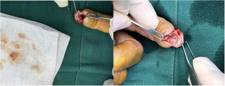
This composite clinical photograph displays an intraoperative view of a surgical procedure on the distal phalanx of a human finger. The image depicts the excision and curettage of an intraosseous epidermoid cyst. A 'fish mouth' flap incision is visible at the fingertip, revealing a window opened through the thinned phalangeal cortex. Surgical instruments, including forceps and a small retractor, are used to expose the cystic cavity within the bone. The content of the cyst, which has been removed and placed on a surgical gauze for inspection, is characterized by a creamy, yellowish-white, and clumpy texture consistent with keratinized material. There is evidence of mild surrounding inflammation and surgical bleeding. This visual demonstrates the classic surgical findings of an intraosseous epidermoid cyst, highlighting the cortical thinning and the pathognomonic sebaceous-like or keratinous material found within the lytic bone lesion. It serves as an educational resource for orthopedic surgery and pathology, illustrating the clinical presentation of rare bone cysts in the hand.
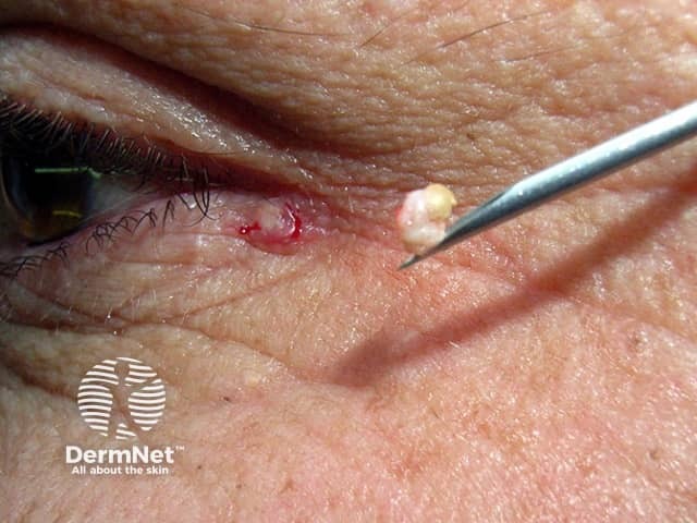
This is a high-resolution clinical photograph of a solitary epidermoid cyst on the periorbital skin (eyelid). Imaging modality: Clinical photography, macro close-up, captured during a minor incision. The focal area shows a small, skin-colored to slightly erythematous nodule with a superficial punctum. An incision with a sterile needle (green-handled device) has been made to evacuate the cyst contents. Exudate or keratinous material is partially expressed, appearing pale yellow to ivory and granular, consistent with laminated keratin debris typical of epidermoid inclusion cysts. The surrounding dermis shows minimal inflammation, with a small amount of blood at the incision margin. The cyst wall may be partially intact or collapsed, with the content partially expressed and not fully extruded in this frame. This image documents a common benign eyelid lesion managed with simple excision or drainage. In clinical practice, differential diagnoses include chalazion, milium, sebaceous cyst, or dermoid cyst; histopathology would reveal a stratified squamous epithelium-lined cyst containing lamellated keratin. Diagnostic significance lies in recognizing a benign, slow-growing eyelid mass that can be reduced by incision and expression, with definitive treatment via complete cyst excision to prevent recurrence. Clinical relevance spans ophthalmology, dermatology, and oculoplastic practices.
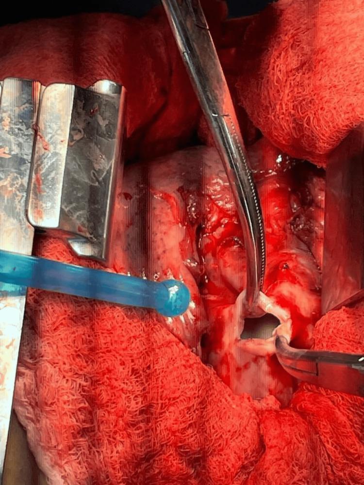
This clinical photograph shows an intraoperative surgical field during a right posterolateral thoracotomy for the treatment of pulmonary hydatid disease. The image captures the drainage and excision of a pulmonary hydatid cyst. A small incision has been made into the white, fibrous cyst wall, through which clear, 'rock water' fluid is draining. Surgical instruments, including a retractor to expose the thoracic cavity and forceps or surgical clamps to secure the cyst wall edges, are visible. The surrounding lung tissue is hyperemic and erythematous, indicating acute inflammation and compression. Red surgical drapes and gauze surround the operative site. A blue suction tip is present in the field to manage fluid drainage and maintain visualization. The procedure illustrates the surgical management of Echinococcus granulosus infection, specifically focusing on controlled cyst decompression to prevent anaphylaxis and daughter cyst spillage before total pericystectomy or lobectomy.
Treatment of Infected Sebaceous Cyst
Note on terminology: These lesions are more precisely called epidermoid cysts (their contents are keratin-based, not sebaceous). "Sebaceous cyst" persists in clinical usage and is retained here.
Clinical Presentation
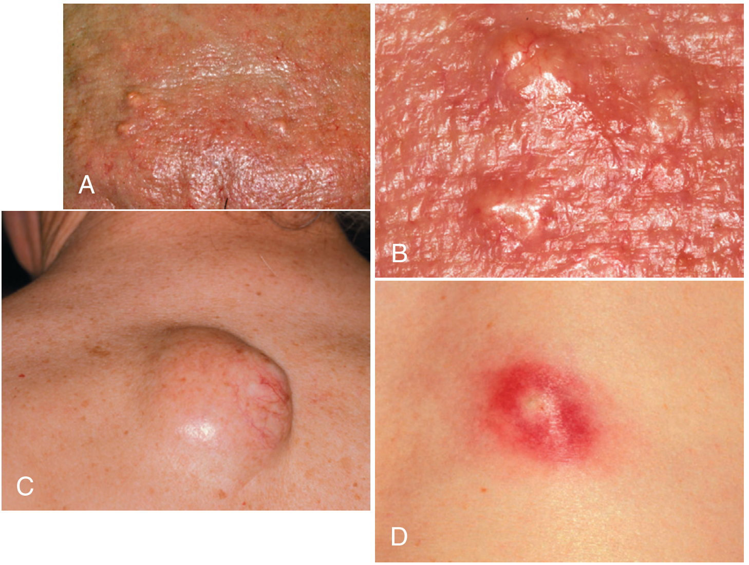
Key Distinction from a Simple Abscess
Treatment Approach
Phase 1: Acute Infection - Incision & Drainage (I&D)
- Anesthesia: Local infiltration (field block)
- Incision: Make an adequate incision over the cyst - the loop drainage technique is not applicable here, because the thick sebaceous material cannot drain spontaneously and the capsule needs removal
- Expression: The thick, cheesy contents must be manually expressed (too viscous to drain passively)
- Wound care: Pack the cavity and arrange timely follow-up, as you would any abscess cavity
- Capsule excision at this stage is optional - if the surgeon can identify the pearly-white capsule and excise it cleanly through the inflamed tissue, this is ideal; otherwise, defer to Phase 2
Roberts and Hedges' Clinical Procedures in Emergency Medicine notes that traditional I&D followed by capsule removal at follow-up is the conventional approach when inflammation is severe.
Phase 2: Definitive Capsule Removal (Prevents Recurrence)
- Field block with local anesthetic
- Make a small fusiform (elliptical) incision in the direction of skin lines, centered on the punctum; length just less than the cyst diameter
- Incise lightly - the overlying skin is often thin
- Dissect deeply at both ends first (curved Metzenbaum scissors work well), being careful not to rupture the capsule
- Use an Allis clamp to grasp the skin wedge over the cyst for traction - this lifts the cyst upward
- Dissect until the cyst is free
- If the cyst ruptures, express all contents then carefully dissect out the entire sac
- Irrigate the cavity with saline
- Close with interrupted sutures (intermediate closure technique)
Minimal Incision Technique (Non-inflamed or Mildly Inflamed)
- Make a 5-6 mm incision with a No. 11 blade (or 3-4 mm dermal punch) directly into the cyst
- Express all contents with external pressure
- Grasp the sac with hemostats and tease it free with a rocking motion
- Use a 3-4 mm dermal curette to remove any residual sac tissue
- No suture closure needed for small incisions
Primary Resection (Single-Stage)
- Faster healing
- Fewer days of pain
- Less scarring
Antibiotics
- Surrounding cellulitis extends beyond the immediate cyst
- Patient is immunocompromised
- Signs of systemic infection (fever, lymphangitis)
- MRSA suspected - use trimethoprim-sulfamethoxazole or doxycycline empirically
Precautions Before Excision
| Location | Concern |
|---|---|
| Preauricular area | Rule out parotid tumor (adenoma/adenocarcinoma) - consider needle biopsy or CT first |
| Infants/children, nasal bridge, scalp, neck, postauricular | Possible dermoid cyst with intracranial connection - MRI before excision; may need neurosurgical consult |
| Any unusual-looking "cyst" | Metastatic melanoma or other malignancy can mimic a sebaceous cyst - excise to confirm diagnosis |
Summary
| Stage | Action |
|---|---|
| Acute infection | I&D, manual expression, wound packing |
| Follow-up (inflammation resolved) | Complete capsule excision to prevent recurrence |
| Non-inflamed cyst | Minimal incision technique (5-6 mm), express + remove sac |
| Large (>2 cm), previously infected, or scarred | Formal excision with suture closure |
| Antibiotics | Only if cellulitis, systemic signs, or immunocompromise |