
History taking of patient according to Russian university
history taking patient Russian medical university format scheme
http://vnmed3.kharkiv.ua/wp-content/uploads/2014/02/Case-his…
History Taking (Anamnesis) - Russian Medical University Format
TITLE PAGE
- University & Department name
- Chief of Department / Assistant Professor names
- Patient's full name (Surname, First name, Patronymic)
- Principal diagnosis
- Complications
- Concomitant (concurrent) diseases
- Curator (student's year, faculty, group, and full name)
- Date of admission / discharge
I. INQUIRY (INTERROGATION / РАССПРОС)
1. Patient's Identifying Data (Паспортная часть)
| Field | Details |
|---|---|
| Full name | Surname, first name, patronymic |
| Age / Date of birth | |
| Gender | |
| Marital status | |
| Occupation | |
| Home address | |
| Date of admission | |
| Source of history | Patient, family member, medical records |
2. Chief Complaints (Жалобы / Главные жалобы)
- Record the patient's own words exactly as stated (e.g., "My stomach hurts and I feel awful.")
- If no complaints: note the reason for admission (e.g., "admitted for routine check-up")
- After chief complaints, screen all other body systems to uncover secondary complaints
- Secondary complaints arise from a systems review and may be unrelated to the main illness
- Location - where exactly
- Quality - character (sharp, dull, burning, pressing)
- Quantity/Severity - how intense (scale 1-10)
- Timing - onset, duration, frequency, course
- Setting - when/where does it occur (at rest, on exertion, at night)
- Aggravating/Relieving factors - what makes it worse or better
- Associated manifestations - accompanying symptoms
3. History of Present Illness (Анамнез настоящего заболевания / История настоящего заболевания)
- Onset - when and how the illness began (sudden vs. gradual)
- Initial symptoms - first signs noticed
- Setting of development - circumstances at the time of onset
- Progression - how symptoms evolved over time
- Previous investigations and results - any labs, imaging, or tests done before
- Previous treatments and response - medications taken, procedures done, effect
- Hospitalizations - prior admissions for this condition
- Reason for current admission - what prompted this visit
Document the patient's response to symptoms and the effect the illness has had on their daily life and function.
4. Past Medical History (Анамнез жизни / История жизни)
a) Biographical History
- Place and date of birth
- Development in childhood (physical and mental)
- Schooling and education
b) Past Illnesses & Surgical Procedures
- Diabetes mellitus
- Hepatitis (A, B, C)
- Tuberculosis
- Hypertension
- Myocardial infarction
- Stroke
- Asthma
- Blood transfusions (when, why, reactions)
- Any operations or injuries
c) Epidemiological History (Эпидемиологический анамнез)
- Contact with infectious patients
- Recent travel
- Animal contact
- Insect bites
- Food sources (water, raw meat)
- Vaccinations history
d) Allergological History (Аллергологический анамнез)
- Drug allergies (specify the drug and nature of reaction)
- Food allergies
- Environmental allergies
- Intolerance to any substances
e) Medications Currently Taken
- Drug name, dose, frequency, duration
f) Gynecological History (for female patients)
- Age of menarche
- Menstrual cycle regularity
- Number of pregnancies, deliveries, abortions
- Menopause (if applicable)
g) Occupational History (Профессиональный анамнез)
- All occupations held (chronologically)
- Occupational hazards - chemicals, dust, radiation, noise
- Duration of exposure
h) Social History (Социальный анамнез)
- Living conditions (housing quality, number of occupants)
- Nutrition and diet
- Physical activity
- Marital status and family situation
i) Bad Habits (Вредные привычки)
- Smoking: number of pack-years (packs per day × years smoked)
- Alcohol: frequency, amount, type
- Drug use: type, frequency, route
j) Family History (Семейный анамнез)
- Health of parents and siblings (if deceased - cause and age of death)
- Hereditary diseases in the family:
- Cardiovascular disease
- Diabetes mellitus
- Malignancies (cancer)
- Tuberculosis
- Mental illness
- Allergic diseases
Logical Structure Summary
CASE HISTORY
│
├── I. INQUIRY (РАССПРОС)
│   ├── 1. Identifying Data (Паспортная часть)
│   ├── 2. Chief Complaints (Жалобы)
│   ├── 3. History of Present Illness (Анамнез morbi)
│   └── 4. Past Medical / Life History (Анамнез vitae)
│       ├── Biographical history
│       ├── Past illnesses & surgeries
│       ├── Epidemiological history
│       ├── Allergological history
│       ├── Medications
│       ├── Gynecological history (F)
│       ├── Occupational history
│       ├── Social history
│       ├── Bad habits
│       └── Family history
│
└── II. PHYSICAL EXAMINATION (Объективный статус)
    ├── General condition, temperature, posture, consciousness
    ├── Constitutional type, height, weight, BMI
    ├── Skin, mucous membranes, lymph nodes
    ├── Musculoskeletal system
    ├── Respiratory system
    ├── Cardiovascular system
    ├── Digestive system
    ├── Urinary system
    ├── Nervous system
    └── Endocrine system
Key Differences from Western History Taking
| Feature | Russian/CIS System | Western (SOAP/Clerking) |
|---|---|---|
| Patronymic name | Always included | Not used |
| Epidemiological history | Separate, mandatory section | Rarely formal |
| Occupational history | Detailed chronological list | Brief mention |
| Life history (Anamnez vitae) | Comprehensive biography | Abbreviated |
| Family history | Detailed, multi-generational | Brief |
| Physical exam structure | Organ-system by system | Variable |
| Written format | Formal long-form document | SOAP notes / clerking |
Tips for Russian University Examinations
- The history is written in third person, past tense (e.g., "The patient complained of...")
- Chief complaints must be listed in order of importance
- Anamnez morbi and Anamnez vitae are always written as separate sections - never combine them
- Every complaint in the chief section must be elaborated in the History of Present Illness
- The document must be signed with the curator's details and dated
- At Russian universities, students are expected to write the full case history (История болезни) which is typically 20-30 pages
What is percussion types of percussion
types of percussion physical examination Russian medical university classification
https://pressbooks.library.torontomu.ca/ippa/chapter/percuss…
Percussion (Перкуссия) - Physical Examination
Definition
- Size, shape, and borders of organs
- Consistency of underlying tissue (air, fluid, solid)
- Presence of pathological changes (fluid, consolidation, cavities)
- Position and displacement of organs
Classification of Percussion
A. By Technique (Method of Performance)
1. Direct Percussion (Непосредственная / Прямая перкуссия)
- Definition: Tapping directly on the patient's skin with one or two fingers, without a pleximeter
- How: Use the tip of the middle or index finger to tap directly on the body surface
- Used for: Assessing sinuses (frontal, maxillary), children's examination, areas where indirect percussion is difficult
- Advantage: Simple, quick
- Disadvantage: Produces a less distinct sound; not suitable for deep structures
2. Indirect (Mediate) Percussion (Опосредованная / Медиатная перкуссия)
- Definition: Tapping is performed through a pleximeter (an intermediary)
- Two subtypes:
- Finger-to-finger percussion (most common): The middle finger of the left hand acts as the pleximeter, placed firmly on the skin; the middle finger of the right hand (the plexor) strikes it
- Hammer-and-pleximeter percussion: A percussion hammer strikes a pleximeter (rubber/ivory plate) - used historically, less common now
- How (finger-to-finger):
- Place the left middle finger (pleximeter) flat and firmly on the skin
- Only the middle phalanx should contact the skin
- Strike with the tip of the right middle finger (plexor) using a quick, wrist-flicking motion
- Two strikes per location, then move
- Strike perpendicular to the pleximeter finger
- Used for: Lungs, heart, liver, spleen, abdomen - most clinical percussion
- Advantage: Most precise, widely used, reproducible
3. Blunt (Fist) Percussion (Перкуссия кулаком / Поколачивание)
- Definition: Using the ulnar edge of a fist (or the fist itself) to strike the body - this is actually a percussion-palpation technique
- Used for:
- Kidney tenderness (Pasternatsky's sign / симптом Пастернацкого) - fist strikes over the costovertebral angle
- Liver pain assessment
- Spine tenderness
- Note: In Russian schools, this is classified as a separate form; in Western schools it may be called "blunt percussion" or "indirect fist percussion"
B. By Purpose (Goal of Percussion)
1. Comparative Percussion (Сравнительная перкуссия)
- Goal: Compare sounds over symmetrical areas of the body (left vs. right)
- Used for: Lungs - comparing right and left, upper vs. lower zones
- Technique: Strike with equal force over mirror-image locations
- Detects: Unilateral dullness (fluid, consolidation), unilateral hyperresonance (pneumothorax, emphysema)
2. Topographic Percussion (Топографическая перкуссия)
- Goal: Determine the boundaries, size, and position of organs
- Technique: Percuss from resonant area toward dull area; the border is where the sound changes
- Rule: Always move from clear (resonant) → dull; mark the border on the dull side
- Used for:
- Upper and lower borders of the lungs
- Cardiac borders (right, left, upper border of cardiac dullness)
- Liver size and borders
- Spleen borders
- Bladder (when distended)
C. By Force of Strike
1. Loud (Deep) Percussion (Громкая / Глубокая перкуссия)
- Strong blows; sound penetrates 7-8 cm deep
- Used for: Localizing deep structures, fluid in the pleural cavity, deep masses
2. Quiet (Threshold) Percussion (Тихая / Пороговая перкуссия)
- Light blows; sound penetrates 4-5 cm deep
- Used for: Determining organ borders more precisely (e.g., heart borders, liver)
3. Quietest Percussion (Тишайшая перкуссия)
- Very gentle taps; penetrates only 1-2 cm
- Used for: Superficial structures, determining absolute cardiac dullness (area directly in contact with chest wall without lung overlap)
Percussion Sounds (Перкуторные звуки)
| Sound | Russian Term | Pitch | Duration | Quality | Normal Location |
|---|---|---|---|---|---|
| Resonance | Ясный легочный звук | Low | Long | Hollow | Normal lung |
| Hyperresonance | Коробочный звук | Lower than normal | Very long | Booming | Emphysema, pneumothorax |
| Tympanic | Тимпанический звук | High | Moderate | Drum-like | Stomach, intestines, pneumothorax |
| Dull | Тупой звук | High | Short | Thud-like | Liver, spleen, solid tumors, fluid |
| Flat | Абсолютно тупой звук | Very high | Very short | Dead | Muscle, bone, massive pleural effusion |
Pneumonic for Sounds (Loud → Quiet):
Topographic Percussion of the Lungs (Clinical Example)
Lower Borders of the Right Lung:
| Line | Border |
|---|---|
| Midclavicular line | 6th rib |
| Anterior axillary line | 7th rib |
| Midaxillary line | 8th rib |
| Posterior axillary line | 9th rib |
| Scapular line | 10th rib |
| Paravertebral line | Level of T11 spinous process |
Lung Mobility (Respiratory Excursion):
- Normally 6-8 cm along the midaxillary line
Rules of Percussion Technique (Russian University Exam Standards)
- The pleximeter finger must lie parallel to the expected border
- Tap with the tip of the plexor finger, not the pad - use a wrist snap, not elbow motion
- Two equal taps per site, then move
- Always move from resonant → dull when determining borders
- Mark the border on the dull side (the side from which you are approaching)
- The patient must be in the correct position - standing or sitting for lung percussion
- Room must be quiet
- Apply equal force for comparative percussion
Summary Table
| Classification | Types |
|---|---|
| By technique | Direct, Indirect (finger-finger, hammer-pleximeter), Blunt (fist) |
| By purpose | Comparative, Topographic |
| By force | Loud (deep), Quiet, Quietest (threshold) |
| Sounds produced | Resonant, Hyperresonant, Tympanic, Dull, Flat |
Topographic percussion of lungs
topographic percussion of lungs upper lower borders respiratory excursion Russian medical
https://repo.knmu.edu.ua/bitstreams/c66a4198-3dae-40c5-af32-…
Topographic Percussion of the Lungs (Топографическая перкуссия лёгких)
- Determination of the upper borders (apices)
- Determination of the lower borders
- Determination of the excursion (mobility) of the lower borders
General Rules of Topographic Percussion
- Always move from resonant (clear) sound → dull sound
- Mark the border on the dull side (the side toward which you are moving)
- Use quiet (threshold) percussion - lighter force for more precise borders
- The pleximeter finger lies parallel to the expected border
- Perform on both right and left sides
- Patient position: standing or sitting (upright)
I. Upper Borders of the Lungs (Apices / Верхушки лёгких)
Anterior (Front):
- Place pleximeter parallel to the clavicle
- Percuss from the middle of the clavicle upward and medially
- Move until dullness appears
Normal: Apex is 3-4 cm above the clavicle (both sides)
Posterior (Back):
- Place pleximeter parallel to the spine of the scapula
- Percuss from the middle of the scapular spine upward toward C7
- Move until dullness appears
Normal: Apex is at the level of the spinous process of C7 (7th cervical vertebra)
Width of Kronig's Fields (Поля Кренига):
- The band of resonance over the apex (on the trapezius muscle) measured in cm
- Percuss medially and laterally from the middle of the trapezius
- Normal: 4-8 cm on each side
II. Lower Borders of the Lungs (Нижние границы)
Technique:
- Start above the expected border (in the area of clear resonance)
- Percuss downward along each topographic line, one intercostal space at a time
- Mark the border where the sound changes from resonant → dull
Normal Lower Borders - Table (Normosthenic Person)
| Topographic Line | Right Lung | Left Lung |
|---|---|---|
| Parasternal line | 5th intercostal space | - (not determined) |
| Midclavicular line | 6th intercostal space | - (not determined) |
| Anterior axillary line | 7th intercostal space | 7th intercostal space |
| Midaxillary line | 8th intercostal space | 8th intercostal space |
| Posterior axillary line | 9th intercostal space | 9th intercostal space |
| Scapular line | 10th intercostal space | 10th intercostal space |
| Paravertebral line | Spinous process of T11 | Spinous process of T11 |
Note: The left lung borders are not determined along the parasternal and midclavicular lines because the heart occupies that area (cardiac dullness). Left lung borders are assessed from the anterior axillary line downward.
Memory Aid (Right Lung):
III. Excursion (Mobility) of the Lower Lung Border
Technique:
- Find the lower border at quiet (normal) breathing - mark it (Point A)
- Ask the patient to take a deep breath and hold - percuss down again, mark new border (Point B)
- Ask the patient to exhale fully and hold - percuss up from below until dullness, mark border (Point C)
- Total excursion = distance from Point B to Point C
Normal Values of Respiratory Excursion:
| Topographic Line | Right Lung | Left Lung | ||||
|---|---|---|---|---|---|---|
| Inspiration | Expiration | Total | Inspiration | Expiration | Total | |
| Midclavicular | 2-3 cm | 2-3 cm | 4-6 cm | - | - | - |
| Midaxillary | 3-4 cm | 3-4 cm | 6-8 cm | 3-4 cm | 3-4 cm | 6-8 cm |
| Scapular | 2-3 cm | 2-3 cm | 4-6 cm | 2-3 cm | 2-3 cm | 4-6 cm |
The midaxillary line has the greatest excursion (6-8 cm) and is the most clinically important.
IV. Changes in Borders - Clinical Significance
Bilateral Lowering of Lower Border:
- Asthenic body type (tall, thin)
- Acute pulmonary dilation - bronchial asthma attack
- Chronic pulmonary dilation - emphysema
Bilateral Elevation of Lower Border:
- Hypersthenic body type (stocky)
- Pleural effusion (bilateral)
- Ascites (pushes diaphragm up)
- Acute bloating/meteorism
Unilateral Lowering of Lower Border:
- Unilateral emphysema
- Compensatory emphysema (opposite lung after pneumonectomy)
- Unilateral pneumothorax
Unilateral Elevation of Lower Border:
- Pleural effusion (on affected side - dull sound, border rises)
- Pneumonia / consolidation (lobe involved)
- Atelectasis (lung collapse)
- Diaphragmatic paralysis
Decreased Excursion (Mobility):
- Inflammatory processes in the lung (pneumonia)
- Decreased elasticity - emphysema (most common cause)
- Pleural effusion or pleural adhesions
- Diaphragmatic fixation/obstruction
Increased Excursion:
- Compensatory (opposite lung after lobectomy)
- Athletic/deep breathing individuals
V. Changes in Upper Borders
| Change | Cause |
|---|---|
| Apices higher than normal (>4 cm above clavicle) | Emphysema, bronchial asthma (increased airiness) |
| Apices lower than normal | Tuberculosis, fibrosis, shrinkage of apex (decreased airiness) |
| Narrowing of Kronig's fields | Tuberculosis, fibrosis, inflammatory scarring of apex |
| Widening of Kronig's fields | Emphysema |
Summary Diagram
TOPOGRAPHIC PERCUSSION OF LUNGS
│
├── UPPER BORDERS
│   ├── Anterior: 3-4 cm above clavicle
│   ├── Posterior: level of C7
│   └── Kronig's fields: 4-8 cm
│
├── LOWER BORDERS (Right)
│   Parasternal → 5th ICS
│   Midclavicular → 6th ICS
│   Ant. axillary → 7th ICS
│   Midaxillary → 8th ICS
│   Post. axillary → 9th ICS
│   Scapular → 10th ICS
│   Paravertebral → T11
│
└── EXCURSION
    Midaxillary: 6-8 cm (most important)
    Midclavicular / Scapular: 4-6 cm
Вопросы к зачету Вопросы к зачету ✓ 1. История болезни: структура, правила заполнения и ведения (medical record: structure, rules of filling in and keeping). ✓ 2. Собирание анамнеза, его значение для диагностики. Роль Г. А. Захарьина в развитии анамнестического метода исследования (anamnesis taking, its significance for diagnosis. The role of G.A. Zaharyin in the development of anamnestic analysis). ✓ 3. Понятие о симптомах и синдромах. Симптомокомплексы. (Notions of symptoms and syndromes. Symptom complexes.). ◦ 4. Порядок общего осмотра в терапии (Order of general inspection in therapy). ✓ 5. Нарушение сознания в клинике внутренних болезней: виды, клиническая характеристика коматозного состояния. (Disturbance of consciousness in the clinical picture of internal diseases: types, clinical characteristics of the comatose state.). ✓ 6. Положение больного: диагностическое значение. (Position of a patient: diagnostic significance.) ✓ 7. Исследование кожных покровов: методика, симптомы, клиническое значение. (Examination of the skin integument: methods, symptoms, clinical significance.). ✓ 8. Осмотр кожи, волос, ногтей: методика, симптомы, клиническое значение. (Visual examination (inspection) of the skin, hair, nails: methods, symptoms, clinical significance.). ✓ 9. Исследование подкожной клетчатки: методика, симптомы, клиническое значение (examination of the hypoderm: methods, symptoms, clinical significance). ◦ 10. Температура тела. Виды лихорадок. Типы температурных кривых и их диагностическое значение. (Body temperature. Types of fever. Types of temperature curves and their diagnostic significance). ◦ 11. Общий осмотр: типы конституции (general visual examination (inspection): types of body build). ◦ 12. Общий осмотр: характерные выражения лица (general visual examination (inspection): common facial expressions). ◦ 13. Ожирение: классификация. Расчет индекса массы тела. (Obesity: classification. Body mass index calculation). ◦ 14. Метод перкуссии: история, физическое обоснование, правила (method of percussion: history, physical basis, rules). ◦ 15. Метод аускультации: история, физическое обоснование, правила (method of auscultation: history, physical basis, rules). ◦ 16. Расспрос при заболеваниях системы дыхания. Жалобы, их детализация, механизм, диагностическое значение. (Interviewing patients with respiratory diseases. Complaints, their specification, mechanism, diagnostic significance). ◦ 17. Одышка: клинические особенности и механизм её возникновения при различных заболеваниях органов дыхания. (Dyspnea: clinical peculiarities and mechanism of its development in different respiratory diseases). ◦ 18. Кашель: характеристика, клинические особенности, механизм его возникновения при различных заболеваниях органов дыхания и диагностическое значение. (Cough: characteristics, clinical peculiarities, mechanism of its development in different respiratory diseases and diagnostic significance). ◦ 19. Мокрота и её характер при различных заболеваниях органов дыхания (sputum and its characteristics in different respiratory diseases). ◦ 20. Диагностическое значение кровохарканья и легочного кровотечения (diagnostic significance of hemoptysis and pulmonary hemorrhage). ◦ 21. Осмотр грудной клетки: патологические формы, определение дыхательной экскурсии грудной клетки. Форма грудной клетки в норме. (Visual examination (inspection) of the thorax: pathological forms, determination of respiratory excursion of the thorax. Shape of the thorax in normal state). ◦ 22. Нарушение ритма дыхания: дыхание типа Куссмауля , Чейн-Стокса, Биота. (respiratory dysrhythmia: Kussmaul respiration, Cheyne-Stokes respiration, Biot respiration). ◦ 23. Пальпация при заболеваниях органов дыхания: голосовое дрожание (методика, диагностическое значение). (palpation in respiratory diseases: vocal fremitus (methods, diagnostic significance)). ◦ 24. Перкуссия легких. Виды перкуссии. Варианты перкуторного звука. Диагностическое значение изменений перкуторного звука при заболеваниях органов дыхания. (percussion of the lungs. Types of percussion. Variants of percussion sound. Diagnostic significance of changes of percussion sound in respiratory diseases). ◦ 25. Сравнительная перкуссия легких (comparative percussion of the lungs). ◦ 26. Топографическая перкуссия легких (topographic percussion of the lungs). ◦ 27. Аускультация легких. Звуковые феномены в норме и при заболеваниях. (auscultation of the lungs. Sound phenomena in norm and diseases). ◦ 28. Везикулярное дыхание: механизм возникновения, диагностическое значение. Качественное и количественное изменение везикулярного дыхания (Vesicular respiration: mechanism of formation, diagnostic significance. Qualitative and quantitative changes of vesicular respiration). ◦ 29. Бронхиальное дыхание: механизм возникновения, диагностическое значение, разновидности. (Bronchial respiration: mechanism of formation, diagnostic significance, variants). ◦ 30. Сухие и влажные хрипы: механизм возникновения, классификация, диагностическое значение. Отличие влажных хрипов от крепитации, шума трения плевры и их диагностическое значение. (Dry and moist rales: mechanism of development, classification, diagnostic significance. The difference between moist rales, crepitation and pleural friction rub and their diagnostic significance). ◦ 31. Крепитация: механизм возникновения, классификация, диагностическое значение. Отличие влажных хрипов от крепитации, шума трения плевры и их диагностическое значение. Crepitation: mechanism of development, classification, diagnostic significance. The difference between moist rales, crepitation and pleural friction rub and their diagnostic significance. ◦ 32. Шум трения плевры: механизм возникновения, классификация, диагностическое значение. Отличие влажных хрипов от крепитации, шума трения плевры и их диагностическое значение. (Рleural friction rub: mechanism of development, classification, diagnostic significance. The difference between moist rales, crepitation and pleural friction rub and their diagnostic significance). ◦ 33. Бронхофония: определение, методика, диагностическое значение (вronchophony: definition, methods, diagnostic significance). ◦ 34. Метод определения функции внешнего дыхания (method of determination of respiratory function). ◦ 35. Экссудаты и транссудаты: причины, отличие экссудатов от транссудатов. Еxudates and transudates: causes, difference between exudates and transudates. ◦ 36. Исследование мокроты: микроскопическое и бактериологическое исследование, диагностическое значение (еxamination of the sputum: microscopic and bacteriological examination, diagnostic significance). ◦ 37. Современные дополнительные методы диагностики заболеваний легких (modern additional methods of diagnostics of pulmonary diseases). ◦ 38. Синдром легочного уплотнения: причины, клинические признаки, данные объективного обследования, зависимость клинических проявлений от локализации очага уплотнения, верифицирующее и дополнительные обследования. (Syndrome of pulmonary consolidation (induration): causes, clinical signs, data of objective examination, dependence of clinical manifestations on localization of the focus of consolidation, verifying and additional examinations). ◦ 39. Плевральный синдром: причины, клинические признаки, данные объективного обследования, верифицирующее и дополнительные обследования. (Pleural syndrome: causes, clinical signs, data of objective examination, verifying and additional examinations). ◦ 40. Синдром скопления жидкости в плевральной полости: причины, клинические признаки, данные объективного обследования, верифицирующее и дополнительные обследования. (Syndrome of fluid accumulation in the pleural cavity: causes, clinical signs, data of objective examination, verifying and additional examinations). ◦ 41. Синдром пневмоторакса: причины, клинические признаки, данные объективного обследования, верифицирующее и дополнительные обследования. (Syndrome of pneumothorax: causes, clinical signs, data of objective examination, verifying and additional examinations). ◦ 42. Синдром гидропневмоторакса: причины, клинические признаки, данные объективного обследования, верифицирующее и дополнительные обследования. (Syndrome of hydropneumothorax: causes, clinical signs, data of objective examination, verifying and additional examinations). ◦ 43. Бронхобструктивный синдром: причины, клинические признаки, данные объективного обследования, верифицирующее и дополнительные обследования. (Broncho-obstructive syndrome: causes, clinical signs, data of objective examination, verifying and additional examinations). ◦ 44. Синдром дыхательной недостаточности: причины, клинические признаки, данные объективного обследования, верифицирующее и дополнительные обследования (syndrome of respiratory failure: causes, clinical signs, data of objective examination, verifying and additional examinations). ◦ 45. Ателектаз легкого: причины, клинические признаки, данные объективного обследования, верифицирующее и дополнительные обследования (аtelectasis of the lung: causes, clinical signs, data of objective examination, verifying and additional examinations).
What would you like me to do with these exam questions?
Question 16
interviewing patients respiratory diseases complaints cough dyspnea hemoptysis chest pain Russian propedeutics
https://doctorlib.org/medical/ambulatory/60.html
Question 16: Interviewing Patients with Respiratory Diseases
Расспрос при заболеваниях системы дыхания
Structure of the Interview (Расспрос)
- Chief complaints (главные жалобы) - what brought the patient in
- Detailization of each complaint (детализация жалоб) - full characterization
- History of present illness (анамнез болезни)
- Past history (анамнез жизни) - with emphasis on respiratory risk factors
Main Complaints in Respiratory Diseases
| # | Complaint | Russian |
|---|---|---|
| 1 | Cough | Кашель |
| 2 | Sputum / expectoration | Мокрота |
| 3 | Dyspnea / breathlessness | Одышка |
| 4 | Hemoptysis | Кровохарканье |
| 5 | Chest pain | Боль в грудной клетке |
1. COUGH (Кашель)
Definition:
- Deep inspiration
- Closure of the glottis + rise in intrathoracic pressure
- Explosive opening of the glottis with forceful expiration
Mechanism:
The lung parenchyma itself has no cough receptors - cough from lung disease occurs via pleural or bronchial involvement.
Detailization (Детализация кашля):
| Parameter | Questions to ask |
|---|---|
| Character | Dry (non-productive) or wet (productive with sputum)? |
| Timing | Morning? Night? Constant? Paroxysmal? |
| Duration | Acute (<3 weeks), subacute (3-8 weeks), chronic (>8 weeks) |
| Sound | Barking? Hoarse? Whooping? Silent? |
| Intensity | Mild, moderate, exhausting |
| Accompanying symptoms | Fever, pain, breathlessness? |
| What relieves it | Position change, medications, warmth? |
Diagnostic Significance:
| Type of Cough | Disease |
|---|---|
| Dry, hacking | Early pneumonia, pleurisy, laryngitis, lung tumor |
| Morning productive ("toilet cough") | Chronic bronchitis, bronchiectasis |
| Night cough | Postnasal drip, GERD, heart failure (cardiac cough) |
| Paroxysmal, whooping | Whooping cough (pertussis), foreign body |
| Barking, hoarse | Laryngitis, laryngeal tumor, compression of recurrent laryngeal nerve |
| Bitonal cough | Mediastinal tumor pressing on trachea |
| Silent ("mute") cough | Vocal cord paralysis, extreme weakness |
| Productive with large amounts of sputum | Bronchiectasis, lung abscess ("full-mouth" emptying in certain positions) |
| Cough worsened lying flat | Bronchiectasis, postnasal drip |
2. SPUTUM (Мокрота)
Definition:
Detailization:
| Parameter | Details |
|---|---|
| Amount | Daily volume in ml (scant <50 ml, moderate 50-200 ml, large >200 ml) |
| Color | White/gray, yellow-green (purulent), rusty, red (bloody), black |
| Consistency | Liquid, viscous, frothy |
| Odor | Odorless or fetid/putrid |
| Layering | Does it separate into layers when left standing? |
| Time of production | Morning, after position change, at any time |
Diagnostic Significance:
| Sputum Character | Disease |
|---|---|
| Mucous, scanty, viscous | Acute bronchitis, early pneumonia |
| Mucopurulent (yellow-green) | Chronic bronchitis, purulent bronchiectasis |
| Purulent, large amounts, fetid | Lung abscess, bronchiectasis |
| Rusty sputum | Lobar (croupous) pneumonia - classic sign |
| Frothy, pink | Pulmonary edema |
| 3-layer sputum (foam + mucus + pus) | Bronchiectasis, lung abscess |
| "Raspberry jelly" | Lung cancer (necrotic) |
| Black sputum | Anthracosis (coal miners) |
3. DYSPNEA (Одышка)
Definition:
Mechanism:
- Hypoxia (↓ PaO₂)
- Hypercapnia (↑ PaCO₂)
- Acidosis (↓ pH)
Detailization:
| Parameter | Questions |
|---|---|
| Type | Inspiratory? Expiratory? Mixed? |
| Onset | Sudden or gradual? |
| Connection to exertion | At rest? On walking? Climbing stairs? |
| Severity | Degree - how many steps before stopping? |
| Relieving factors | Position, rest, bronchodilators, oxygen? |
| Associated symptoms | Wheezing, cough, chest pain? |
Types by Phase:
| Type | Mechanism | Disease |
|---|---|---|
| Inspiratory dyspnea | Upper airway obstruction | Laryngeal edema, foreign body, croup, tracheal stenosis |
| Expiratory dyspnea | Lower airway obstruction (bronchospasm) | Bronchial asthma, COPD |
| Mixed dyspnea | Both phases affected - lung parenchyma or pleural | Pneumonia, pleural effusion, heart failure |
Diagnostic Significance by Disease:
| Condition | Character of Dyspnea |
|---|---|
| Bronchial asthma | Expiratory, paroxysmal, with audible wheezing, relieved by bronchodilators |
| COPD | Expiratory, chronic, progressive, on exertion |
| Pneumonia | Mixed, acute onset, with fever |
| Pleural effusion | Mixed, positional (worse lying on healthy side) |
| Pneumothorax | Sudden severe mixed dyspnea + chest pain |
| Pulmonary embolism | Sudden onset, severe, with chest pain + tachycardia |
| Lung cancer | Progressive, worsening over months |
| Pulmonary edema | Mixed, orthopnea, pink frothy sputum (cardiac cause) |
4. HEMOPTYSIS (Кровохарканье)
Definition:
Mechanism:
- Erosion of bronchial/pulmonary vessels (tumor, inflammation, abscess)
- Rupture of dilated vessels (bronchiectasis, tuberculosis)
- Increased pulmonary capillary pressure (mitral stenosis - "cardiac hemoptysis")
- Infarction of lung tissue (pulmonary embolism)
Detailization:
| Parameter | Questions |
|---|---|
| Amount | Streaks in sputum? Tablespoons? Frank hemorrhage (>100 ml)? |
| Color | Bright red (arterial), dark red, rusty |
| Mixed with | Pure blood? Mixed with sputum? Frothy? |
| Associated symptoms | Pain? Fever? Weight loss? |
| First episode or recurrent? |
Distinguish from hematemesis (vomiting blood):
- Hemoptysis: bright red, frothy, alkaline pH, mixed with sputum, preceded by cough
- Hematemesis: dark brown, acid pH, mixed with food, preceded by nausea
Diagnostic Significance:
| Character | Disease |
|---|---|
| Streaks of blood in sputum | Bronchitis, pneumonia, any irritation |
| Frank bright red blood | Bronchiectasis, tuberculosis, lung abscess |
| "Rusty" blood-tinged sputum | Lobar pneumonia |
| Recurrent moderate hemoptysis | Tuberculosis (classic), bronchiectasis |
| Massive hemoptysis (>600 ml/24h) | Tuberculosis cavity, bronchiectasis, lung cancer |
| Frothy pink blood | Pulmonary edema (mitral stenosis, LV failure) |
| Dark blood + pleuritic pain | Pulmonary infarction (embolism) |
| Blood in sputum + weight loss + night sweats | Lung cancer or tuberculosis - must rule out |
5. CHEST PAIN (Боль в грудной клетке)
Mechanism:
The lung parenchyma has NO pain receptors. Chest pain in respiratory disease is caused by involvement of the parietal pleura (richly innervated), chest wall, or referred pain.
- Parietal pleura - most common in respiratory pain (intercostal nerves)
- Chest wall (ribs, muscles, costochondral junctions)
- Trachea and large bronchi (limited pain sensation)
Detailization (SOCRATES approach):
| Parameter | Questions |
|---|---|
| Site | Where exactly? Can you point to it? |
| Onset | Sudden or gradual? |
| Character | Sharp, stabbing, dull, burning, pressure? |
| Radiation | Does it spread anywhere? |
| Associated symptoms | Cough, fever, dyspnea? |
| Timing | Constant or intermittent? |
| Exacerbating factors | Deep breathing, coughing, movement, lying on side? |
| Severity | 1-10 scale |
Diagnostic Significance:
| Type of Pain | Disease |
|---|---|
| Pleuritic pain - sharp, stabbing, worsened by deep inspiration and coughing, relieved by lying on affected side | Pleuritis (dry), pneumonia with pleural involvement, pulmonary infarction |
| Sudden severe pain + dyspnea | Pneumothorax |
| Dull aching, not related to breathing | Mediastinal mass, tumor invading chest wall |
| Pain worsened by palpation over costochondral junction | Tietze syndrome (costochondritis) |
| Unilateral burning pain in a dermatomal distribution | Herpes zoster |
| Central chest pain + dyspnea + tachycardia | Pulmonary embolism |
Summary: Complaints and Their Most Characteristic Diseases
| Complaint | Most Characteristic Disease |
|---|---|
| Productive morning cough + large sputum | Bronchiectasis, chronic bronchitis |
| Rusty sputum + fever + chest pain | Lobar pneumonia |
| Paroxysmal expiratory dyspnea + wheezing | Bronchial asthma |
| Hemoptysis + night sweats + weight loss | Tuberculosis / Lung cancer |
| Sudden chest pain + dyspnea | Pneumothorax / Pulmonary embolism |
| Pleuritic pain + friction rub | Dry pleuritis |
| Frothy pink sputum + orthopnea | Pulmonary edema (cardiac) |
Anamnesis Features to Specifically Ask in Respiratory Diseases
- Smoking history - pack-years (главный фактор риска ХОБЛ и рака легкого)
- Occupational hazards - dust (silicosis, asbestosis, coal miners' pneumoconiosis), chemicals, fumes
- Contact with TB patients - epidemiological history
- Travel history - endemic mycoses, parasites
- Family history - lung cancer, asthma, cystic fibrosis, alpha-1-antitrypsin deficiency
- Prior lung diseases - pneumonia, TB, asthma, surgery
- Allergies - particularly inhaled allergens
Answer it for the exam just a specific information mentioned in the questions
Question 16 — Exam Answer
Main Complaints (Главные жалобы)
- Cough (кашель)
- Sputum (мокрота)
- Dyspnea (одышка)
- Hemoptysis (кровохарканье)
- Chest pain (боль в грудной клетке)
1. COUGH (Кашель)
- Character: dry or wet (productive)
- Time: morning, night, constant, paroxysmal
- Sound: barking, hoarse, whooping, silent
- Duration: acute / chronic
| Cough type | Disease |
|---|---|
| Dry | Pleuritis, early pneumonia, tumor |
| Morning productive | Chronic bronchitis, bronchiectasis |
| Paroxysmal whooping | Pertussis, foreign body |
| Barking/hoarse | Laryngitis, vocal cord damage |
| Large sputum at once | Lung abscess, bronchiectasis |
2. SPUTUM (Мокрота)
- Amount (ml/day)
- Color: white, yellow-green, rusty, pink, black
- Consistency: liquid, viscous, frothy
- Odor: odorless or fetid
| Sputum type | Disease |
|---|---|
| Rusty | Lobar pneumonia |
| Mucopurulent, large amounts, fetid | Lung abscess, bronchiectasis |
| Frothy pink | Pulmonary edema |
| Mucous, scanty | Acute bronchitis |
3. DYSPNEA (Одышка)
- Type: inspiratory / expiratory / mixed
- Onset: sudden or gradual
- Relation to exertion: at rest, on walking, on climbing
- Relieving factors: position, bronchodilators
| Type | Mechanism | Disease |
|---|---|---|
| Inspiratory | Upper airway obstruction | Laryngeal edema, foreign body, croup |
| Expiratory | Bronchospasm (lower airways) | Bronchial asthma, COPD |
| Mixed | Parenchymal/pleural | Pneumonia, pleural effusion, pulmonary edema |
| Sudden severe | Pneumothorax, pulmonary embolism |
4. HEMOPTYSIS (Кровохарканье)
- Amount: streaks / tablespoons / massive (>100 ml)
- Color: bright red, dark, rusty
- Mixed with: pure blood / sputum / frothy
- Hemoptysis: bright red, frothy, alkaline, mixed with sputum, preceded by cough
- Hematemesis: dark brown, acid pH, mixed with food, preceded by nausea
| Hemoptysis type | Disease |
|---|---|
| Rusty sputum | Lobar pneumonia |
| Recurrent, bright red | Tuberculosis, bronchiectasis |
| Massive | TB cavity, lung cancer |
| Frothy pink | Pulmonary edema (mitral stenosis) |
| Blood + pleuritic pain | Pulmonary infarction (embolism) |
5. CHEST PAIN (Боль в грудной клетке)
- Site and radiation
- Character: sharp/stabbing vs. dull/aching
- Relation to breathing: worsened by deep inspiration and coughing = pleuritic
- Relation to movement/palpation
| Pain type | Disease |
|---|---|
| Sharp, worsened by breathing/coughing | Dry pleuritis, pneumonia with pleural involvement |
| Sudden + dyspnea | Pneumothorax |
| Dermatomal burning | Herpes zoster |
| Dull, not related to breathing | Mediastinal tumor, chest wall invasion |
| + dyspnea + tachycardia | Pulmonary embolism |
Diagnostic significance of hemoptysis and pulmonary hemorrhage
hemoptysis pulmonary hemorrhage diagnostic significance causes classification Russian propedeutics
Question 20 — Exam Answer
Definitions
Classification by Volume
| Grade | Amount | Clinical term |
|---|---|---|
| Streaks of blood in sputum | <5 ml | Minor hemoptysis |
| Moderate | 5-100 ml/day | Hemoptysis (кровохарканье) |
| Massive | >100 ml/day or ≥100 ml/h | Pulmonary hemorrhage |
| Catastrophic | >600 ml/day | Life-threatening hemorrhage |
The tracheobronchial tree holds only ~150 ml total - even "moderate" bleeding can be fatal.
Distinguish Hemoptysis from Hematemesis (Important!)
| Feature | Hemoptysis | Hematemesis |
|---|---|---|
| Color | Bright red, frothy | Dark brown/coffee-ground |
| pH | Alkaline | Acid |
| Mixed with | Sputum, mucus | Food particles |
| Preceded by | Cough | Nausea, vomiting |
| Reaction | Foamy | Non-foamy |
| After event | Patient feels tickling/irritation in chest | Nausea remains |
Mechanism of Bleeding
Causes and Diagnostic Significance
By System:
| Disease | Character of hemoptysis | Key features |
|---|---|---|
| Tuberculosis | Recurrent, bright red, moderate to massive | + night sweats, weight loss, low fever |
| Bronchiectasis | Recurrent, large amounts, purulent blood | Morning, postural, fetid sputum |
| Lung cancer | Streaks → moderate, persistent | + weight loss, hoarse voice, chest pain |
| Lung abscess | Massive, fetid, purulent blood | + high fever, putrid sputum |
| Pneumonia | Rusty/blood-tinged sputum | + fever, consolidation signs |
| Foreign body | Acute onset | History of aspiration |
| Pulmonary infarction (PE) | Dark blood + pleuritic chest pain | + tachycardia, DVT risk factors |
| Disease | Character | Mechanism |
|---|---|---|
| Mitral stenosis | Frothy pink blood | Elevated pulmonary capillary pressure → rupture of bronchial veins |
| Left ventricular failure | Frothy pink sputum | Pulmonary edema |
| Pulmonary embolism | Dark blood + sudden chest pain + dyspnea | Lung infarction |
- Coagulation disorders (anticoagulant therapy)
- Vasculitis (Goodpasture syndrome, Wegener's granulomatosis)
- Thoracic endometriosis (catamenial hemoptysis - linked to menstrual cycle)
- Trauma, post-procedural
Diagnostic Significance Summary Table
| Hemoptysis character | Most likely diagnosis |
|---|---|
| Rusty sputum (ржавая мокрота) | Lobar pneumonia |
| Recurrent bright red, moderate | Tuberculosis |
| Large amounts + fetid + purulent | Lung abscess / Bronchiectasis |
| Persistent streaks + weight loss | Lung cancer |
| Frothy pink | Pulmonary edema (mitral stenosis, heart failure) |
| Sudden dark blood + chest pain + dyspnea | Pulmonary embolism |
| Cyclic (with menstruation) | Thoracic endometriosis |
| Massive, sudden | TB cavity, bronchiectasis, fungal (aspergillosis) |
Pulmonary Hemorrhage - Clinical Danger Signs
- Volume >100 ml/day = emergency
- Asphyxiation is the main cause of death (not hypovolemia)
- Signs: rapid large volume fresh blood, respiratory distress, cyanosis, hemodynamic instability
- High-risk patients: active cancer, aspergillosis, alcoholism, pulmonary artery involvement, mechanical ventilation
Exam Key Points
- Hemoptysis = <100 ml/day; pulmonary hemorrhage = >100 ml/day
- Blood originates below the vocal cords - distinguish from epistaxis and hematemesis
- Most common cause worldwide = tuberculosis
- In developed countries = cancer, bronchiectasis, pneumonia
- Massive hemoptysis source = bronchial arteries (high pressure, direct aortic branches)
- Death from hemorrhage = asphyxiation, not exsanguination
- Frothy pink = cardiac (pulmonary edema); bright red frothy = bronchial/pulmonary vessel
Crimson colour indicate which disease in hemoptysis
Blood Color in Hemoptysis — Diagnostic Significance
| Color | Russian term | Disease |
|---|---|---|
| Crimson / Bright scarlet red | Алая кровь | Tuberculosis (classic), bronchiectasis, lung abscess |
| Rusty / Brown-red | Ржавая мокрота | Lobar (croupous) pneumonia |
| Dark red / Cherry | Тёмно-красная | Pulmonary infarction (pulmonary embolism) |
| Frothy pink | Розовая пенистая | Pulmonary edema (mitral stenosis, left heart failure) |
| Streaks of blood in sputum | Прожилки крови | Bronchitis, mild irritation, early cancer |
| Raspberry jelly / dark mixed | Малиновое желе | Lung cancer (necrotic tumor) |
Why Crimson = Tuberculosis?
Key Rule for the Exam
Crimson (алая) blood = tuberculosis until proven otherwise
- Crimson blood + night sweats + weight loss + low-grade fever + upper lobe infiltrate = Tuberculosis
- Rusty sputum = Pneumonia
- Frothy pink = Pulmonary edema / cardiac cause
- Dark blood + sudden chest pain = Pulmonary embolism
Generate a differential diagnosis flowchart based on hemoptysis character
A clean medical differential diagnosis flowchart titled "Hemoptysis - Differential Diagnosis by Character". The flowchart starts at the top with a box labeled "HEMOPTYSIS" and branches down based on blood character. Five main branches: 1. "CRIMSON / BRIGHT RED (Алая кровь)" → leads to box "Tuberculosis (classic), Bronchiectasis, Lung Abscess" with note "Bronchial artery erosion, Rasmussen aneurysm in TB" 2. "RUSTY / BROWN-RED (Ржавая мокрота)" → leads to box "Lobar (Croupous) Pneumonia" with note "RBC breakdown + fibrin in alveoli" 3. "FROTHY PINK (Розовая пенистая)" → leads to box "Pulmonary Edema, Mitral Stenosis, Left Heart Failure" with note "Elevated pulmonary capillary pressure" 4. "DARK RED (Тёмно-красная)" → leads to box "Pulmonary Infarction (Pulmonary Embolism)" with note "Lung infarction, venous blood" 5. "BLOOD STREAKS IN SPUTUM (Прожилки крови)" → leads to box "Bronchitis, Early Lung Cancer, Pneumonia" with note "Capillary disruption" 6. "RASPBERRY JELLY / MIXED DARK (Малиновое желе)" → leads to box "Lung Cancer (Necrotic Tumor)" with note "Tumor necrosis + blood" Bottom section shows a summary table with columns: Color | Disease | Key Associated Symptoms - Crimson red | Tuberculosis | Night sweats, weight loss, upper lobe cavity - Rusty | Pneumonia | Fever, consolidation, pleuritic pain - Frothy pink | Pulmonary edema | Orthopnea, cardiac history - Dark red | Pulmonary embolism | Sudden chest pain, tachycardia, DVT - Blood streaks | Bronchitis / Cancer | Chronic cough, smoking history - Raspberry jelly | Lung cancer | Weight loss, hoarseness Professional medical style, white background, color-coded branches matching blood color descriptions, clear arrows, easy to read fonts.
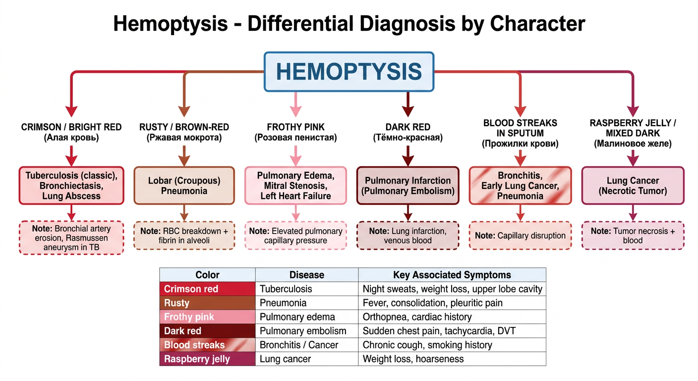
| Blood Color | Disease | Key Clues |
|---|---|---|
| 🔴 Crimson / Bright red | Tuberculosis, Bronchiectasis, Lung abscess | Night sweats, weight loss, upper lobe cavity, Rasmussen aneurysm |
| 🟤 Rusty / Brown-red | Lobar Pneumonia | Fever, consolidation, pleuritic pain |
| 🩷 Frothy Pink | Pulmonary edema, Mitral stenosis, LV failure | Orthopnea, cardiac history, bilateral crackles |
| 🟣 Dark red | Pulmonary Embolism (infarction) | Sudden chest pain, tachycardia, DVT risk |
| 🔸 Streaks in sputum | Bronchitis, Early lung cancer | Chronic cough, smoking history |
| 🍓 Raspberry jelly | Lung cancer (necrotic) | Weight loss, hoarseness, mass on CXR |
Q21
thorax inspection pathological forms barrel emphysematous funnel keel chest Russian propedeutics respiratory excursion
https://repo.knmu.edu.ua/bitstreams/c66a4198-3dae-40c5-af32-…
Question 21 — Exam Answer
I. Normal Shape of the Thorax (Норма)
| Parameter | Normal value |
|---|---|
| Anteroposterior (AP) diameter : Transverse diameter | 1 : 2 (AP is smaller) |
| Epigastric angle | ~90° (normosthenic) |
| Ribs angle to spine | ~45° |
| Intercostal spaces | Moderately visible |
| Supraclavicular fossae | Slightly visible |
| Scapulae | Fit closely to chest wall |
Normal Types by Body Build (Конституция):
| Type | AP:Transverse | Epigastric angle | Shape |
|---|---|---|---|
| Normosthenic | 1:2 | ~90° | Conical, proportional |
| Hypersthenic | Almost 1:1 | >90° (obtuse) | Wide, flat, short |
| Asthenic | <1:2 | <90° (acute) | Narrow, long, flat; prominent clavicles & scapulae |
II. Pathological Forms of the Thorax (Патологические формы)
1. Emphysematous (Barrel) Chest — Бочкообразная грудная клетка
- Appearance: Barrel-shaped; AP diameter ≈ transverse (nearly circular cross-section)
- Features:
- Ribs run horizontally (not obliquely)
- Epigastric angle >90° (obtuse)
- Intercostal spaces widened
- Supraclavicular fossae bulging
- Chest appears fixed in inspiration position
- Cause: Pulmonary emphysema (COPD) - air trapping causes chronic lung overinflation
- Respiratory excursion: Severely decreased
2. Paralytic Chest — Паралитическая грудная клетка
- Appearance: Extreme asthenic type - flat, narrow, elongated
- Features:
- AP diameter greatly reduced
- Prominent clavicles, wide intercostal spaces
- Asymmetric, scapulae "winged" (отстают)
- Epigastric angle sharply acute (<60°)
- Cause: Pulmonary tuberculosis, cachexia, chronic debilitating diseases, connective tissue disorders
- Respiratory excursion: Decreased
3. Rachitic (Keeled / Chicken) Chest — Килевидная (куриная) грудная клетка
- Appearance: Sternum protrudes anteriorly like a ship's keel (prow)
- Features:
- AP diameter increased
- Lateral (transverse) diameter decreased
- Sternum pushed forward
- Ribs curve sharply from sternum laterally ("rachitic beads" at costochondral junctions)
- Cause: Rickets (childhood vitamin D deficiency)
- Respiratory excursion: Reduced
4. Funnel Chest — Воронкообразная грудная клетка (Pectus Excavatum)
- Appearance: Lower sternum and adjacent costal cartilages are sunken inward (funnel-shaped depression)
- Features:
- Depression in lower third of sternum
- Can compress heart and great vessels
- Cause: Congenital malformation (developmental), Marfan syndrome
- Diagnostic significance: Can cause cardiac displacement, murmurs, reduced lung capacity
5. Scaphoid (Boat-shaped) Chest —Ладьевидная грудная клетка
- Appearance: Hollowing in the upper and middle parts of the sternum (boat-shaped)
- Cause: Syringomyelia (spinal cord disease)
- Note: Rare, specifically associated with syringomyelia in Russian propedeutics
6. Asymmetric Chest — Асимметричная грудная клетка
| Finding | Meaning |
|---|---|
| One side enlarged/bulging | Pleural effusion, pneumothorax (pushes out) |
| One side retracted/flattened | Fibrosis, atelectasis, post-TB scarring, lobectomy (pulls in) |
| One side lagging in breathing | Pathology on that side (pneumonia, pleuritis, pneumothorax) |
III. Determination of Respiratory Excursion of the Thorax
Method:
- Axillary level — tape at the level of the 4th rib anteriorly / below the scapular angle posteriorly (most important)
- Upper chest — level of the 2nd rib
- Lower chest — level of the 10th rib
Technique:
- Measure chest circumference at quiet (normal) breathing
- Ask patient to take a maximum deep breath — measure again
- Ask patient to do a maximum full expiration — measure again
- Total excursion = inspiration measurement − expiration measurement
Normal Values:
| Level | Total excursion |
|---|---|
| Axillary (main) | 6-8 cm |
| Upper | 4-6 cm |
| Lower | 4-6 cm |
Normal excursion at axillary level = 6-8 cm
Decreased Excursion — Causes:
- Emphysema (most common - chest fixed in inspiration)
- Pleural effusion or adhesions
- Pneumothorax
- Obesity
- Ankylosing spondylitis (rib joints fused)
- Severe pneumonia (pain limits breathing)
Increased Excursion:
- Compensatory on the healthy side after lobectomy/pneumonectomy
IV. Other Parameters Assessed During Thorax Inspection
| What to assess | What to look for |
|---|---|
| Symmetry | One side lagging = pathology on that side |
| Breathing type | Thoracic (women), abdominal (men), mixed |
| Respiratory rate (RR) | Normal 16-20/min; tachypnea >20, bradypnea <12 |
| Breathing rhythm | Regular or Cheyne-Stokes / Biot / Kussmaul |
| Depth | Superficial or deep |
| Accessory muscles | Use of neck/shoulder muscles = increased work of breathing |
| Retraction of intercostal spaces | Inspiratory = upper airway obstruction |
| Bulging of intercostal spaces | Expiratory = bronchial obstruction (asthma) |
| Position of trachea | Midline or deviated? |
Summary Table — Pathological Chest Forms
| Form | Shape | Cause | Key Feature |
|---|---|---|---|
| Barrel (emphysematous) | AP = Transverse | Emphysema | Horizontal ribs, obtuse angle, fixed inspiration |
| Paralytic | Flat, narrow, asymmetric | TB, cachexia | Winged scapulae, acute angle |
| Rachitic (keeled) | Keel/prow anteriorly | Rickets | Sternum protrudes forward |
| Funnel | Sunken sternum | Congenital, Marfan | Depression lower sternum |
| Scaphoid | Upper sternal hollow | Syringomyelia | Boat-shaped upper sternum |
| Asymmetric (enlarged) | One side bigger | Effusion, pneumothorax | Intercostal spaces widened |
| Asymmetric (retracted) | One side smaller | Fibrosis, atelectasis | Intercostal spaces narrowed |
Use this as a source
python3 -c " import pdfplumber files = [ '/tmp/workspace/attachments/4163dc99-b927-4c90-90e3-995cd90ebb1b/1000128702.pdf', '/tmp/workspace/attachments/4163dc99-b927-4c90-90e3-995cd90ebb1b/1000189922.pdf' ] for f in files: print(f'=== FILE: {f} ===') with pdfplumber.open(f) as pdf: print(f'Pages: {len(pdf.pages)}') # Print first 3 pages to understand content for i, page in enumerate(pdf.pages[:3]): text = page.extract_text() if text: print(f'--- Page {i+1} ---') print(text[:1500]) print() "
python3 -c " import pdfplumber # Search in book 1 (310 pages) for thorax/chest inspection content f1 = '/tmp/workspace/attachments/4163dc99-b927-4c90-90e3-995cd90ebb1b/1000128702.pdf' with pdfplumber.open(f1) as pdf: for i, page in enumerate(pdf.pages): text = page.extract_text() if text and any(kw in text.lower() for kw in ['thorax', 'chest shape', 'barrel', 'inspection of the thorax', 'forms of the thorax', 'respiratory excursion of the thorax']): print(f'--- Book1 Page {i+1} ---') print(text[:2000]) print() " 2>&1 | head -300
python3 -c " import pdfplumber f1 = '/tmp/workspace/attachments/4163dc99-b927-4c90-90e3-995cd90ebb1b/1000128702.pdf' with pdfplumber.open(f1) as pdf: # Pages 63-75 should cover pathological forms + dynamic inspection + respiratory excursion for i in range(62, 80): page = pdf.pages[i] text = page.extract_text() if text: print(f'--- Page {i+1} ---') print(text) print() " 2>&1
Question 21 — Exam Answer
I. Static Chest Inspection (Статический осмотр)
- Supraclavicular and infraclavicular fossae (pronounced, smooth, or bulging)
- Position of clavicles and ribs (oblique or horizontal)
- Intercostal spaces (width)
- Epigastric angle (angle of Louis)
- Position of scapulae
- Symmetry and ratio of anteroposterior (AP) to transverse dimensions (thoracic index)
II. Normal Forms of the Chest (Норма)
| Form | AP : Transverse | Epigastric angle | Ribs | Other features |
|---|---|---|---|---|
| Normosthenic (mesomorph) | 0.65-0.75 | ~90° | Directed obliquely downward | Supraclavicular fossa expressed, subclavian smooth; scapulae tightly against chest |
| Asthenic (ectomorph) | < 0.6 | < 90° (acute) | Nearly vertical, oblique | Wide intercostal spaces; supraclavicular & infraclavicular fossae expressed; "winged" scapulae (scapulae alatae); X rib free end not joined to costal arch |
| Hypersthenic (endomorph) | > 0.75 | > 90° (obtuse) | Almost horizontal | Narrow intercostal spaces; fossae smooth; scapulae tight to chest; well-developed muscles |
Normal chest - regardless of form - is symmetrical, with both halves equal in size.
III. Pathological Forms of the Chest
1. Emphysematous (Barrel-like) Chest
- Resembles hypersthenic but with wide intercostal spaces (unlike hypersthenic)
- AP diameter ≈ transverse; thoracic index may exceed 1.0
- Supra- and subclavian fossae smoothed or inflated (increased airiness of lung apex)
- Chest fixed in inspiration position
- Cause: Pulmonary emphysema (decreased lung elasticity, increased airiness)
2. Paralytic Chest
- Resembles a modified asthenic chest - flat, reduced AP diameter
- Can be asymmetric (one half smaller than the other)
- Lungs shrunken in size
- Cause: Severe malnutrition, long-term pulmonary tuberculosis
3. Rachitic (Pigeon / Keeled) Chest
- Sternum protrudes anteriorly (like a keel)
- AP diameter exceeds transverse diameter
- Cause: Rickets (vitamin D deficiency in childhood)
4. Funnel (Foveated) Chest — Pectus Excavatum
- Depression in the lower part of the sternum
- Also called "shoemaker's chest" (from prolonged pressure on the sternum)
- Cause: Congenital abnormality or chronic pressure
5. Scaphoid (Boat-shaped) Chest
- Depression in the upper and middle part of the sternum
- Cause: Syringomyelia (rare spinal cord disease)
6. Kyphoscoliotic Chest
- Develops from kyphoscoliosis (combined lateral + posterior curvature of the spine)
- Leads to impaired lung and heart function; predisposes to bronchitis, pneumonia, early respiratory and heart failure
IV. Asymmetric Chest Changes
| Finding | Cause |
|---|---|
| Retraction (one side smaller/flattened) | Decreased lung volume: tuberculosis, bronchiectasis, lung abscess, atelectasis, lung resection, pleural obliteration |
| Protrusion (one side enlarged/bulging) | Air or fluid in pleural cavity: hydrothorax, pneumothorax |
V. Dynamic Chest Inspection (Динамический осмотр)
Types of Respiration:
| Type | Main muscles | Normal for |
|---|---|---|
| Thoracic (costal) | Intercostal muscles | Women |
| Abdominal (diaphragmatic) | Diaphragm | Men |
| Mixed | Both | Elderly, some pathological conditions |
Symmetry of Respiratory Movements:
- Normal: movements are symmetrical
- Unilateral lag (one side moves less) → pathology on that side:
- Pleurisy, fluid or gas in pleural cavity, pleural adhesions, pneumofibrosis, obturator atelectasis, post-surgery
Respiratory Rate (RR):
- Normal: 16-20 per minute at rest
- Tachypnea (>20/min): bronchospasm, reduced respiratory surface (pneumonia, atelectasis, lung infarction), sharp chest pain (dry pleurisy, broken ribs)
- Bradypnea (<16/min): depression of the medullary respiratory center (cerebral hemorrhage, brain edema, brain tumor, toxic agents)
VI. Chest Excursion (Дыхательная экскурсия грудной клетки)
Method:
- In men: under the inferior corners of the scapulae (posteriorly) and at the lower segment of the nipple (anteriorly)
- In women: at the level of attachment of IV rib to the sternum (anteriorly)
Measurement:
- Measure circumference at maximum inspiration
- Measure circumference at maximum expiration
- Excursion = inspiration circumference − expiration circumference
Normal Values:
| Parameter | Normal |
|---|---|
| Chest circumference (men) | 88-92 cm |
| Chest circumference (women) | 83-85 cm |
| Chest excursion | 6-8 cm (average person) |
| Athletes | Up to 12-15 cm |
Decreased Excursion - Causes:
- Pulmonary emphysema (most common - reduced lung elasticity)
- Hydrothorax, pleurisy with effusion
- Pneumothorax
- Pleural adhesions
- Ossification of costal cartilages
- Rib fractures
- Severe obesity
Summary Table
| Chest form | Cause | Key sign |
|---|---|---|
| Barrel (emphysematous) | Emphysema | AP ≈ transverse; wide ICS; fixed in inspiration |
| Paralytic | TB, cachexia | Flat, narrow; may be asymmetric |
| Rachitic (keeled) | Rickets | Sternum protrudes forward |
| Funnel | Congenital / pressure | Lower sternum depressed |
| Scaphoid | Syringomyelia | Upper/mid sternum depressed |
| Kyphoscoliotic | Kyphoscoliosis | Spinal curvature deforms chest |
| Asymmetric (bulging) | Pneumothorax, effusion | One side enlarged |
| Asymmetric (retracted) | TB, atelectasis, fibrosis | One side smaller |
Answer Q22: Abnormal breathing patterns (Kussmaul, Cheyne-Stokes, Biot)
Question 22 — Exam Answer
Normal Breathing (Норма)
- Rate: 16-20 per minute
- Rhythm: regular
- Depth: moderate
- Both phases (inspiration + expiration) smooth and equal
Abnormal Breathing Patterns — Overview

| Pattern | Russian name | Key feature |
|---|---|---|
| Kussmaul | Дыхание Куссмауля | Deep, noisy, rapid - no pauses |
| Cheyne-Stokes | Дыхание Чейна-Стокса | Crescendo-decrescendo + apnea |
| Biot | Дыхание Биота | Equal depth cycles + sudden apnea |
| Grocco (Undulant) | Волнообразное дыхание Грокко | Like Cheyne-Stokes but no apnea |
1. Kussmaul's Respiration (Дыхание Куссмауля)
Character:
- Deep, noisy, frequent breathing
- Sometimes described as "breathing of a hunted animal"
- No pauses between cycles
- Very large tidal volume - audible from a distance
- Rhythm is regular but rate is fast
Mechanism:
Causes:
- Diabetic hyperglycemic-hyperketonemic coma (most classic cause - DKA)
- Uremic coma (renal failure - accumulation of acid metabolites)
- Severe metabolic acidosis of any origin
Key phrase for exam:
"Deep, noisy, frequent breathing in decompensated acidosis - diabetic or uremic coma"
2. Cheyne-Stokes Respiration (Дыхание Чейна-Стокса)
Character:
- Apnea (respiratory pause) lasting up to 1 minute
- Then breathing resumes as very shallow breaths
- Breaths gradually increase in depth (crescendo) - reaching maximum at 5th-7th breath
- Then gradually decrease in depth (decrescendo)
- Returns to apnea → cycle repeats
Apnea → shallow → deeper → DEEP → deeper → shallow → Apnea → ...
          ↑_____________crescendo-decrescendo____________↑
Mechanism:
Causes:
- Acute insufficiency of cerebral circulation (brain stroke) - classic cause per textbook
- Severe heart failure (cardiac Cheyne-Stokes)
- Brain edema, tumors
- Uremia, severe intoxication
- Can be physiological in newborns and elderly during sleep
Key phrase for exam:
"Crescendo-decrescendo breathing with prolonged apnea (up to 1 minute) - seen in acute cerebrovascular accident (stroke)"
3. Biot's Respiration (Дыхание Биота)
Character:
- Series of rhythmic, deep respiratory movements of equal amplitude
- Interrupted by sudden respiratory pauses (apnea)
- Unlike Cheyne-Stokes: amplitude does NOT change between pauses - all breaths are equal depth
- Pauses appear and disappear abruptly (not gradually)
deep-deep-deep-deep → PAUSE → deep-deep-deep-deep → PAUSE → ...
(equal depth throughout)
Mechanism:
Causes:
- Inflammatory diseases of the brain and meninges:
- Meningitis
- Encephalitis
- Increased intracranial pressure
- Brain tumor pressing on medulla
- Severe brain injury
Key phrase for exam:
"Equal-depth breaths with sudden apnea - seen in meningitis and encephalitis"
4. Grocco's (Undulant / Wavy) Respiration — Bonus
Character:
- Similar to Cheyne-Stokes but NO complete apnea
- Breathing only becomes very superficial at its minimum (never stops)
- Considered a prelude to Cheyne-Stokes respiration
Cause:
- Early stages of the same conditions causing Cheyne-Stokes
- Less severe depression of the respiratory center
Comparison Table (Exam Summary)
| Feature | Kussmaul | Cheyne-Stokes | Biot |
|---|---|---|---|
| Apnea | No | Yes (up to 1 min) | Yes (sudden) |
| Depth of breaths | Very deep, noisy | Crescendo-decrescendo | Equal, uniform |
| Rhythm | Regular | Periodic waves | Periodic, sudden stops |
| Rate | Fast | Slow-fast-slow | Normal within bursts |
| Mechanism | Metabolic acidosis → hyperventilation | Impaired CO₂ sensitivity of resp. center | Organic damage to resp. center |
| Classic cause | Diabetic/uremic coma | Stroke, brain ischemia | Meningitis, encephalitis |
| Level of damage | Metabolic (no structural damage) | Cerebral circulation failure | Medulla/brainstem inflammation |
Memory Aid
| Pattern | Remember by |
|---|---|
| Kussmaul | Ketoacidosis → Kussmaul (deep, fast, no pause) |
| Cheyne-Stokes | Stroke → Stokes (waves + long pause) |
| Biot | Brain infection (meningitis) → Biot (equal bursts + sudden pause) |