
Myopia
Reading File
Myopia (nearsightedness) is a refractive error where distant objects appear blurry because light focuses in front of the retina rather than on it. It's the most common refractive error worldwide, with a rapidly rising global prevalence - now affecting nearly 30% of the world's population.
Searching sources...
Finding Sources
Searching PubMed
myopia epidemiology management
Reading File
Reading File
Reading File
Finding Sources
Reading File
Reading File
Reading File
Reading File
Loading Image
Here is a thorough overview of myopia, drawing from multiple authoritative textbooks:
Myopia (Nearsightedness)
Definition and Mechanism
Myopia is a refractive error in which parallel rays of light focus anterior to the retina, producing a blurred image of distant objects. It most commonly results from excessive axial elongation of the eye (axial myopia), though increased corneal or lenticular curvature can also contribute. The normal axial length is 20-24 mm; in pathologic myopia it frequently exceeds 26 mm and can reach 40 mm in severe cases.
- Goldman-Cecil Medicine, p. 4120-4121
- Kanski's Clinical Ophthalmology, p. 631
Classification
| Type | Refractive Error | Key Features |
|---|---|---|
| Low myopia | Up to -3 D | Very common, usually stable after age 25 |
| Moderate myopia | -3 to -6 D | LASIK-correctable |
| High / pathologic myopia | > -6 D (axial length >26 mm) | Progressive posterior scleral changes, risk of complications |
Epidemiology and Risk Factors
-
Myopia usually presents during childhood (school age), progressing until age 20-25 years.
-
High myopia affects >2% of adult Western/American populations and up to 10% in East Asians.
-
Risk factors include prolonged near-work (reading, screens) and reduced time outdoors.
-
Strong hereditary component; it results from "complex hereditary and environmental factors."
-
Rapidly progressing myopia after age 25 should prompt evaluation for diabetes mellitus (crystalline lens metabolic changes), trauma/cataract, or corticosteroid use.
-
Goldman-Cecil Medicine, p. 4121 | Kanski's Clinical Ophthalmology, p. 631
Pathologic / Degenerative Myopia
Pathologic myopia is characterized by progressive anteroposterior scleral elongation leading to mechanical stretching of the entire posterior segment. It is a significant cause of legal blindness, with maculopathy being the most common cause of visual loss.
Fundus Changes (Diagnostic Features)
- Tessellated (tigroid) fundus: Diffuse RPE attenuation with visible choroidal vessels
- Focal chorioretinal atrophy: Patchy areas with visible choroidal vessels or bare sclera
- Tilted / anomalous optic disc: Oblique insertion, peripapillary crescent (white sclera or choroidal vessels adjacent to the disc)
- Lacquer cracks: Fine, irregular yellow lines crossing the posterior pole - ruptures in the RPE-Bruch-choriocapillaris complex (in ~5% of highly myopic eyes)
- Fuchs spot: Raised, circular, pigmented macular lesion after subretinal hemorrhage resolves
- Posterior staphyloma: Peripapillary or macular ectasia due to focal scleral thinning, present in ~1/3 of pathologic myopia cases
- Lattice degeneration
The fundus image below demonstrates high myopia with macular hemorrhage:
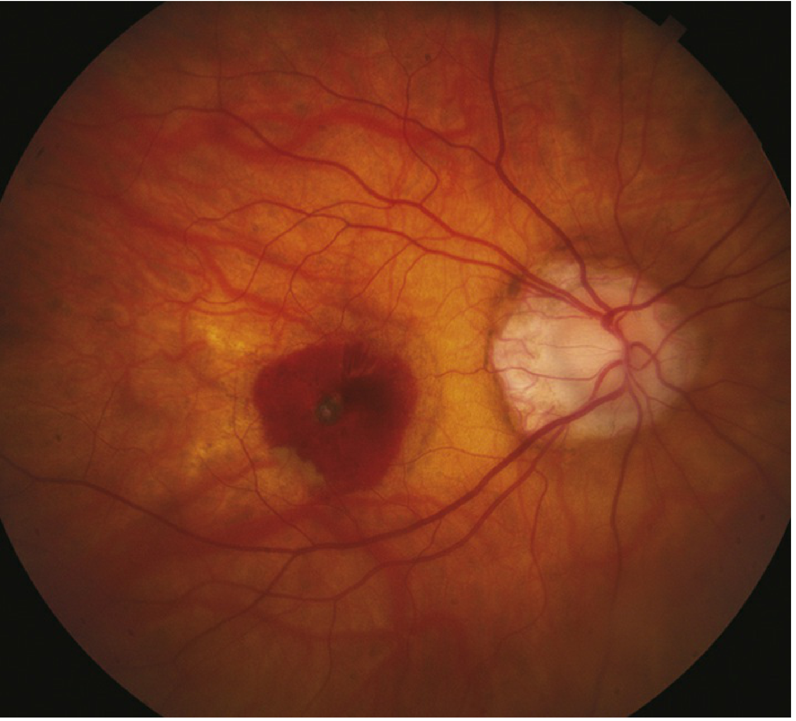
Fundus photograph: High myopia with macular hemorrhage - note the large dark hemorrhage at the macula and pale disc with peripapillary atrophy. - Wills Eye Manual, p. 856
Systemic Associations of High Myopia
- Down syndrome
- Stickler syndrome
- Marfan syndrome
- Prematurity
- Noonan syndrome
- Ehlers-Danlos syndrome
- Pierre-Robin syndrome
- Cornelia de Lange syndrome
Complications of Pathologic Myopia
| Complication | Notes |
|---|---|
| Myopic neovascularization (MNV) | Occurs in 10% of highly myopic eyes |
| Rhegmatogenous retinal detachment (RRD) | Increased risk due to PVD, lattice degeneration, atrophic holes |
| Macular hole | Associated with posterior staphyloma |
| Choroidal neovascularization (CNV) | From lacquer cracks; treatment with anti-VEGF |
| Glaucoma | Visual field loss can mimic glaucoma; IOP measurement may be underestimated with Schiotz tonometry |
| Cataract | Posterior subcapsular opacity; early nuclear sclerosis can paradoxically increase myopic error |
- Kanski's Clinical Ophthalmology, p. 631 | Wills Eye Manual, p. 857-858
Correction and Treatment
Optical Correction
- Spectacles and contact lenses: Correct non-pathologic myopia fully; achieve 20/40 or better in ≥95% of patients.
- Refractive procedures are less successful in pathologic myopia due to severity of error and posterior segment abnormalities.
Surgical Correction
| Procedure | Indication |
|---|---|
| Surface ablation (PRK) | Low-moderate myopia |
| LASIK | Moderate to high myopia (limited by corneal thickness); complications include glare, dry eye, undercorrection, epithelial ingrowth, diffuse keratitis, flap dislocation |
| SMILE (refractive lenticule extraction) | Myopia and myopic astigmatism, small incision technique |
| Clear lens exchange | Very high myopia; small risk of retinal detachment |
| Iris clip (lobster claw) implant | Anterior chamber phakic IOL; complications: subluxation, endothelial loss, cataract, glaucoma, retinal detachment |
| ICL (implantable contact lens) | Posterior chamber phakic IOL (-3 D to -20.5 D); complications: uveitis, pupillary block, cataract, retinal detachment |
| Radial keratotomy | Now of historical interest only |
- Kanski's Clinical Ophthalmology, p. 299
Myopia Progression Control (Pediatric)
-
Low-dose atropine 0.01% at bedtime: Significantly slows myopia progression in children. A reasonable approach is to treat children aged 5-15 years whose myopia progresses >1 diopter/year.
-
Time outdoors in sunshine: Should be encouraged alongside atropine.
-
High-add (+2.50 D) center-distance soft multifocal contact lens: Can be considered for children aged 7-11 years with <5 D myopia (BLINK trial evidence; dose-dependent response).
-
Kanski's Clinical Ophthalmology, p. 631
Workup for Pathologic Myopia
- Manifest and/or cycloplegic refraction
- IOP measurement (note: Schiotz/Tono-pen may underestimate IOP in highly myopic eyes)
- Dilated indirect ophthalmoscopy (look for retinal breaks/detachment)
- Slit lamp biomicroscopy with 60/90 D lens for macula, CNV screening
- IVFA for suspected CNV
- OCT: reveals CNV, macular detachment over staphyloma, foveal schisis
Treatment of Complications
- Retinal breaks: Laser photocoagulation, cryotherapy, or scleral buckle surgery
- CNV/MNV: Anti-VEGF agents are first-line; laser photocoagulation may be considered for extrafoveal/juxtafoveal lesions only
- Glaucoma: Serial visual fields; progression in absence of progressive myopia indicates true glaucoma requiring treatment
- Wills Eye Manual, p. 857-858
Note: A recent 2025 systematic review on pediatric refractive error prevalence in Europe (PMID: 41223238) reinforces that myopia is increasingly common in children - clinicians should be vigilant about early identification and progression monitoring.
This is a shared conversation. Sign in to Orris to start your own chat.