FUNCTIONAL ANATOMY OF MUSCLES OF RESPIRATION, EXTERNAL & INTERNAL RESPIRATION, PATHOPHYSIOLOGY & MANAGEMENT OF HYPOXAEMIA
Exam-Oriented | Postgraduate Emergency Medicine | References: Tintinalli's Emergency Medicine & Harrison's Principles of Internal Medicine 22e
PART 1: FUNCTIONAL ANATOMY OF MUSCLES OF RESPIRATION
(Fishman's Pulmonary Diseases & Disorders, Chapter 3 - Respiratory Muscle Anatomy)
KEY CONCEPT (High-Yield Exam Point)
The respiratory muscles form a complex pump system. They are unique skeletal muscles that must contract rhythmically and continuously - the only skeletal muscles on which life directly depends. They are adapted for fatigue resistance with:
- High oxidative capacity
- Greater capillary density
- Greater maximal blood flow
A. INSPIRATORY MUSCLES
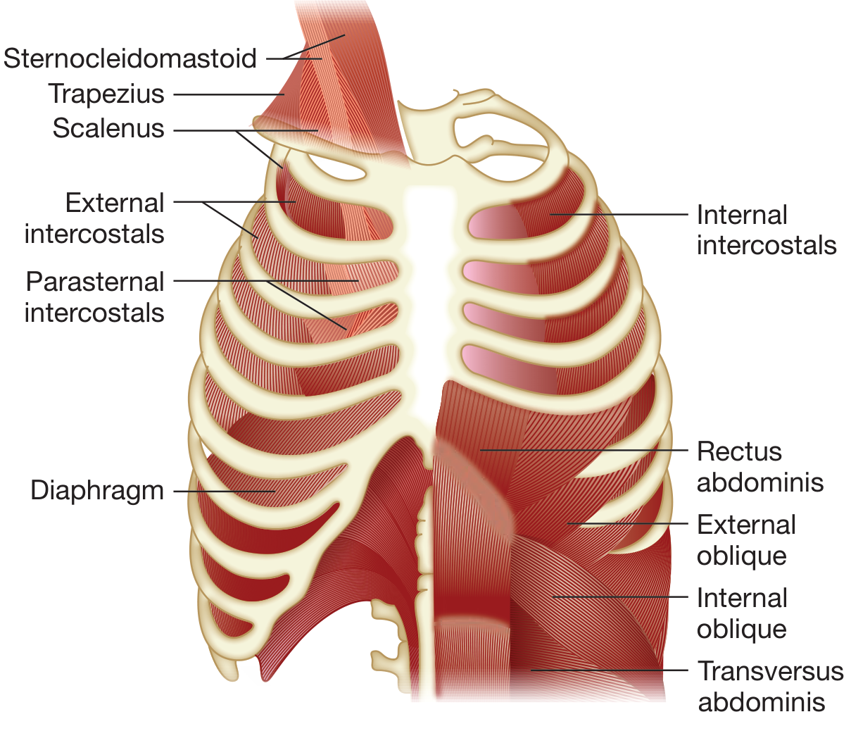
1. THE DIAPHRAGM (Most Important)
- Innervation: Phrenic nerve (C3, C4, C5) - "C3, 4, 5 keeps the diaphragm alive"
- Fiber composition: ~55% Type I (slow oxidative, fatigue-resistant), ~21% Type IIa, ~23% Type IIx
- Actions during contraction (3 components):
- Piston action: The dome descends, increasing vertical thoracic diameter and increasing abdominal pressure
- Appositional action: The zone of apposition (where diaphragm is apposed to inner rib cage) transmits abdominal pressure to lower rib cage, expanding it outward
- Insertional action: Diaphragmatic fibers pull on lower ribs in an axial direction causing cephalad motion and outward rotation of lower ribs, expanding lower rib cage
Exam Fact: When the diaphragm acts in isolation (as in high quadriplegia with only SCM intact), it paradoxically exerts an expiratory effect on the upper rib cage during inspiration. This demonstrates that normal quiet inspiration requires coordinated activity of diaphragm + rib cage inspiratory muscles + abdominal muscles.
- Accounts for ~60% of muscular work of breathing (intercostals ~40%)
2. INTERCOSTAL MUSCLES
Between chondral portions (anterior): Only parasternal intercostals present (one layer).
Between osseous portions (lateral): TWO layers:
| Muscle | Fiber Direction | Action |
|---|
| External intercostals | Obliquely downward and FORWARD | Inspiratory - elevates ribs (bucket handle + pump handle) |
| Internal intercostals | Obliquely downward and BACKWARD | Expiratory (interosseous portion) |
| Parasternal intercostals | Same orientation as internal | Inspiratory (act as inspiratory muscles despite same fiber direction as internal) |
Exam Point - Mnemonic "EXILE": EXternal = Inspiration, Internal = Expiration (for interosseous portion). Exception: Parasternal intercostals are inspiratory despite same fiber orientation as internal intercostals.
- Levator costae: Fusiform muscle from transverse processes of thoracic vertebrae to rib below - assists inspiration.
3. ACCESSORY INSPIRATORY MUSCLES
| Muscle | Nerve Supply | Action |
|---|
| Sternocleidomastoid (SCM) | CN XI (spinal accessory) + C2 | Elevates sternum, increases AP diameter |
| Scaleni (anterior, middle, posterior) | Cervical plexus (C3-C8) | Elevates and fixes ribs 1 & 2, stabilizes thoracic inlet |
| Trapezius | CN XI | Stabilizes shoulder girdle |
| Pectoralis major | Medial + lateral pectoral n. | Elevates ribs when arms fixed |
| Serratus anterior | Long thoracic n. (C5-C7) | Protracts scapula, assists thoracic expansion |
Clinical Exam Point: Recruitment of SCM and scaleni at rest = sign of respiratory distress. SCM becomes the dominant inspiratory muscle in high cervical spinal cord injury (C3-5 injury spares SCM via CN XI).
B. EXPIRATORY MUSCLES (Active in forced expiration, coughing, Valsalva)
Quiet expiration is passive (elastic recoil). Forced expiration / coughing requires active muscle contraction:
| Muscle | Action |
|---|
| Internal intercostals (interosseous) | Depresses ribs, decreases thoracic volume |
| Abdominals (rectus, external oblique, internal oblique, transversus abdominis) | Increase intra-abdominal pressure, push diaphragm up, decrease thoracic volume; also flex trunk |
High-Yield Exam Fact: Transversus abdominis is the most effective expiratory muscle. Abdominal muscles also facilitate inspiration by:
- Lengthening the diaphragm during expiration (keeping it on optimal length-tension curve)
- Acting as a "pre-tensioner" at onset of inspiration
- Pressing abdominal contents against the diaphragm, augmenting the appositional component
C. FIBER TYPES IN RESPIRATORY MUSCLES
| Type | Name | Speed | Fatigue | Enzyme | Role |
|---|
| Type I | Slow oxidative | Slow | Fatigue-resistant | Oxidative | Sustained quiet breathing |
| Type IIa | Fast oxidative-glycolytic | Fast | Fatigue-resistant | Mixed | Sustained vigorous breathing |
| Type IIx | Fast glycolytic | Fast | Intermediate | Glycolytic | Bursts of effort |
- Diaphragm: ~55% Type I, 21% IIa, 23% IIx
- Other respiratory muscles (intercostals, SCM, scalenes): At least 60% highly oxidative fibers
Why This Matters Clinically: High proportion of fatigue-resistant fibers suits the diaphragm for continuous work. In critical illness/ICU: diaphragmatic atrophy from disuse (mechanical ventilation) impairs weaning - this is "ventilator-induced diaphragmatic dysfunction (VIDD)."
D. EFFECT OF PATHOLOGIC STATES ON RESPIRATORY MUSCLES
Hyperinflation (COPD):
- Diaphragm flattened - loses its dome shape, shortens by 30-40% from FRC to TLC
- Appositional zone reduced - decreases appositional contribution
- Paradoxical inward movement of lower chest wall (Hoover's sign) - the inertial component becomes expiratory
- Pressure-generating capacity falls
- Compensatory recruitment of scalenes and SCM to maintain ventilation
- Inspiratory intercostals impaired as well
- Force-generating capacity of neck muscles (scalenes, SCM) is relatively preserved with hyperinflation
PART 2: EXTERNAL AND INTERNAL RESPIRATION
A. EXTERNAL RESPIRATION (Pulmonary Gas Exchange)
Definition: Gas exchange between alveolar air and pulmonary capillary blood across the alveolar-capillary membrane.
The Alveolar Gas Equation (PAO2):
PAO2 = FiO2 × (PB - PH2O) - PaCO2/R
Where:
- FiO2 = fraction of inspired oxygen (0.21 at sea level)
- PB = barometric pressure (760 mmHg at sea level)
- PH2O = water vapour pressure at 37°C (47 mmHg)
- R = respiratory quotient (~0.8)
Normal values (sea level, room air):
- PAO2 ≈ 100 mmHg (alveolar)
- PaO2 ≈ 95 mmHg (arterial)
- A-a gradient = PAO2 - PaO2 = 5-15 mmHg (increases with age)
- PaCO2 ≈ 40 mmHg
- SpO2 ≈ 98%
Age-adjusted normal PaO2 formula (Harrison's):
PaO2 = 109 - 0.43 × (age in years) [SD ± 4.10 mmHg]
Mechanics of Ventilation (Prerequisite for External Respiration)
During spontaneous inspiration:
- Respiratory muscles contract (primarily diaphragm + intercostals)
- Intrathoracic pressure falls below atmospheric pressure
- Air flows from high (atmospheric) to low (alveolar) pressure
- Lung volume increases
- Alveoli expand, thinning the blood-gas interface
During passive expiration:
- Elastic recoil of lungs and chest wall
- Intrathoracic pressure rises above alveolar pressure
- Air flows out
- FRC is the resting equilibrium point where lung elastic recoil inward = chest wall elastic recoil outward
Work of breathing:
- 70-year-old: ~70% of elastic work on chest wall (vs 40% in a 20-year-old) due to stiffened rib articulations/kyphosis from osteoporosis
B. INTERNAL RESPIRATION (Tissue Gas Exchange)
Definition: Gas exchange between systemic capillary blood and tissue cells at the mitochondrial level.
Oxygen Transport to Tissues:
Oxygen Content of Blood (CaO2):
CaO2 = (Hb × 1.34 × SaO2) + (PaO2 × 0.003)
Normal: ~20 mL O2/dL
Oxygen Delivery (DO2):
DO2 = Cardiac Output (CO) × CaO2
Normal: ~1000 mL O2/min
Oxygen Consumption (VO2):
VO2 = CO × (CaO2 - CvO2) [Fick's Principle]
Normal: ~250 mL O2/min
Oxygen Extraction Ratio (OER):
OER = VO2 / DO2 = ~25% (normally tissues extract ~25% of delivered O2)
CO2 Transport from Tissues:
- ~70% as bicarbonate (CO2 + H2O → H2CO3 → H+ + HCO3- via carbonic anhydrase in RBCs)
- ~23% as carbaminohaemoglobin (CO2 + protein -NH2)
- ~7% dissolved in plasma
Oxyhemoglobin Dissociation Curve
| Factor | Curve Shift | Effect on O2 Release |
|---|
| ↑ Temperature | Right | ↑ O2 release to tissues |
| ↑ PaCO2 | Right | ↑ O2 release (Bohr effect) |
| ↑ H+ (↓ pH) | Right | ↑ O2 release |
| ↑ 2,3-DPG | Right | ↑ O2 release |
| Fetal Hb, CO-Hb, Met-Hb | Left | ↓ O2 release |
P50 (PaO2 at which Hb is 50% saturated) = 26.6 mmHg normally.
PART 3: PATHOPHYSIOLOGY OF HYPOXAEMIA
Definition (Harrison's 22e)
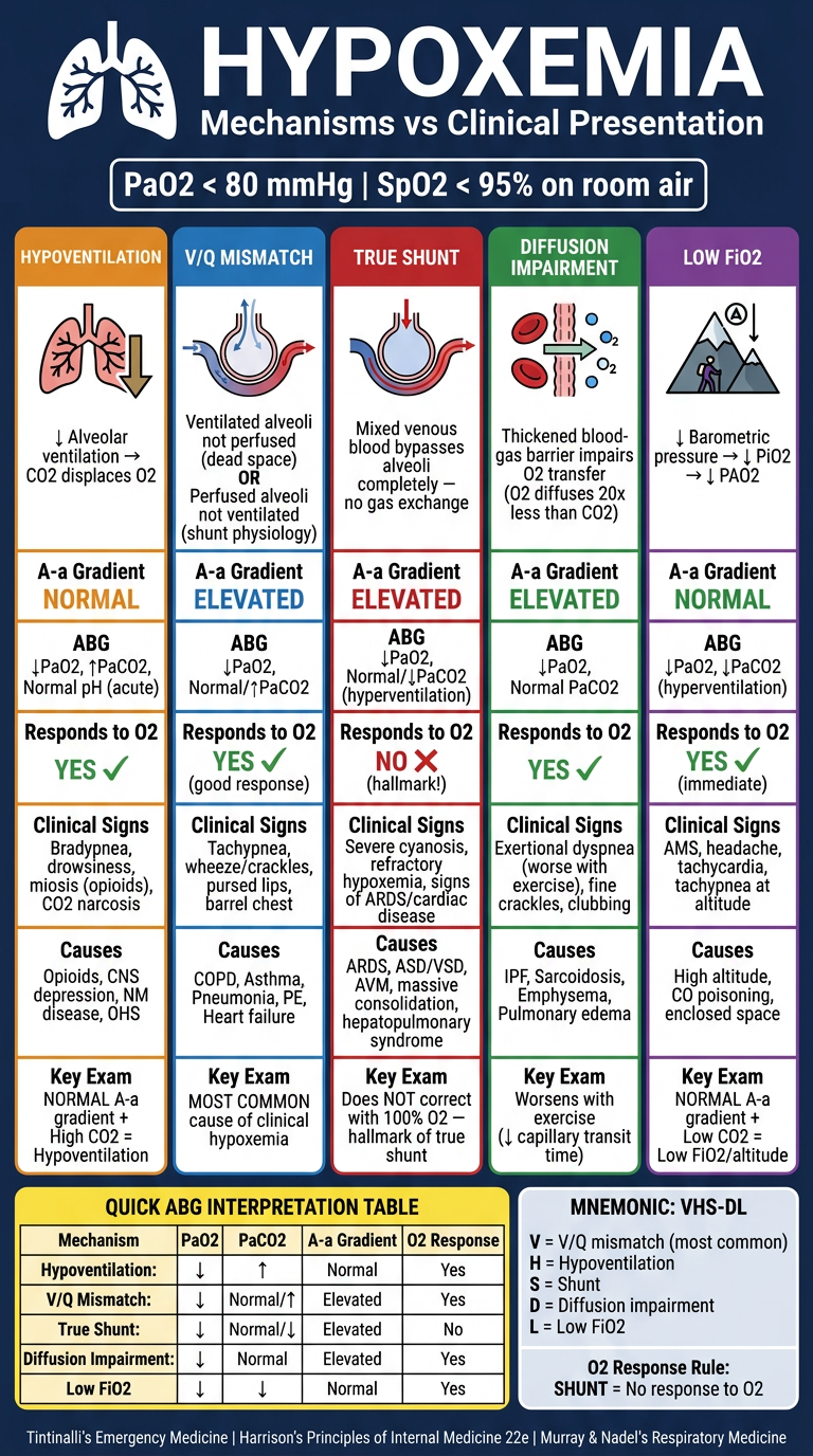
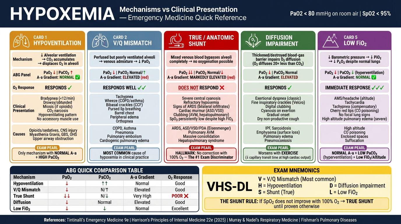
- Hypoxaemia: PaO2 < 80 mmHg or SpO2 < 95% on room air
- Clinically significant hypoxaemia: SpO2 < 90% (PaO2 ~60 mmHg) - on the steep part of oxyhemoglobin dissociation curve
- Hypoxia: Inadequate O2 delivery/utilization at tissue level (hypoxaemia can cause hypoxia but they are distinct)
FRAMEWORK: A-a Gradient-Based Classification
A-a Gradient = PAO2 - PaO2
- Normal (young adult): 5-10 mmHg
- Age-adjusted formula: A-a gradient = Age/4 + 4 (or roughly Age/4 + 4)
-
15-20 mmHg = elevated (suggests intrinsic lung disease)
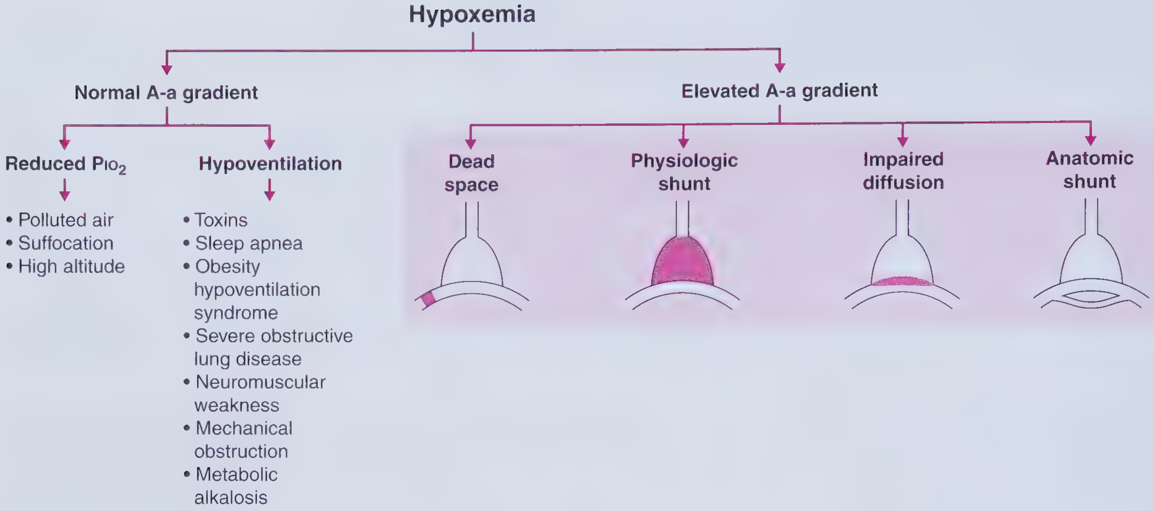
FIVE MECHANISMS OF HYPOXAEMIA
(Harrison's 22e + Fishman's Pulmonary + Murray & Nadel's)
MECHANISM 1: HYPOVENTILATION
- Pathophysiology: Inadequate alveolar ventilation → CO2 accumulates → CO2 displaces O2 in alveoli → ↓ PAO2 → ↓ PaO2
- Key feature: A-a gradient is NORMAL (lung parenchyma is intact)
- ABG: ↑ PaCO2 + ↓ PaO2 (hypercapnic hypoxaemia)
- Responds well to supplemental O2
- Causes:
- CNS depression (opioids, sedatives, stroke, head injury)
- Neuromuscular disease (GBS, myasthenia gravis, ALS)
- Chest wall deformity (kyphoscoliosis)
- Severe COPD (advanced disease)
- Obesity hypoventilation syndrome (OHS/Pickwickian)
- Upper airway obstruction
MECHANISM 2: V/Q MISMATCH (Most Common Cause in Clinical Practice)
Murray & Nadel: "Perfusion to areas with low ventilation in relation to perfusion (V/Q) is the most common cause of hypoxemia in clinical practice."
Dead space (High V/Q > 1):
- Ventilation with reduced/absent perfusion
- Wasted ventilation
- Causes: PE, pulmonary hypertension, positive pressure ventilation
- Results in: hypoxaemia + hypercapnia (raised PaCO2 if severe)
- Normal physiologic dead space = anatomic dead space (~150 mL, 1 mL/pound body weight) + alveolar dead space
Physiologic Shunt / Low V/Q < 1 (Most common V/Q mismatch):
- Perfusion with reduced ventilation
- Blood passes through poorly ventilated alveoli
- A-a gradient ELEVATED
- PaO2 improves with supplemental O2 (because some ventilation exists)
- Causes: COPD, asthma, pneumonia, atelectasis, heart failure, pleural effusion
Key Formula (Bohr Dead Space Equation): VD/VT = (PaCO2 - PeCO2) / PaCO2
Normal VD/VT = 0.3 (30%)
MECHANISM 3: ANATOMIC / TRUE SHUNT
- Mixed venous blood completely bypasses alveoli - no gas exchange occurs
- A-a gradient ELEVATED
- Hallmark: Does NOT respond to 100% O2 (shunted blood cannot be oxygenated regardless of FiO2)
- The 100% oxygen test: If SpO2 fails to normalize on 100% FiO2 → true shunt likely
- Normal physiologic shunt = 2-3% (bronchial + Thebesian vessels)
- Shunt fraction (Qs/Qt) = (CcO2 - CaO2) / (CcO2 - CvO2)
Causes:
- Intracardiac: ASD, VSD, PDA with Eisenmenger's, PFO (patent foramen ovale - opened by high right-sided pressures in PE/ARDS)
- Intrapulmonary: AVM, hepatopulmonary syndrome, ARDS (consolidated alveoli)
- Massive consolidation (pneumonia), massive atelectasis
MECHANISM 4: DIFFUSION IMPAIRMENT
- Thickening of blood-gas barrier OR loss of alveolar surface area
- Impairs O2 diffusion from alveolus to capillary (O2 diffuses 20x less readily than CO2)
- A-a gradient ELEVATED
- Worsens with exercise (reduced transit time in capillary)
- Responds to supplemental O2
Causes:
- Interstitial lung disease (IPF, sarcoidosis)
- Emphysema (loss of alveolar surface area)
- Pulmonary edema (increased diffusion distance)
- Pulmonary hypertension (impaired diffusion + dead space)
Exam Fact: CO2 is rarely affected by diffusion impairment because it is ~20 times more diffusible than O2.
MECHANISM 5: LOW INSPIRED PO2 (Reduced FiO2)
- A-a gradient NORMAL
- At high altitude: ↓ barometric pressure → ↓ PiO2
- No intrinsic lung disease
- Responds rapidly to supplemental O2
- Also occurs in: enclosed spaces (fires, CO poisoning), closed circuit breathing
ADDITIONAL MECHANISMS (High-Yield Points)
Mixed Venous PO2 (PvO2) and Hypoxaemia:
- Low PvO2 (↓ mixed venous saturation) worsens hypoxaemia when shunt or V/Q mismatch is present
- Causes of low PvO2: ↓ cardiac output, anaemia, ↑ O2 consumption (fever, sepsis, exercise)
- Formula: CvO2 = CaO2 - VO2/CO (Fick)
Carbon Monoxide Poisoning:
- SpO2 falsely normal (pulse oximetry cannot distinguish HbCO from OxyHb)
- True SaO2 measured by co-oximetry
- Requires high-flow O2 to displace CO (half-life of HbCO: 4-5h on room air → 60-90 min on 100% O2 → 20-30 min on hyperbaric O2)
Methemoglobinaemia:
- Ferrous (Fe2+) → Ferric (Fe3+) state → cannot carry O2
- SpO2 reads falsely ~85% regardless of true saturation
- Treatment: Methylene blue 1-2 mg/kg IV (ROSEN's Emergency Medicine)
CLINICAL ALGORITHM: DISTINGUISHING CAUSES (Murray & Nadel)
HYPOXAEMIA
     |
Does it respond to supplemental O2?
     |                    |
   YES                   NO
     |                    |
V/Q mismatch or       True Shunt
Hypoventilation            |
     |              Check CXR for
ABG: PaCO2?         ARDS, massive
  Normal or ↑        consolidation,
  |        |         AVM, cardiac
Normal ↑PaCO2
V/Q     Hypoventil-
mismatch  ation
PART 4: MANAGEMENT OF HYPOXAEMIA
IMMEDIATE GOALS (Tintinalli's Emergency Medicine + Harrison's 22e)
- Identify and treat the underlying cause simultaneously
- Target SpO2 94-98% (avoid hyperoxia in most patients)
- SpO2 88-92% in COPD/hypercapnic respiratory failure (to preserve hypoxic drive)
- Correct contributing factors: anaemia (Hb), cardiac output (CO), ventilation
STEP 1: SUPPLEMENTAL OXYGEN DELIVERY SYSTEMS
| Device | FiO2 | Flow Rate | Indication |
|---|
| Nasal cannula | 24-44% | 1-6 L/min | Mild hypoxaemia, awake patients |
| Simple face mask | 40-60% | 5-10 L/min | Moderate hypoxaemia |
| Non-rebreather mask (NRB) | 60-100% | 10-15 L/min | Severe hypoxaemia, emergency |
| Venturi mask | 24-60% (precise) | Variable | COPD, precise FiO2 needed |
| High-flow nasal cannula (HFNC) | 21-100% | Up to 60 L/min | Hypoxaemic respiratory failure |
Harrison's (STEMI example): "In patients whose arterial O2 saturation is normal, supplemental O2 is not recommended. However, when hypoxemia is present (O2 saturation <90%), O2 should be administered to correct the hypoxemia; the patient should then be reassessed to determine if there is a continued need for such treatment."
STEP 2: HIGH-FLOW NASAL CANNULA (HFNC)
- Delivers humidified O2 at high flow (up to 60 L/min) with precise FiO2
- Generates low-level PEEP (~1-2 cmH2O per 10 L/min)
- Reduces nasopharyngeal dead space
- ROX Index (SpO2/FiO2 / RR): ROX ≥ 4.88 at 12h predicts HFNC success
- Indication: Acute hypoxaemic respiratory failure (AHRF) with SpO2 < 94% on conventional O2
STEP 3: NON-INVASIVE VENTILATION (NIV)
CPAP (Continuous Positive Airway Pressure):
- Maintains constant positive pressure throughout respiratory cycle
- Recruits collapsed alveoli, reduces shunt fraction, improves FRC
- Best for: Cardiogenic pulmonary oedema (CPAP reduces preload + afterload), obstructive sleep apnoea
BiPAP (Bilevel Positive Airway Pressure):
- IPAP: provides inspiratory support (reduces WOB)
- EPAP: provides expiratory support (recruits alveoli, similar to PEEP)
- Best for: COPD exacerbation with hypercapnia + hypoxaemia, mild-moderate ARDS
Contraindications to NIV: Apnoea, inability to protect airway, haemodynamic instability, facial trauma, vomiting risk, high secretion load
STEP 4: ENDOTRACHEAL INTUBATION AND MECHANICAL VENTILATION
Indications for Intubation in Hypoxaemia:
- Failure of non-invasive methods (SpO2 < 88% despite max O2 + NIV)
- Inability to protect airway
- Haemodynamic instability
- GCS ≤ 8 / obtundation
- Impending respiratory arrest
- Severe acidosis (pH < 7.25)
Ventilator Settings (Lung-Protective for ARDS - Harrison's/Tintinalli's):
- Tidal Volume: 6 mL/kg ideal body weight (lung-protective strategy)
- PEEP: 5-20 cmH2O (titrate to FiO2 needed)
- FiO2: reduce to ≤ 0.6 as soon as possible
- Plateau pressure: < 30 cmH2O
- Target: SpO2 88-95%, PaO2 55-88 mmHg
STEP 5: ADJUNCT THERAPIES FOR REFRACTORY HYPOXAEMIA (ARDS)
| Therapy | Mechanism | Evidence |
|---|
| Prone positioning | Recruits dorsal lung, reduces V/Q mismatch, reduces shunt | Reduces mortality in severe ARDS (PROSEVA trial) |
| Neuromuscular blockade (NMBA) | Reduces ventilator asynchrony, decreases O2 consumption | ACURASYS trial (controversial) |
| Recruitment maneuvers | Transiently increase airway pressure to open collapsed alveoli | Evidence mixed |
| Inhaled Nitric Oxide (iNO) | Selectively vasodilates ventilated alveoli, reduces shunt | Improves oxygenation, no mortality benefit |
| Inhaled Prostacyclin (epoprostenol) | Similar to iNO | Bridge therapy |
| ECMO (VV-ECMO) | Extracorporeal gas exchange bypasses lungs | Last resort in severe ARDS with refractory hypoxaemia |
STEP 6: DISEASE-SPECIFIC MANAGEMENT
| Cause | Specific Treatment |
|---|
| COPD exacerbation | Controlled O2 (target SpO2 88-92%), bronchodilators, steroids, NIV if PaCO2 > 45 + acidosis |
| Cardiogenic pulmonary oedema | CPAP/BiPAP, diuresis, nitrates |
| Pneumonia/ARDS | Antibiotics, lung-protective ventilation, prone if P/F < 150 |
| PE | Anticoagulation, thrombolytics if haemodynamically unstable |
| Tension pneumothorax | Needle decompression 2nd ICS MCL, followed by chest tube |
| Haemothorax | Chest tube insertion |
| Methemoglobinaemia | Methylene blue 1-2 mg/kg IV (Rosen's Emergency Medicine) |
| CO poisoning | 100% O2 via NRB, consider hyperbaric O2 |
| Anaphylaxis | IM adrenaline, O2, IV fluids |
STEP 7: TREAT CONTRIBUTING FACTORS TO HYPOXIA
Even with corrected hypoxaemia, tissue hypoxia may persist if:
- Anaemia: Transfuse to Hb > 7-8 g/dL (higher threshold in ACS: Hb > 10)
- Low cardiac output: Treat heart failure, optimize volume, vasopressors/inotropes
- Sepsis: Source control, antibiotics, resuscitation (Surviving Sepsis Guidelines)
- Right-to-left shunting in heart: Cardiology/cardiothoracic surgery consultation
SUMMARY TABLE: EXAM QUICK-REVIEW
| Mechanism | PaO2 | PaCO2 | A-a Gradient | Response to O2 |
|---|
| Hypoventilation | ↓ | ↑ | Normal | Good |
| Low V/Q | ↓ | Normal/↑ | Elevated | Good |
| True shunt | ↓ | Normal/↓ | Elevated | Poor |
| Diffusion impairment | ↓ | Normal | Elevated | Good |
| Low FiO2 (altitude) | ↓ | ↓ (hyperventilation) | Normal | Good |
REFERENCES
- Tintinalli JE et al. Tintinalli's Emergency Medicine: A Comprehensive Study Guide, 9th ed. McGraw-Hill. [Cyanosis, Oxygen Delivery, Emergency Airway Management]
- Longo DL et al. Harrison's Principles of Internal Medicine, 22nd ed. (2025). McGraw-Hill Medical. Chapter on Respiratory Function, STEMI management, oxygen supplementation. [ISBN: 9781265977061]
- Fishman AP et al. Fishman's Pulmonary Diseases and Disorders, 2-Volume Set. Chapter 3: Respiratory Muscle Anatomy and Function. [ISBN: 9781260473940]
- Murray J, Nadel JA et al. Murray & Nadel's Textbook of Respiratory Medicine, 2-Volume. Chapter 44: Mechanisms of Hypoxemia. [ISBN: 9780323655873]
- Rosen's Emergency Medicine: Concepts and Clinical Practice (latest ed.) - Cyanosis, Methemoglobinemia management. [ISBN: 9780323757898]
- Frameworks for Internal Medicine - Hypoxemia Framework, A-a Gradient. [ISBN: 9781496359308]
High-Yield Exam Mnemonic - Causes of Hypoxaemia: "VHS DL"
- V - V/Q mismatch (most common)
- H - Hypoventilation
- S - Shunt (anatomic/true)
- D - Diffusion impairment
- L - Low FiO2