
Basics of surgery
Reading File
The query "Basics of surgery" is broad. Let me clarify what would be most useful before diving in.
Asking for Preferences
What aspect of surgical basics are you interested in? · What is your level / purpose?
Finding Sources
Searching the Web
basics of surgery medical student overview wounds healing asepsis pre-op post-op
Good - Bailey and Love's is the classic general surgery textbook. Let me search for key surgical basics topics in parallel.
Finding Sources
Finding Sources
Finding Sources
Reading File
Reading File
Finding Sources
Finding Sources
Finding Sources
Finding Sources
Finding Sources
Reading File
Reading File
Reading File
Loading Image
Reading File
I now have excellent material from multiple authoritative surgical textbooks. Let me compile the comprehensive answer.
Basics of Surgery - Medical Student Overview
Sources: Bailey and Love's Short Practice of Surgery (28th ed.), Schwartz's Principles of Surgery (11th ed.), Mulholland & Greenfield's Surgery (7th ed.), Pye's Surgical Handicraft (22nd ed.)
1. Surgical Terminology & Wound Classification
Wounds are classified by mechanism and contamination level:
| Class | Description | Infection Risk |
|---|---|---|
| Clean (Class I) | Elective, no GI/GU/respiratory entry, no break in technique | <2% |
| Clean-contaminated (Class II) | GI/GU/respiratory entry under controlled conditions | 5-10% |
| Contaminated (Class III) | Open fresh traumatic wound, major break in technique | 15-20% |
| Dirty/Infected (Class IV) | Old traumatic wound, perforated viscus, pus encountered | >30% |
Intention of healing:
- Primary intention (1st intention): Wound edges are approximated immediately (e.g., surgical incision closed with sutures). Clean wounds with no tissue loss. Best cosmetic outcome.
- Secondary intention (2nd intention): Wound left open, heals by granulation tissue formation from the base upward. Used for infected or heavily contaminated wounds.
- Tertiary / Delayed primary intention: Wound left open initially, then closed 4-5 days later after infection risk decreases (e.g., perforated bowel repair).
2. Phases of Wound Healing
Wound healing is a dynamic, overlapping four-phase process spanning up to 1 year:
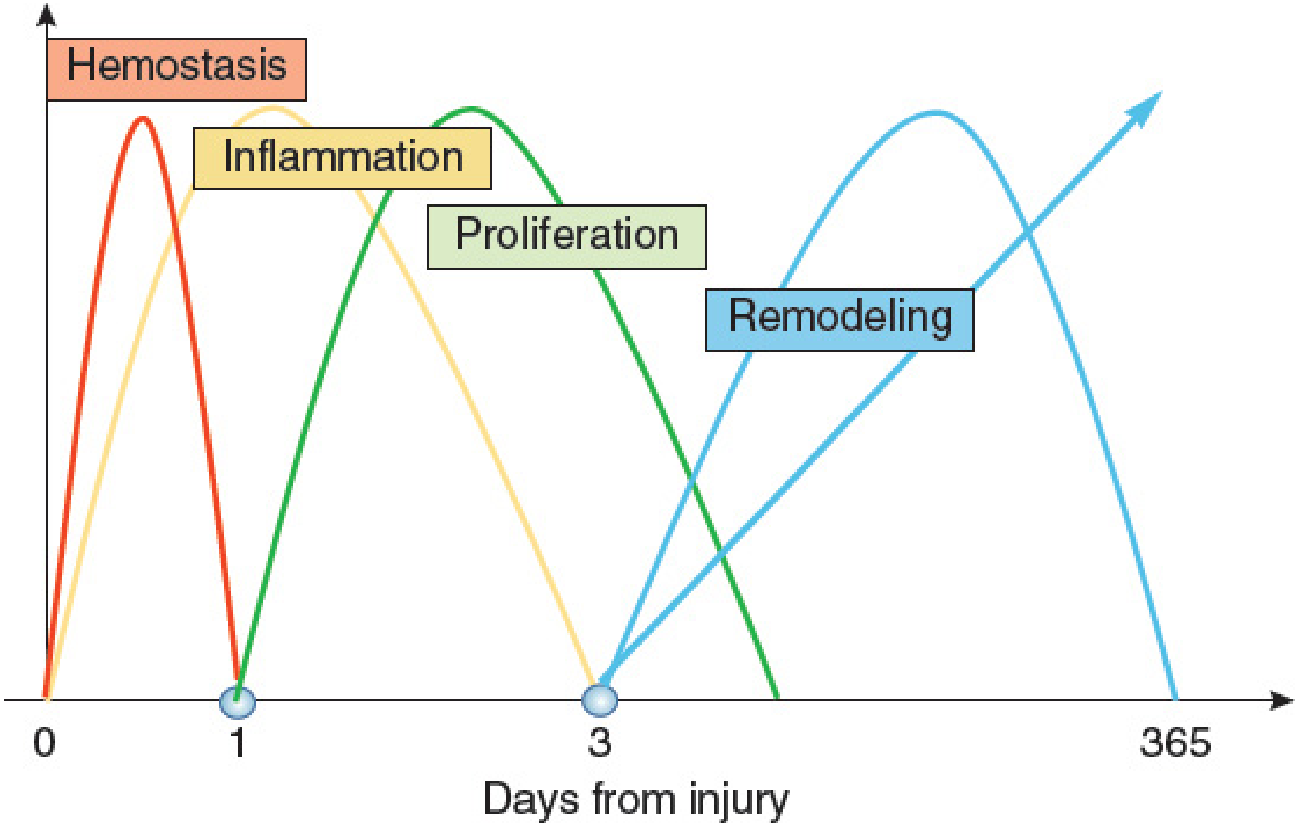
Figure: Four overlapping phases of wound healing (Mulholland & Greenfield's Surgery, 7th ed.)
Phase 1 - Hemostasis (minutes to hours)
- Injured vessels constrict; platelets adhere to exposed collagen via GP Ia/IIa complex
- Platelet aggregation forms a primary platelet plug; coagulation cascade activates -> fibrin clot
- Platelets release PDGF, TGF-β, VEGF - signaling molecules that drive subsequent healing
- The fibrin clot serves as a scaffold for migrating inflammatory cells
Phase 2 - Inflammation (days 1-4)
- PMNs (neutrophils) are the first cells recruited, peaking at 24-48 hours; they phagocytose bacteria/debris and release cytokines (TNF-α, IL-1)
- Macrophages (from monocytes) dominate by 48-96 hours - they are essential to healing; they release TGF-β, VEGF, IGF, EGF to regulate proliferation, matrix synthesis, and angiogenesis
- T-lymphocytes peak at ~1 week; CD8+ suppressor cells downregulate healing (their depletion enhances healing), while CD4+ helpers have minimal direct effect
- Clinical signs: redness, warmth, swelling, pain around wound
Phase 3 - Proliferation (days 4-12)
- Fibroblasts migrate in (chemotaxis by PDGF) -> proliferate -> synthesize collagen (types I and III) and extracellular matrix
- Lactate accumulation (~10 mmol) in the wound potently regulates collagen synthesis via ADP-ribosylation
- Angiogenesis: endothelial cells form new capillaries under VEGF, TNF-α, TGF-β stimulation
- Re-epithelialization: keratinocytes migrate from wound edges and skin appendages
- Wound contraction occurs via myofibroblasts (transformed fibroblasts)
Phase 4 - Maturation & Remodeling (weeks to 1 year)
- Collagen remodels: type III collagen is replaced by stronger type I collagen
- Collagen cross-linking increases wound tensile strength
- Maximum wound tensile strength = 80% of unwounded skin (never reaches 100%)
- Excess collagen deposition -> hypertrophic scar (stays within wound margins) or keloid (extends beyond wound margins, more common in dark skin, deltoid/presternal regions)
Factors impairing wound healing:
- Local: infection, foreign body, ischemia, radiation, poor technique
- Systemic: diabetes, malnutrition (esp. Vitamin C, zinc), corticosteroids, chemotherapy, jaundice, uremia, advanced age
3. Surgical Asepsis & Infection Prevention
Key concepts:
| Term | Definition |
|---|---|
| Asepsis | Absence of pathogenic organisms in a field |
| Antisepsis | Use of chemical agents on living tissue to destroy/inhibit organisms |
| Sterilization | Complete destruction of ALL microorganisms (including spores) |
| Disinfection | Destruction of most pathogens (not necessarily spores) |
Methods of sterilization:
- Autoclaving (steam under pressure): 134°C for 3 min or 121°C for 15 min - gold standard for heat-stable instruments
- Dry heat: 160°C for 60 min - for heat-stable items damaged by moisture
- Ethylene oxide gas: For heat-sensitive instruments (scopes, plastics)
- Gamma irradiation: Industrial sterilization of single-use items
Surgical scrub technique: 3-5 minute scrub with antiseptic (chlorhexidine or povidone-iodine) from fingertips to elbows, followed by gowning and gloving in a sterile manner.
Surgical site infection (SSI) prevention:
- Prophylactic antibiotics given within 60 minutes before incision (30 min for vancomycin)
- Hair removal with clippers (not razor shaving) if needed
- Maintain normothermia, normoglycemia intraoperatively
- Gentle tissue handling; avoid dead space and haematomas
4. Preoperative Assessment & Preparation
History: full surgical, medical, drug, allergy, family, and social history. Focus on cardiovascular, respiratory, renal, hepatic, and haematological status.
Examination: general, cardiorespiratory, and system-specific exam.
Investigations (tailored by risk):
| Investigation | Indication |
|---|---|
| FBC | All major surgery |
| U&E, Creatinine | Renal disease, diuretics, ACEi/ARB use, major surgery |
| LFTs, Coagulation | Liver disease, jaundice, anticoagulation |
| ECG | Age >40, cardiac disease |
| Chest X-ray | Cardiorespiratory symptoms, major thoracic surgery |
| Pulmonary function tests (FEV1, FVC, PFR) | COPD, respiratory disease |
| Group & Save / Crossmatch | Any surgery with blood loss risk |
Optimisation before elective surgery (Pye's Surgical Handicraft):
- Treat respiratory infections; avoid elective surgery during acute illness
- Stop smoking 3 weeks preoperatively to reduce bronchial hypersecretion
- Carious teeth removed 3 weeks before (risk of lung abscess from dental sepsis)
- Obese patients encouraged to diet/lose weight before elective surgery
- Physiotherapy teaching of breathing exercises preoperatively
Fasting (NBM): "2-4-6 rule"
- Clear fluids: 2 hours before
- Breast milk: 4 hours before
- Solids/formula/cow's milk: 6 hours before
Consent: informed, voluntary, and patient must have capacity. Covers procedure, alternatives, material risks, and expected outcomes.
5. Surgical Instruments & Sutures
Suture Classification
Absorbable sutures (for deep tissues, subcutaneous layers):
- Natural: Plain catgut (absorbed in 7-10 days), Chromic catgut (absorbed in 21 days)
- Synthetic: Vicryl (polyglactin), Dexon (polyglycolic acid), PDS (polydioxanone) - longer absorption, less tissue reaction, preferred
Non-absorbable sutures (for skin and permanent repairs):
- Braided (silk, braided nylon): easier to handle and knot, tension adjustable - but cause drag and may cause stitch abscess via capillary action
- Monofilament (nylon, polypropylene/Prolene): less tissue reaction, less drag - but harder to knot and tension harder to adjust
Needles:
- Cutting needles - for skin
- Round-bodied needles - for bowel, muscle, deep layers
- Fine slim-line cutting needles for facial work (4/0 suture)
- 3/0 gauge on stout cutting needle for most minor surgical procedures
Other wound closure methods:
- Staples: Fast; avoid on face and hands
- Adhesive strips (Steri-strips): Painless; useful for small superficial lacerations
- Tissue glue (cyanoacrylate): Clean lacerations with good apposition
Basic Instruments
- Needle holder (e.g., Gillies combined needle holder/scissors)
- Retractors: Catspaw retractors, skin hooks
- Haemostatic forceps: Mosquito/artery forceps
- Scissors: Mayo scissors (curved), Metzenbaum scissors (tissue dissection)
- For abscesses: Sinus forceps, Volkmann spoon
Drains
Used to prevent accumulation of blood, pus, bile, or serous fluid:
- Corrugated (open) drains - passive, drain by gravity
- Closed suction drains (Redivac, Jackson-Pratt) - fine perforated plastic tubes connected to negative-pressure reservoir; reduce infection risk
6. Postoperative Care & Complications
Immediate (0-24 hours)
- Airway: Ensure muscle relaxants fully reversed; suction upper airways clear before extubation
- Oxygen: Give routinely after major surgery to counteract arterial desaturation
- Analgesia: Adequate pain control enables deep breathing; narcotics given judiciously (respiratory depression risk)
- "Stir-up" regime: Patient encouraged to breathe deeply, cough, and move every hour
Common Postoperative Complications by Timing
| Time | Complication |
|---|---|
| 0-24 hrs | Reactionary haemorrhage, airway obstruction, hypotension, pain |
| 24-72 hrs | Secondary haemorrhage, wound infection starts, pulmonary atelectasis |
| Day 3-5 | Pulmonary complications (pneumonia, PE), UTI |
| Day 5-10 | Wound dehiscence, anastomotic leak, deep vein thrombosis |
| >10 days | Chronic wound infection, incisional hernia (months later) |
"The 4 Ws" of Postoperative Fever (mnemonic)
- Wind (day 1-2): Atelectasis/pneumonia
- Water (day 3-5): UTI
- Wound (day 5-7): Surgical site infection
- Walking (day 5+): DVT/PE
DVT/PE Prevention
- Early mobilisation
- TED stockings
- LMWH (low molecular weight heparin) prophylaxis
- Hydration
7. Shock in Surgical Practice
Shock = inadequate tissue perfusion. Classification critical for exams:
| Type | Mechanism | Example |
|---|---|---|
| Hypovolaemic | Low circulating volume | Haemorrhage, burns, GI losses |
| Distributive | Maldistribution of flow | Septic, anaphylactic, neurogenic |
| Cardiogenic | Pump failure | MI, cardiac tamponade |
| Obstructive | Outflow obstruction | Tension pneumothorax, PE |
Haemorrhagic shock classes (ATLS):
| Class | Blood loss | HR | BP | RR |
|---|---|---|---|---|
| I | <750 mL (<15%) | <100 | Normal | 14-20 |
| II | 750-1500 mL (15-30%) | 100-120 | Normal | 20-30 |
| III | 1500-2000 mL (30-40%) | 120-140 | Decreased | 30-40 |
| IV | >2000 mL (>40%) | >140 | Very low | >35 |
Management: 2 large-bore IV cannulae, crystalloid/blood resuscitation, treat source.
8. Surgical Anatomy Principles
Key concepts tested in exams:
- Langer's lines (relaxed skin tension lines): Incisions made along these lines produce least tension and best scars
- Anatomical planes: Surgeons work in tissue planes (e.g., fascial planes) to minimize blood loss and identify structures
- Surgical triangles & landmarks: e.g., Triangle of Calot (cystic artery, cystic duct, hepatic duct), Hesselbach's triangle (inguinal hernias)
- Nerve identification: Preserving motor/sensory nerves (e.g., facial nerve in parotid surgery, recurrent laryngeal nerve in thyroid surgery)
Quick Exam Summary
| Topic | High-Yield Points |
|---|---|
| Wound healing phases | Hemostasis -> Inflammation -> Proliferation -> Remodeling |
| Key cells | PMNs first (24-48h), Macrophages essential (48-96h), Fibroblasts in proliferation |
| Max wound strength | 80% of original (never 100%) |
| Keloid vs hypertrophic | Keloid extends beyond wound margins; hypertrophic does not |
| Sterilization | Autoclave = gold standard (134°C, 3 min) |
| Prophylactic antibiotics | Within 60 min before incision |
| Absorbable sutures | Vicryl, PDS, catgut (deep layers) |
| Shock classes | Class III (30-40% blood loss) = first drop in BP |
| Post-op fever | Wind-Water-Wound-Walking mnemonic |
Sources: Bailey and Love's Short Practice of Surgery (28th ed.); Schwartz's Principles of Surgery (11th ed.); Mulholland & Greenfield's Surgery (7th ed.); Pye's Surgical Handicraft (22nd ed.)
This is a shared conversation. Sign in to Orris to start your own chat.