
Steroids in ophthamology Pg question Classification of steroids short intrermediate long working Use in ophthal Main indication Main side effects in eye due to steroids
classification of ophthalmic steroids short intermediate long acting duration of action
ocular corticosteroid glaucoma cataract
Steroids in Ophthalmology
1. Classification by Duration of Action
| Category | Biological t½ | Drugs |
|---|---|---|
| Short-acting | < 12 h | Hydrocortisone, Cortisone |
| Intermediate-acting | 18-36 h | Prednisolone, Prednisone, Methylprednisolone, Triamcinolone |
| Long-acting | 36-54 h | Dexamethasone, Betamethasone, Fluocinolone acetonide |
- High-potency ("hard") steroids - penetrate the cornea well and control inflammation strongly, but carry the highest risk of IOP rise: Prednisolone acetate 1%, Dexamethasone 0.1%, Difluprednate 0.05%
- Low-potency ("soft") steroids - designed to be inactivated rapidly once they reach the anterior chamber, giving good anti-inflammatory effect with less IOP rise: Fluorometholone 0.1%, Loteprednol etabonate 0.5%, Rimexolone 1%
- Topical drops/ointment - prednisolone acetate/phosphate, dexamethasone, fluorometholone, loteprednol, rimexolone, difluprednate
- Periocular (subconjunctival/subtenon/peribulbar) - triamcinolone, dexamethasone, betamethasone
- Intravitreal - triamcinolone acetonide (short-lasting, months); sustained-release implants - dexamethasone implant (Ozurdex, 4-6 months), fluocinolone acetonide implant (Retisert/Iluvien, up to 3 years)
- Systemic (oral/IV pulse) - prednisolone, methylprednisolone (used for posterior segment/optic nerve/orbital disease and systemic autoimmune disease with ocular involvement)
2. Uses in Ophthalmology / Main Indications
- Allergic conjunctivitis (vernal/atopic keratoconjunctivitis) - short pulses
- Episcleritis and scleritis
- Anterior uveitis/iritis (mainstay of treatment along with cycloplegics)
- Post-operative inflammation - after cataract surgery, corneal transplant, glaucoma surgery, LASIK/PRK (to reduce haze/inflammation)
- Disciform (stromal) herpetic keratitis - always under antiviral cover
- Immune-mediated corneal graft rejection - topical/periocular steroids to suppress rejection
- Posterior uveitis, intermediate uveitis, panuveitis
- Cystoid/diabetic macular edema and macular edema following retinal vein occlusion - intravitreal triamcinolone or dexamethasone/fluocinolone implants
- Optic neuritis - IV methylprednisolone pulse therapy
- Sympathetic ophthalmia and other autoimmune posterior segment disease
- Thyroid eye disease (Graves' orbitopathy) - IV pulse steroids for active moderate-severe disease
- Idiopathic orbital inflammation (orbital pseudotumor)
- Giant cell arteritis (urgent high-dose systemic steroids to prevent irreversible visual loss)
- Ocular myasthenia, ocular cicatricial pemphigoid, Cogan syndrome, Behçet disease with ocular involvement
3. Main Ocular Side Effects of Steroids
-
Steroid-induced (steroid-response) glaucoma / ocular hypertension
- About 1 in 3 individuals are "steroid responders." IOP rise typically occurs 2-4 weeks after starting potent topical/periocular/intravitreal steroid (can be faster with intravitreal triamcinolone, up to hours with systemic ACTH/steroid).
- Mechanism: increased outflow resistance from altered trabecular meshwork extracellular matrix/endothelial function.
- Risk factors: pre-existing POAG/family history, high myopia, young age (children especially), connective tissue disease (e.g., RA), diabetes, African ancestry.
- Management: stop or taper steroid (IOP usually normalizes in days-weeks); switch to a "softer" steroid (fluorometholone, loteprednol, rimexolone); aqueous suppressants if needed; rarely glaucoma surgery if IOP elevation persists (~3% of cases) - Kanski's Clinical Ophthalmology, p. 407-408; The Wills Eye Manual, p. 587-588.
-
Posterior subcapsular cataract (PSC) - with prolonged use (weeks to months), dose- and duration-dependent, more common with systemic and long-term topical use.
-
Increased susceptibility to/exacerbation of infection
- Reactivation or worsening of herpes simplex keratitis (stromal/dendritic)
- Predisposition to fungal keratitis (classically after inappropriate steroid use in a corneal ulcer)
- Can mask signs of infection, delaying diagnosis.
-
Delayed corneal/wound healing - impaired epithelialization after surgery or injury.
-
Corneal and scleral thinning/melt - risk of perforation, especially in eyes with pre-existing thinning (e.g., after pterygium excision, rheumatoid scleritis).
-
Ptosis and periocular skin atrophy - with prolonged periocular injections.
-
Mydriasis and accommodation changes - occasionally with certain preparations.
-
Route-specific risks: intravitreal injections carry additional risks of endophthalmitis, vitreous hemorrhage, and retinal detachment from the injection procedure itself; systemic steroids add the classic systemic Cushingoid effects (osteoporosis, hyperglycemia, growth suppression in children, HPA-axis suppression).
Quick exam-oriented summary table
| Feature | Point to remember |
|---|---|
| Most potent topical | Prednisolone acetate 1%, Difluprednate |
| Least IOP-raising ("safe") topical | Fluorometholone, Loteprednol, Rimexolone |
| Longest duration device | Fluocinolone implant (Retisert - up to 3 years) |
| Classic triad of ocular steroid side effects | Glaucoma, Cataract (PSC), Infection (esp. fungal/HSV) |
| Time to steroid-response IOP rise | 2-4 weeks (topical/periocular), months (intravitreal triamcinolone), hours (rare, systemic) |
Scleritis episcleritis Ophthalmology Pictures wuth Classificatin pathogenesis sign symtomps treatment
scleritis eye clinical photograph
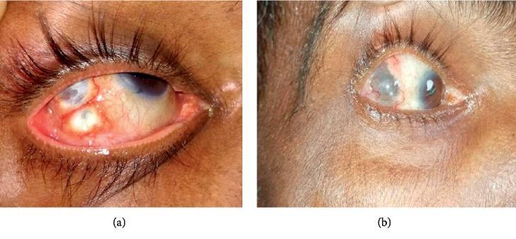
This comparison clinical photograph depicts a case of anterior nodular scleritis of the right eye before and after treatment. Image (a) shows the eye in an acute inflammatory state, characterized by significant scleral and conjunctival injection (redness) and the presence of multiple, well-defined, yellowish-white elevated nodules located on the temporal sclera. Engorged episcleral blood vessels are clearly visible. Image (b) shows the same eye after two months of antitubercular therapy (ATT) and highly active antiretroviral therapy (HAART). The photograph demonstrates a marked clinical improvement, with a significant reduction in inflammation and conjunctival hyperaemia. The previously prominent nodules have largely regressed, leaving behind slight scleral thinning and residual grayish discoloration, indicative of healing and resolution of the granulomatous process. This visual comparison illustrates the efficacy of targeted systemic therapy in treating infectious nodular scleritis associated with tuberculosis and HIV coinfection.
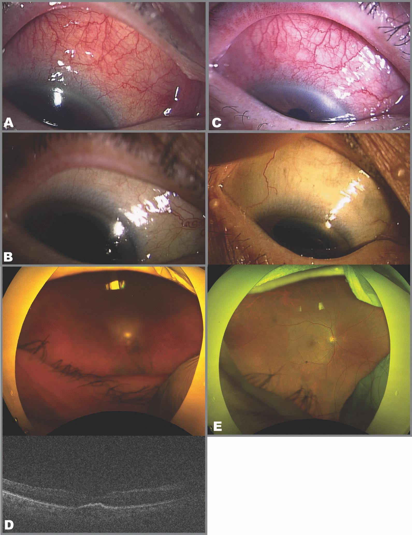
This composite of ophthalmological images documents a clinical case of intraocular inflammation (IOI) following intravitreal brolucizumab injection. Panels A, B, and C are external clinical photographs of the right eye focusing on the superior sclera. Image A shows active, diffuse superior scleritis with intense vascular congestion and episcleral injection. Image B demonstrates clinical resolution of the scleritis following topical therapy, showing a white, quiet sclera. Image C illustrates a recurrence of the scleritis with marked conjunctival and episcleral hyperemia. Panel D includes a wide-field fundus photograph and a corresponding Optical Coherence Tomography (OCT) scan. The fundus image in D shows severe vitreous opacification (haze), obscuring retinal details, while the OCT B-scan displays poor signal-to-noise ratio due to vitreous media opacity, though a macular profile is visible. Panel E shows a follow-up wide-field fundus photograph demonstrating significant clearing of the vitreous opacity, allowing for a detailed view of the optic nerve head, retinal vasculature, and macula. This series illustrates the correlation between clinical scleritis and internal ocular inflammation (vitritis) as complications of intravitreal pharmacotherapy.

This composite diagnostic image presents a side-by-side comparison of the right eye using clinical photography and Optical Coherence Tomography (OCT) to illustrate the progression of anterior scleritis. Image A (Day 22) demonstrates active pathology: the clinical photograph shows significant scleral injection and localized redness, while the corresponding anterior segment OCT reveals structural disruption. Specifically, dashed arrows indicate separated scleral fibers, solid arrows highlight hyper-reflective inflammatory infiltrates, and an asterisk marks a region of irregular reflectivity within the scleral lamellae. Image B (Day 29) shows the clinical resolution: the external photograph displays a quiet eye with diminished redness, and the OCT scan demonstrates a more homogenous, compact tissue architecture with the disappearance of the previously noted infiltrates and fiber separation. This comparison serves as an educational tool for ophthalmology, specifically for monitoring the clinical and structural response to treatment in inflammatory ocular conditions such as scleritis.
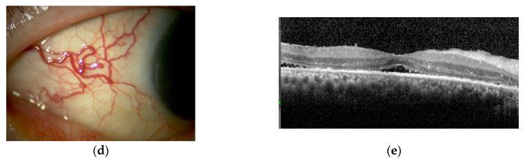
This composite educational image features a clinical photograph and a diagnostic Optical Coherence Tomography (OCT) scan illustrating ocular inflammation.
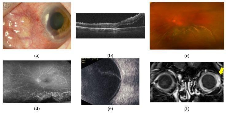
A multi-modal diagnostic panel illustrating anterior and posterior scleritis in a human eye. (a) Clinical photograph of the anterior segment showing hyperemic necrotizing scleritis and corneal marginal infiltration. (b) Optical Coherence Tomography (OCT) cross-section demonstrating a raised, wrinkled neuroepithelial layer with subretinal fluid accumulation. (c) Color fundus image depicting papilledema and extensive serous retinal detachment (SRD) involving the macula. (d) Fundus Fluorescein Angiography (FFA) highlighting retinal vasculitis and leakage consistent with SRD. (e) B-mode ultrasonography showing characteristic 'T-sign' (posterior scleral edema) and scleral thickening associated with extensive SRD. (f) Contrast-enhanced MRI (coronal view) revealing significant contrast enhancement localized to the posterior ocular wall (yellow arrow). This composite illustrates the typical progression and diagnostic findings for combined anterior and posterior inflammatory eye disease, highlighting key clinical features such as scleral melting, subretinal fluid, and optic disc swelling.
episcleritis eye clinical photograph
Clinical photograph (ophthalmic external/anterior segment) of the right eye showcasing anterior segment inflammation with conjunctival injection. Frontal view highlights diffuse bulbar conjunctival redness, greatest inferiorly, with adjacent vascular engorgement and mild episcleral whitening not clearly seen. The cornea shows no obvious defect in this frame, and the eyelids appear unremarkable. In the setting of Behçet disease, ocular involvement most often presents as noninfectious anterior uveitis or episcleritis, and can progress to vision impairment if undertreated. The observed redness and focal vascular congestion are findings compatible with active ocular surface inflammation and possible early keratouveitis, though histologic detail is unavailable in a photograph. This image captures a snapshot of inflammatory activity that informs clinical assessment of BD activity, helps distinguish autoimmune inflammatory processes from infectious conjunctivitis, and supports therapeutic decision-making regarding systemic immunomodulation and local therapy. Clinically, it complements other BD features such as oral/genital ulcers and skin lesions, and may prompt rheumatology consultation. In educational use, it illustrates differential diagnosis of acute ocular surface inflammation and the importance of monitoring response to treatment over time. Keywords: anterior uveitis, episcleritis, noninfectious ocular inflammation, BD activity scoring, immunosuppressants, corticosteroids, pathergy, systemic therapy; educational resource for clinicians, residents, and students. Clinical decision support material.
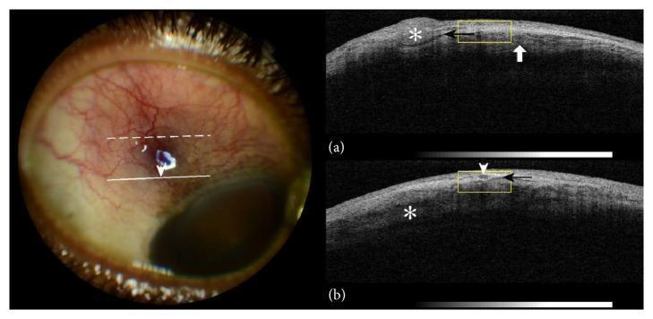
This composite educational image features a color photograph and corresponding Anterior Segment Optical Coherence Tomography (AS-OCT) scans of a right eye, illustrating clinical signs of nodular episcleritis and scleritis. The color photograph reveals marked violaceous ocular injection, superficial blood vessel congestion, and a localized nodular thickening approximately 4mm from the limbus. AS-OCT cross-sections (a) and (b) provide detailed visualization of the underlying tissue architecture. Key findings include nodular episcleral thickening (asterisk), a subepiscleral fluid level presenting as a hyporeflective space (black arrow), and intlamellar scleral edema (white arrow/asterisk). These OCT features correlate with the clinical appearance of vessel dilation and deep tissue inflammation. The image serves as a diagnostic reference for identifying inflammatory eye conditions using both external photography and non-invasive cross-sectional imaging, highlighting the relationship between visible clinical signs and internal structural alterations like scleral lamellae bisection by hyporeflective fluid.
This clinical photograph of a patient's right eye displays intense ocular inflammation and post-surgical complications. The primary findings include significant scleritis or episcleritis characterized by diffuse, deep-seated redness and engorgement of the episcleral and scleral blood vessels, obscuring the normal white appearance of the scleral tissue. The cornea exhibits notable corneal neovascularization, with fine, branching blood vessels invading the peripheral corneal stroma from the limbus. These vascular changes are concentrated adjacent to a visible surgical site, identified as a clear corneal incision typically associated with cataract surgery. The image illustrates educational concepts related to postoperative ophthalmological complications, specifically the inflammatory response and the angiogenic process following intraocular surgery. It serves as a diagnostic reference for identifying severe anterior segment inflammation and pathological vessel growth in a postoperative clinical setting.
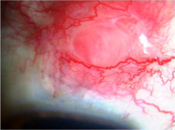
This clinical photograph, captured via slit-lamp biomicroscopy, illustrates a focal scleral nodule in a patient with systemic sarcoidosis. The image focuses on the anterior segment of the eye, specifically the bulbar conjunctiva and underlying sclera. The primary feature is a well-circumscribed, elevated, pinkish-red nodular mass. Surrounding the lesion is intense episcleral and conjunctival injection, characterized by dilated, tortuous blood vessels radiating towards the nodule, indicating significant localized inflammation and vascular engorgement. The central portion of the nodule appears slightly paler than its hyperemic periphery. This finding represents a rare ocular manifestation of sarcoidosis, showcasing granulomatous infiltration of the sclera. Key educational concepts include the identification of inflammatory scleral lesions, differentiating episcleritis from nodular scleritis, and recognizing ocular markers of systemic granulomatous diseases. The image is a valuable diagnostic reference for ophthalmologists and rheumatologists managing multi-system inflammatory conditions.
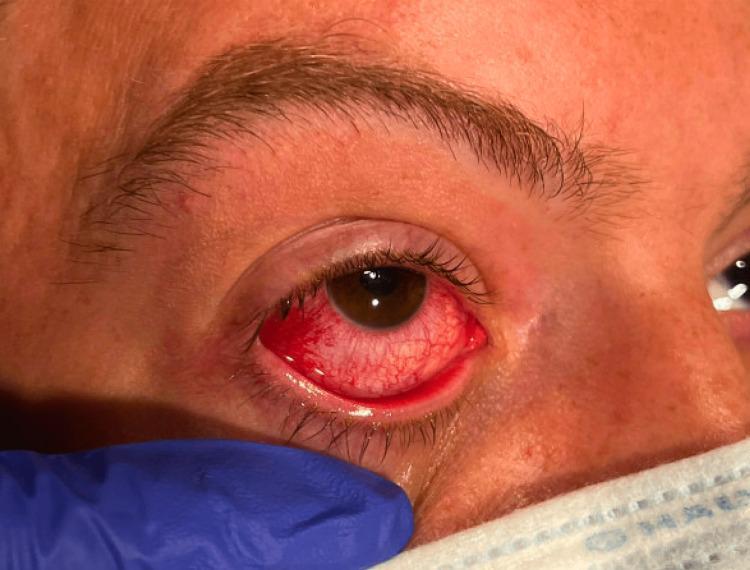
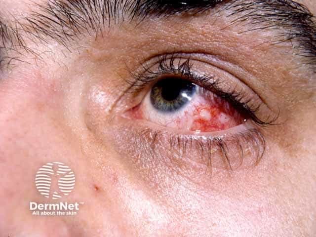
This clinical photograph shows a close-up of a patient's right eye during a medical examination, illustrating significant conjunctival and scleral injection. The primary finding is marked diffuse erythema over the bulbar conjunctiva, characterized by engorged and prominent blood vessels across the scleral surface. The intensity of the vascular injection is most pronounced at the medial canthus (inner corner) and the inferior aspect of the globe. The cornea remains relatively clear with visible iris details, and there is no obvious purulent discharge, suggesting an inflammatory rather than infectious etiology. Surrounding structures, including the eyebrow, eyelashes, and skin tone, appear normal. The lower portion of the frame shows an examiner's gloved hand and surgical mask, providing clinical context. This visual is representative of ocular manifestations associated with systemic inflammatory conditions, such as the extraintestinal manifestations seen in Crohn's disease or other forms of episcleritis/uveitis.
scleritis diagnosis treatment
necrotizing scleritis with scleral thinning
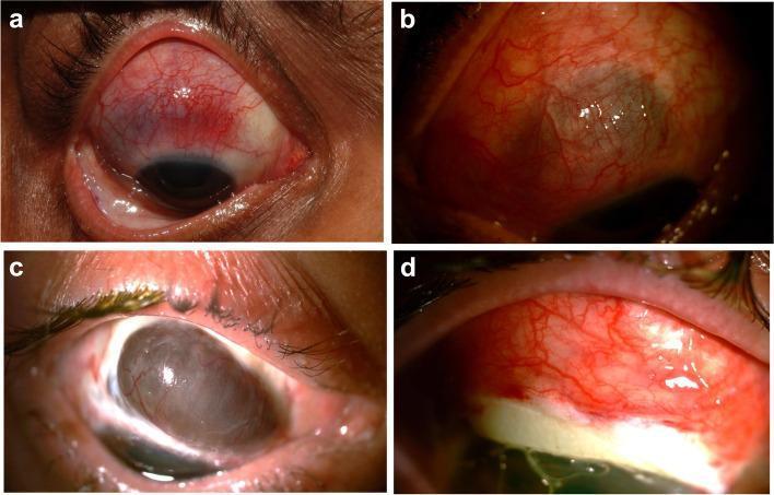
This composite of four clinical photographs illustrates various stages and complications of necrotizing scleritis associated with Granulomatosis with Polyangiitis (GPA). Figure A displays an acute phase of necrotizing scleritis in the right eye, characterized by intense episcleral and scleral vascular congestion and localized areas of bluish discoloration indicating scleral thinning. Figure B shows similar active necrotizing scleritis in a left eye with diffuse hyperemia and haziness. Figure C demonstrates a severe complication of the disease process: significant superior scleral melt with subsequent uveal prolapse, visible as a dark, bulging mass where the scleral integrity has been lost. Figure D shows the postoperative appearance of the same eye following surgical intervention with a scleral patch graft to repair the defect. The graft is visible as a white, opaque tectonic support material integrated into the superior globe, with surrounding conjunctival injection and visible sutures. These images serve as educational examples of the destructive potential of systemic vasculitis on ocular collagenous tissues and the necessary surgical management of scleral perforation.
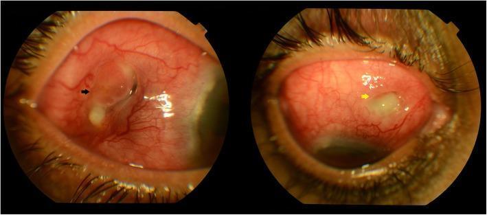
Clinical photograph of the anterior segment of the right eye demonstrating severe necrotizing scleritis. The image shows marked, diffuse episcleral and scleral vascular congestion (hyperemia). Two distinct areas of scleral melt are visible: one in the temporal quadrant indicated by a black arrow, and a second, newer focal area of melting in the superonasal quadrant indicated by a yellow arrow. The areas of melting appear as regions of significant scleral thinning and tissue loss, with irregular borders and associated yellowish-white necrotic debris. This clinical presentation is characteristic of advanced autoimmune-mediated or infectious scleritis, indicating a high risk of ocular perforation. The surrounding conjunctival tissue is intensely injected, reflecting an active, severe inflammatory process.
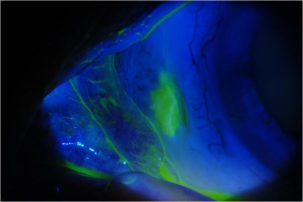
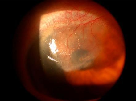
Clinical photograph of an eye under cobalt blue light illumination after fluorescein staining, illustrating conjunctival and scleral pathology. The image displays a focal area of bright green fluorescein uptake, signifying conjunctival erosion and epithelial defect. This irregular, elongated region of fluorescence is located over a section of markedly white, avascular sclera, indicative of scleral thinning or melting. Within the affected zone, small, punctate white spots are visible, consistent with calcium deposits. The surrounding conjunctival tissue appears relatively hypovascular, with visible but sparse perilimbal vessels. This finding is clinically relevant in the context of necrotizing scleritis or surgically induced scleral necrosis (SINS), demonstrating the loss of tissue integrity and the characteristic 'melting' appearance associated with chronic inflammation or ischemia of the ocular surface.
nodular episcleritis close up
Episcleritis vs Scleritis — Ophthalmology PG Notes
Anatomy (why the distinction matters)
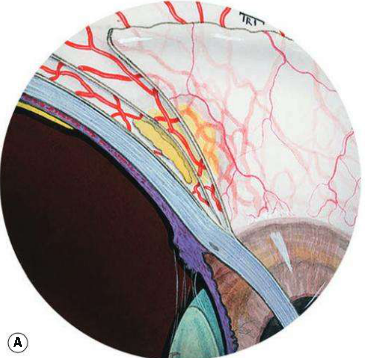
- Conjunctival vessels (most superficial)
- Superficial episcleral plexus — maximal congestion in episcleritis; blanches with topical phenylephrine 2.5-10%
- Deep vascular plexus (superficial sclera) — maximal congestion in scleritis; gives a violaceous/purplish hue (best seen in daylight); does NOT blanch with phenylephrine
EPISCLERITIS
Classification
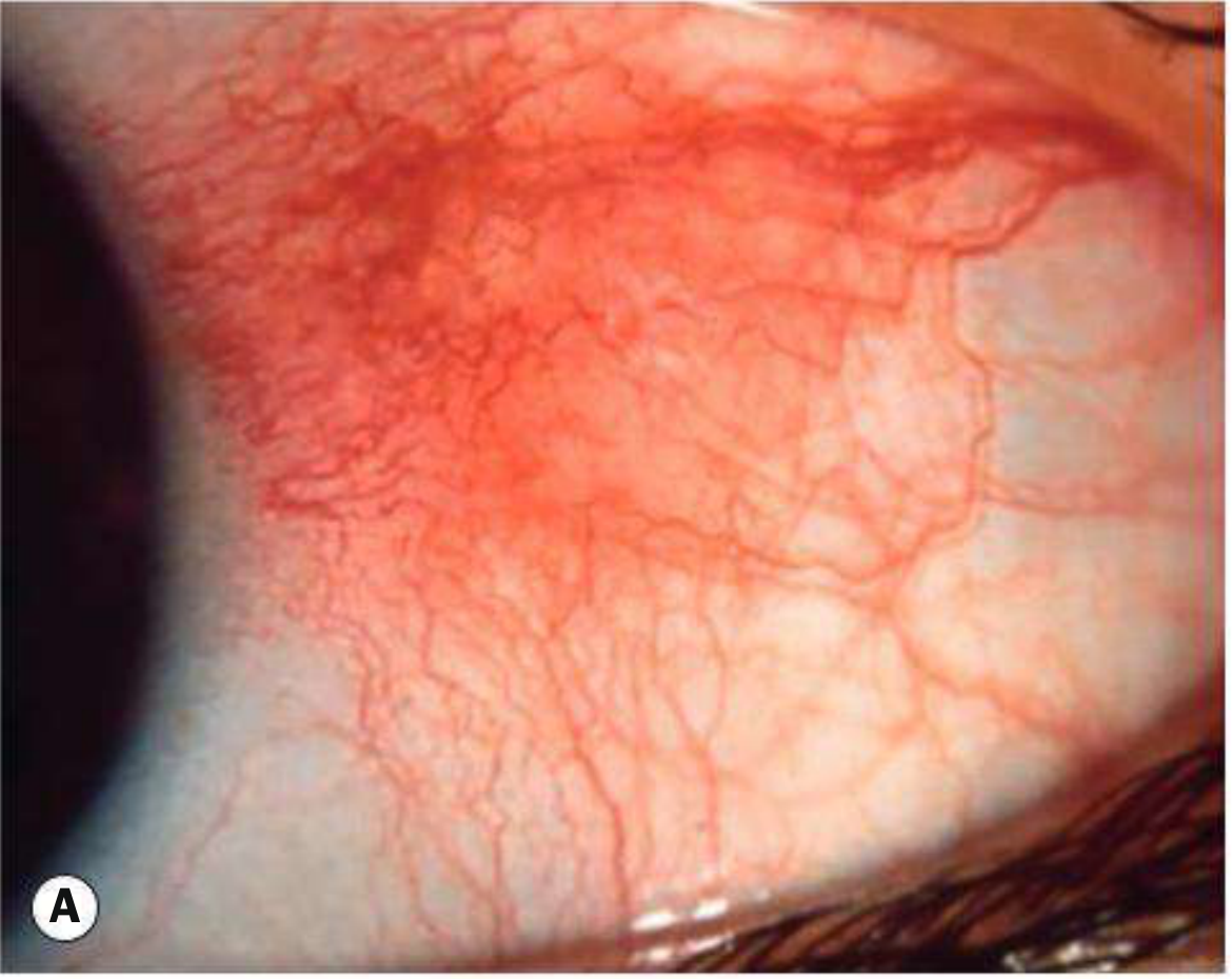
- Simple episcleritis (~75% of cases) — sectoral or diffuse redness, recurs in 60%
- Nodular episcleritis — mobile, tender episcleral nodule
Pathogenesis
- Ocular: dry eye, rosacea, contact lens wear
- Systemic: rheumatoid arthritis, other collagen vascular disease, herpes zoster ophthalmicus, gout, Crohn disease
- Infectious causes are rare
Signs & Symptoms
- Simple: discomfort ranges from none (up to 50%) to moderate grittiness ± photophobia; >50% bilateral simultaneously; normal vision; redness sectoral (triangular, interpalpebral, base at limbus) or diffuse; chemosis/uveitis/keratitis are rare
- Nodular: insidious mobile, tender nodule; slit-lamp shows the episcleral surface is NOT elevated (unlike scleritis); nodule and vessels move with a cotton bud (mobile — a key differentiator from scleral nodules)
Treatment
- Mild: none needed — cool compresses, refrigerated artificial tears
- Weak topical steroid (e.g. fluorometholone) QID for 1-2 weeks, tapered; or topical NSAID (less effective)
- Occasionally an oral NSAID (ibuprofen 200 mg TDS, or indomethacin for resistant cases)
SCLERITis
Classification (Table 9.1, Kanski)
- Non-necrotizing — Diffuse / Nodular
- Necrotizing with inflammation — Vaso-occlusive / Granulomatous / Surgically-induced
- Scleromalacia perforans (necrotizing without inflammation)
Pathogenesis
| Systemic disease | Key features |
|---|---|
| Rheumatoid arthritis (commonest association) | Symmetrical deforming polyarthropathy, RF+ in 80-90%; can cause any type of scleritis, often aggressive; also causes sicca, ulcerative keratitis |
| Granulomatosis with polyangiitis (GPA/Wegener's) | Small-vessel vasculitis of sinuses, lungs, kidneys; cANCA+ >90%; scleritis often rapidly progressive, necrotizing, granulomatous; peripheral ulcerative keratitis |
| Relapsing polychondritis | Cartilage vasculitis — ears, nose, respiratory tract; scleritis often intractable |
| Polyarteritis nodosa | Medium/small vessel aneurysmal vasculitis; poor untreated prognosis |
| SLE | Malar rash, alopecia; can cause scleritis |
| Others | Herpes zoster ophthalmicus (nodular scleritis), gout, porphyria (thin, excavated sclera in sun-exposed area) |
Signs & Symptoms by type
- Vaso-occlusive — associated with RA; patches of scleral oedema with non-perfused overlying episclera → coalesce → necrosis
- Granulomatous — associated with GPA/PAN; rapid (within 24h) raised oedematous sclera/cornea
- Surgically-induced necrotizing scleritis (SINS) — starts within weeks of surgery (trabeculectomy + mitomycin C, pterygium excision, scleral buckle), localizes to surgical site
Treatment of immune-mediated scleritis
- Topical steroids — do NOT alter natural history but relieve symptoms/oedema in non-necrotizing disease only
- Systemic NSAIDs — first line for non-necrotizing disease alone (COX-2 inhibitor if elderly/peptic ulcer history)
- Periocular steroid injection — may help non-necrotizing disease (transient effect); avoid in necrotizing disease
- Systemic steroids — prednisolone 1-1.5 mg/kg/day when NSAIDs inadequate or disease is necrotizing; IV methylprednisolone 0.5-1 g/day for 3 days in severe cases
- Immunosuppressants/biologics — indicated if steroid control incomplete, as steroid-sparing agents, or to treat underlying systemic disease (mandatory immediate immunotherapy for GPA, PAN, RA-associated necrotizing scleritis, relapsing polychondritis): cyclophosphamide, azathioprine, methotrexate, ciclosporin/tacrolimus, and biologics — rituximab is particularly effective in necrotizing scleritis
- Infectious scleritis: targeted antimicrobial/antiviral therapy (± surgical debridement); herpes zoster is the commonest infective cause and necrotizing disease here is notably treatment-resistant
Quick Comparison Table
| Feature | Episcleritis | Scleritis |
|---|---|---|
| Layer affected | Superficial episcleral plexus | Deep scleral vascular plexus |
| Pain | Mild/absent, grittiness | Severe, boring, radiates, wakes at night |
| Vessel blanching with 10% phenylephrine | Yes | No |
| Nodule mobility (if nodular) | Mobile | Immobile |
| Globe tenderness | Absent/mild | Marked |
| Vision | Normal | Can be reduced (esp. posterior/necrotizing) |
| Systemic disease association | Uncommon | Common (RA, GPA, PAN, relapsing polychondritis) |
| Serious complications | Essentially none | Glaucoma, keratitis, perforation, vision loss |
| Treatment | Self-limiting; weak topical steroid/NSAID | Systemic NSAID → systemic steroid → immunosuppression/biologics depending on severity |