
What is Necrotising pancreatitis and how it is differe from Acute necrotic collection or walled off necrosis, how to diagnosed , how to manage, when to suspect in detailed manner
Reading File
Checking Date & Time
Finding Sources
Finding Sources
Searching PubMed
"necrotizing pancreatitis" AND management
Good, I have multiple sources. Now let me read the key textbook sections in parallel.
Reading File
Reading File
Reading File
Reading File
Excellent content. Let me continue reading the key management sections and Harrison's specific content.
Reading File
Reading File
Reading File
Excellent, getting very rich content. Let me read more on management, diagnosis, and the step-up approach.
Reading File
Reading File
Reading File
Now let me get the management section from Current Surgical Therapy more completely, and also get the CT imaging/diagnosis section from Rosen's.
Reading File
Reading File
Loading Image
Loading Image
Loading Image
I now have comprehensive content from multiple authoritative textbooks plus excellent images. Let me compile the full answer.# Necrotising Pancreatitis — A Comprehensive Guide
1. What Is Necrotising Pancreatitis?
Necrotising pancreatitis (NP) is a severe form of acute pancreatitis characterised by irreversible death (necrosis) of pancreatic parenchyma and/or the surrounding peripancreatic fat tissue, caused by failure of the pancreatic microcirculation. It occurs in approximately 5–10% of all acute pancreatitis cases but accounts for the vast majority of its morbidity and mortality.
Pathophysiology
When acinar cells are injured (by gallstones, alcohol, etc.), lysosomal hydrolases (especially cathepsin B) colocalize with zymogen granules, activating trypsinogen intracellularly. Trypsin then activates a cascade of other proteolytic and lipolytic enzymes. Sustained cytosolic calcium rise drives this process. Activated neutrophils release superoxide and proteases; macrophages release TNF-α, IL-1, IL-6, and IL-8. These inflammatory mediators increase pancreatic vascular permeability, leading to:
- Haemorrhage and oedema
- Microthrombus formation in the pancreatic microcirculation
- Hypoperfusion and necrosis of pancreatic parenchyma and peripancreatic fat
The failure of the pancreatic microcirculation — a hallmark feature — distinguishes necrotising pancreatitis from interstitial oedematous pancreatitis, where the blood supply is maintained. — Schwartz's Principles of Surgery, 11th Ed.
Three Anatomical Patterns (Revised Atlanta Classification 2012)
| Pattern | Frequency |
|---|---|
| Pancreatic parenchymal necrosis + peripancreatic necrosis | 75–80% (most common) |
| Peripancreatic fat necrosis alone (no parenchymal necrosis) | ~15–20% |
| Parenchymal necrosis alone | ~5% (least common) |
Extrapancreatic fat necrosis alone fulfils the criteria for NP but carries a better prognosis than cases with parenchymal necrosis. — Grainger & Allison's Diagnostic Radiology
2. The Classification Landscape — NP vs ANC vs WON
The 2012 Revised Atlanta Classification made a critical distinction: collections arising in necrotising pancreatitis are fundamentally different from those in interstitial pancreatitis. They contain both fluid AND solid necrotic material (heterogeneous), not pure fluid.
Full Comparison Table
| Feature | Necrotising Pancreatitis (disease) | Acute Necrotic Collection (ANC) | Walled-Off Necrosis (WON) |
|---|---|---|---|
| What it is | The underlying disease process | Early local complication | Late, matured local complication |
| Timing | Diagnosed within first 1–2 weeks | <4 weeks from symptom onset | >4 weeks from symptom onset |
| Content | Necrotic parenchyma/fat + fluid | Fluid + necrotic debris (heterogeneous) | Mature encapsulated necrosis + fluid + solid debris |
| Wall | No wall (it's the gland itself) | No definitive wall — poorly defined | Well-defined, thick inflammatory wall — completely encapsulating |
| Location | Intrapancreatic ± peripancreatic | Intra- and/or extrapancreatic | Intra- and/or extrapancreatic |
| CT density | Non-enhancing parenchyma | Heterogeneous, non-liquid density | Heterogeneous with liquid AND non-liquid densities, possible loculations |
| Sterile/Infected | Usually sterile initially | Usually sterile; may become infected | May be sterile or infected |
| Comparator (interstitial) | — | Analogous to APFC (but has solid debris) | Analogous to pseudocyst (but has solid debris, not pure fluid) |
The Distinction From Pseudocyst
A pseudocyst (complication of interstitial pancreatitis) contains only fluid — homogeneous, well-defined wall, no solid debris. A WON contains solid necrotic material in addition to fluid — this is critical because pseudocysts can be drained simply, while WON often requires debridement.
3. When to Suspect Necrotising Pancreatitis
Clinical Red Flags
At presentation:
- Acute pancreatitis with persisting or worsening pain despite initial management
- SIRS criteria (≥2 of): temperature <36°C or >38°C, HR >90, RR >20 or PaCO₂ <32 mmHg, WBC >12,000 or <4,000/µL
- Hemoconcentration — haematocrit >44% (marker of third-space loss)
- Elevated admission BUN >20 mg/dL
- BISAP score ≥3: BUN >25, Impaired mental status, SIRS, Age >60, Pleural effusion
- APACHE II ≥8 at 24 h
Risk factors for severe disease:
- Age >60 years
- Obesity (BMI >30)
- Significant comorbidities (Charlson comorbidity index)
- Hypertriglyceridaemia-induced pancreatitis
During hospitalisation — suspect NP specifically when:
- No clinical improvement after 48–72 hours of aggressive supportive care
- Rising or persistently elevated CRP >150 mg/L
- Development of organ failure — renal (creatinine >2.0 mg/dL), respiratory (PaO₂ <60), cardiovascular (SBP <90, HR >130)
- Fever spike after initial defervescence → raises concern for infected necrosis
- Gas bubbles on CT within a peripancreatic collection → strongly suggests infected necrosis
- Deteriorating haemodynamics despite fluid resuscitation
Key clinical principle: Unlike interstitial pancreatitis (which is usually self-limited), NP follows a biphasic course:
- First 1–2 weeks: Dominated by SIRS and multi-organ dysfunction (early phase mortality)
- Weeks 2–4 and beyond: Dominated by infectious complications — infected necrosis accounts for ~80% of deaths from acute pancreatitis
4. Diagnosis
Step 1 — Confirm Acute Pancreatitis
At least two of three criteria:
- Abdominal pain characteristic of acute pancreatitis (epigastric, radiating to back)
- Serum lipase or amylase >3× upper limit of normal
- Characteristic imaging findings
Step 2 — Identify Necrosis (CECT)
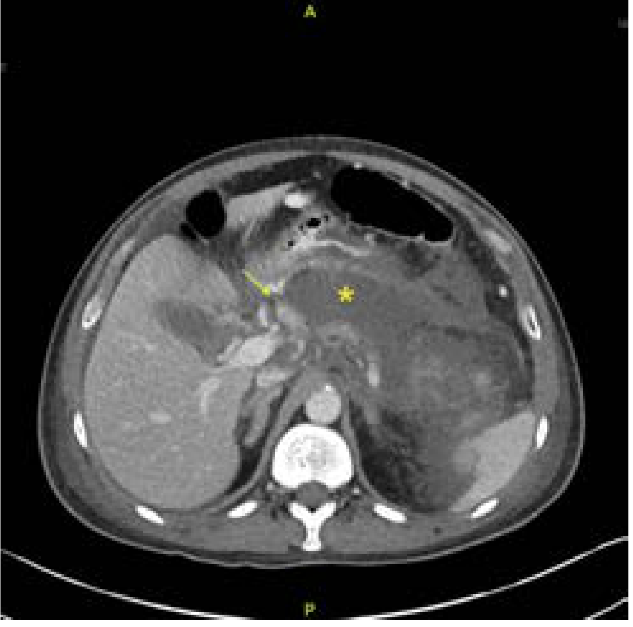
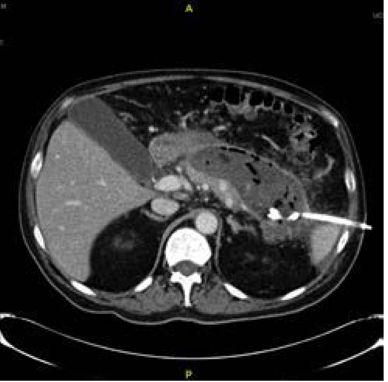
Contrast-Enhanced CT (CECT) is the gold standard for diagnosing and grading necrosis.
- Timing: Not recommended in the first 24–48 hours (early CT underestimates necrosis extent). Optimal window is 48–72 hours after onset, or earlier if diagnosis is unclear.
- Key finding: Non-enhancing pancreatic parenchyma — necrotic tissue fails to take up IV contrast, appearing as a dark hypodense area against enhancing viable pancreas.
- ANC appearance: Heterogeneous, mixed-density collection (fluid + solid debris), no wall
- WON appearance: Heterogeneous collection with a thick, well-defined, enhancing rim; may have loculations
- Infected necrosis: Gas bubbles within the collection (pathognomonic); also rim-enhancement
CT Severity Index (CTSI / Modified CTSI): Grades pancreatic inflammation, necrosis extent (0%, <30%, 30–50%, >50%), and extrapancreatic complications. MCTSI ≥4 = high risk.
MRI / MRCP
- Superior to CT for characterising solid debris within collections (distinguishing pseudocyst from WON)
- Can identify disconnected pancreatic duct (critical for surgical planning)
- Preferred in patients with renal impairment (no gadolinium needed for necrosis assessment)
- Detects choledocholithiasis without radiation
EUS (Endoscopic Ultrasound)
- Best for confirming WON location relative to stomach/duodenum before endoscopic drainage
- Allows aspiration for Gram stain and culture if infection is suspected but CT equivocal
Fine-Needle Aspiration (FNA)
- Image-guided FNA for Gram stain/culture when infected necrosis is suspected but CT is equivocal
- Risk: may introduce infection into a sterile collection — weigh this carefully
- Less commonly needed since gas on CT is sufficient evidence of infection
Laboratory Markers
| Marker | Significance |
|---|---|
| CRP >150 mg/L | Marker of severity during hospitalisation |
| Rising BUN | Inadequate hydration AND increased mortality |
| Procalcitonin elevation | Suggests infected necrosis |
| WBC, fever | Clinical infection markers |
| Haematocrit >44% | Haemoconcentration — severity marker |
Severity Scoring Systems
| Score | Calculated | Threshold for Severe |
|---|---|---|
| Ranson's criteria | On admission + 48 h | ≥3 = predicted severe |
| APACHE II | Any time | ≥8 = severe |
| BISAP | Within 24 h | ≥3 = high risk |
| Modified CT Severity Index (MCTSI) | Based on CT | ≥4 = high risk |
| Revised Atlanta (2013) | Clinical/radiological | Mild / Moderately Severe / Severe |
Scoring systems should augment clinical judgment, but not replace it. — Schwartz's Principles of Surgery, 11th Ed.
5. Management
A. Immediate Supportive Care (First 24–72 Hours)
-
IV fluid resuscitation — the single most important early intervention
- Lactated Ringer's is preferred over normal saline (lowers CRP, reduces systemic inflammation)
- Initial strategy: ~10–15 mL/kg bolus, followed by 1.5–2 mL/kg/h; titrate to urine output >0.5 mL/kg/h
- Recent RCT evidence: overly aggressive hydration (>20 mL/kg) risks fluid overload without improved outcomes
- Monitor: hematocrit and BUN every 8–12 h; rising BUN = inadequate resuscitation and higher mortality
-
Analgesia — IV opioid analgesics for pain control
-
NPO initially — to minimise pancreatic stimulation
-
Supplemental oxygen and monitoring
-
Early enteral nutrition (within 24–48 h if tolerated)
- Strongly preferred over TPN; maintains gut barrier, reduces infectious complications
- Nasojejunal (NJ) or nasogastric (NG) feeding both acceptable
- TPN only if enteral route not tolerated
-
Antibiotics: Do NOT use prophylactic antibiotics routinely in sterile necrosis
- Current guidelines recommend against routine prophylactic carbapenem use
- Antibiotics indicated ONLY when infected necrosis is confirmed (clinical + radiological)
- Use carbapenems, fluoroquinolones, or metronidazole (good pancreatic tissue penetration)
-
ICU admission for persistent organ failure, haemodynamic instability
-
Transfer to tertiary centre early for severe/complicated cases
B. Management of Infected Necrosis
When to intervene: Confirmed infected necrosis (gas on CT or positive FNA), or deteriorating patient despite adequate supportive care.
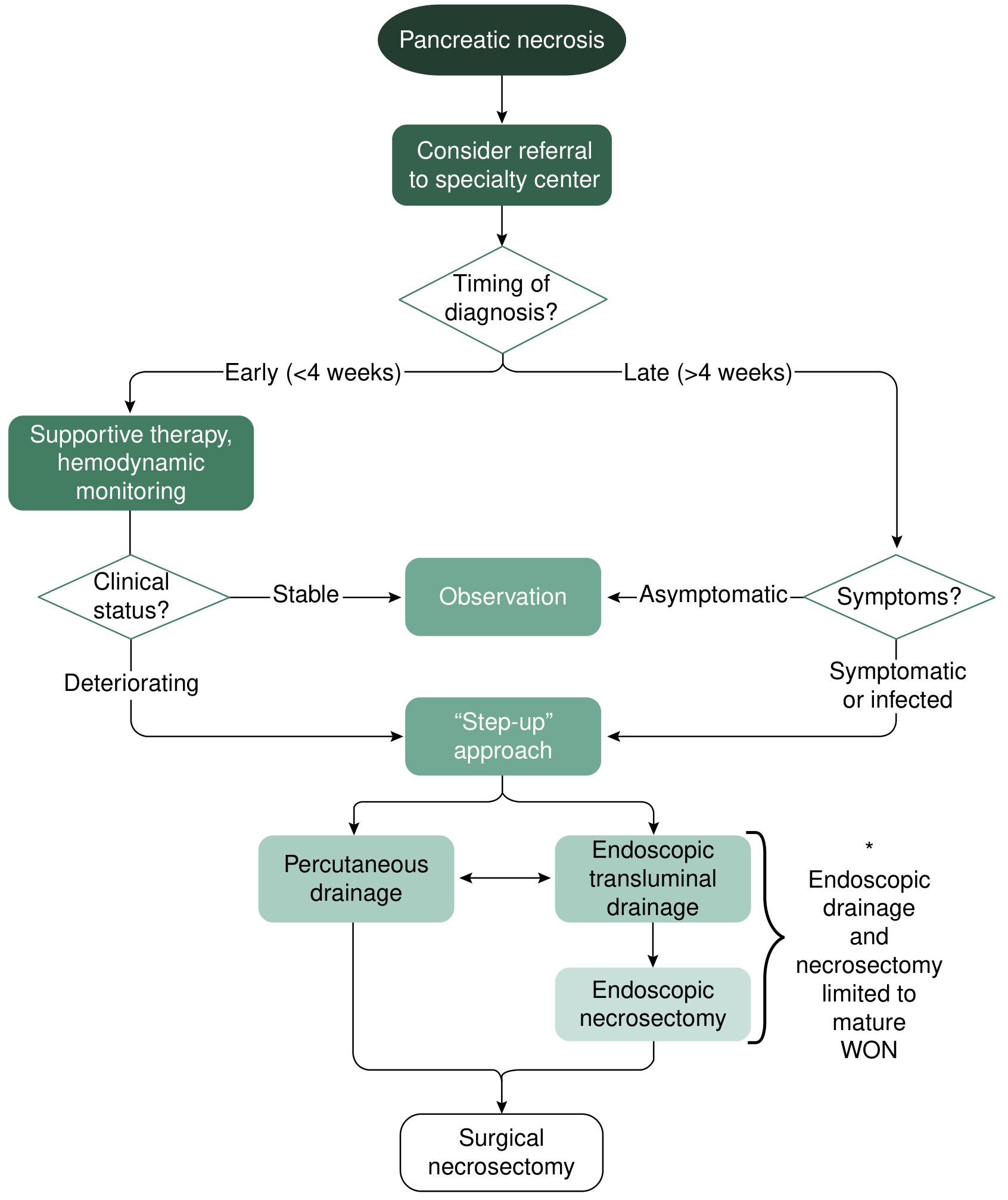
Timing: Delay intervention as long as safely possible — minimum 4 weeks from onset to allow collection to "wall off" and mature into WON. Intervening in the first 2 weeks carries dramatically higher mortality. If clinically unstable earlier than 4 weeks, a temporising percutaneous drain can "bridge" until the necrosis matures.
The Step-Up Approach (Current Standard of Care)
The Dutch PANTER trial established that a step-up approach is superior to primary open necrosectomy — fewer complications, less new-onset organ failure.
Step 1 — Percutaneous Catheter Drainage (PCD)
- First-line intervention for infected/symptomatic necrosis
- Performed by interventional radiology under CT/US guidance
- Retroperitoneal access preferred (left flank) to allow potential VARD if needed
- Serial catheter upsizing + irrigation with saline or H₂O₂ for thick debris
- Up to 50% of patients treated with PCD alone — obviating need for further surgery
- Disadvantage: risk of pancreaticocutaneous fistula, especially if communicating with pancreatic duct
Step 2 — Endoscopic Transluminal Drainage + Necrosectomy (for WON only)
- Best for mature WON (≥4 weeks) within 2 cm of stomach or duodenum
- EGD + EUS to confirm location → needle puncture → balloon dilation of cystgastrotomy → placement of pigtail stents or lumen-apposing metal stent (LAMS)
- Nasocystic catheter for irrigation
- Direct endoscopic necrosectomy: endoscope inserted into cavity; mechanical debridement
- Requires multiple sessions (typically 3–5); specialist centre
- PENGUIN trial: endoscopic necrosectomy had fewer major complications, less organ failure, fewer fistulas than open surgery
- Operative debridement avoided in ~2/3 of patients
Step 3 — Surgical Necrosectomy
- Reserved for failures of percutaneous and endoscopic approaches
- Options: video-assisted retroperitoneal debridement (VARD), minimally invasive retroperitoneal pancreatic necrosectomy (MIRPN), or open necrosectomy
- Open necrosectomy now last resort due to high mortality historically (~45% vs 8.3% with conservative/minimally invasive approach)
6. Complications to Monitor
| Complication | Notes |
|---|---|
| Infected necrosis | Occurs in 20–70% of NP; responsible for ~80% of deaths |
| Disconnected pancreatic duct | May require ERCP stenting or surgical repair |
| Gastrointestinal obstruction | Gastric outlet obstruction from WON mass effect |
| Splenic/portal vein thrombosis | Enzymatic erosion |
| Pseudoaneurysm | Splenic, gastroduodenal artery erosion → life-threatening haemorrhage → angioembolisation |
| Colonic infarction | Extension of necrosis to the colon |
| Pancreaticocutaneous/enteric fistula | Post-drainage |
| Endocrine/Exocrine insufficiency | Long-term — new-onset diabetes, steatorrhoea |
7. Quick Reference Summary
Acute Pancreatitis
       │
       ├── Interstitial Oedematous Pancreatitis (90–95%)
       │         │
       │         ├── <4 wks → APFC (Acute Peripancreatic Fluid Collection)
       │         └── >4 wks → Pseudocyst (pure fluid, thin wall)
       │
       └── NECROTISING PANCREATITIS (5–10%)
                 │
                 ├── <4 wks → ANC (Acute Necrotic Collection)
                 │         • Heterogeneous, NO wall, fluid + solid debris
                 │         • May be sterile or infected
                 │
                 └── >4 wks → WON (Walled-Off Necrosis)
                           • Heterogeneous, THICK WALL, fluid + solid debris
                           • May be sterile or infected
                           • Target for endoscopic/percutaneous drainage
Recent Evidence (2025)
A 2025 Bayesian network meta-analysis (PMID: 40898567) evaluated invasive strategies for NP and a concurrent 2025 systematic review and network meta-analysis on infected NP (PMID: 40675209) both support endoscopic approaches over open surgery as the preferred step-up strategy for infected WON, consistent with the PENGUIN trial findings.
Sources:
- Current Surgical Therapy, 14th Edition — Local Complications of Acute Pancreatitis
- Schwartz's Principles of Surgery, 11th Edition — Management of Acute Pancreatitis
- Harrison's Principles of Internal Medicine, 22nd Edition (2025)
- Grainger & Allison's Diagnostic Radiology — Necrotising Pancreatitis
- Rosen's Emergency Medicine — Necrotizing Pancreatitis
- 2012 Revised Atlanta Classification (Banks PA et al., Gut 2013;62:102–111)Here is a comprehensive breakdown of everything you asked about:
Key Takeaways
Necrotising Pancreatitis (NP) is caused by failure of the pancreatic microcirculation, leading to death of pancreatic parenchyma and/or peripancreatic fat. It complicates 5–10% of acute pancreatitis cases but drives the majority of serious morbidity and death. The 2012 Revised Atlanta Classification defines the entire spectrum clearly.
The Core Distinction: NP vs ANC vs WON
| Necrotising Pancreatitis | ANC | WON | |
|---|---|---|---|
| What | The disease itself | Early complication | Late complication |
| Timing | <2 weeks | <4 weeks | >4 weeks |
| Wall | No wall (it's the gland) | No wall | Thick, complete wall |
| Content | Necrotic parenchyma/fat | Fluid + solid necrotic debris | Fluid + solid debris (loculated) |
| Analogy | — | Like APFC but with solid debris | Like pseudocyst but with solid debris |
The critical point: ANC → WON is a maturation process over 4 weeks. Both are fundamentally heterogeneous (solid + liquid), unlike pseudocysts (pure fluid).
When to Suspect NP
- Acute pancreatitis that does not improve in 48–72 hours
- Persistent SIRS, haemoconcentration (Hct >44%), rising BUN
- BISAP ≥3 or APACHE II ≥8
- Late deterioration or new fever → think infected necrosis (responsible for ~80% of deaths)
- Gas bubbles in a collection on CT = infected until proven otherwise
Diagnosis
- CECT at 48–72 h is gold standard — non-enhancing parenchyma = necrosis
- MRI/MRCP preferred if renal impairment or when characterising solid debris
- EUS to confirm WON position before endoscopic drainage
- FNA only if CT equivocal for infection (risk of seeding sterile necrosis)
Management
- Supportive care: Lactated Ringer's IV fluid resuscitation, early enteral nutrition within 24–48 h, analgesia, ICU
- No prophylactic antibiotics in sterile necrosis
- Infected necrosis: delay intervention to ≥4 weeks if possible (let it wall off), then step-up approach:
- Percutaneous catheter drainage first (~50% cured here alone)
- Endoscopic transluminal drainage + necrosectomy for mature WON near stomach/duodenum
- Surgical necrosectomy only as last resort
This is a shared conversation. Sign in to Orris to start your own chat.