
Food poison information
food poisoning causes symptoms treatment 2025
food poisoning bacteria contaminated food gastroenteritis
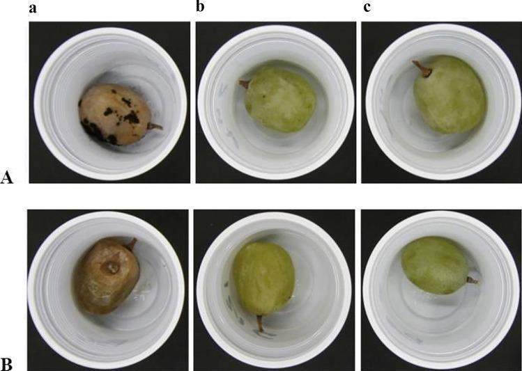
This comparison chart displays experimental specimens of table grapes used as a food model to evaluate the bioprotective efficacy of Lactic Acid Bacteria (LAB) against common fungal pathogens. The image is organized into a 2x3 grid. Row A shows grapes contaminated with Aspergillus niger, while Row B shows grapes contaminated with Botrytis cinerea. Column 'a' represents control samples without LAB treatment, demonstrating significant pathological progression including dark fungal outgrowths (A. niger) and extensive shriveling with brownish discoloration (B. cinerea) after 14 days at 4°C. Columns 'b' and 'c' demonstrate the inhibitory effects of Fructobacillus fructosus AREP6 and Lactobacillus plantarum MEP3, respectively. In these treated columns, the grapes maintain their green color, structural integrity, and smooth surface texture, illustrating the potential of these LAB strains as natural preservatives or biocontrol agents in food safety and preservation. The visual focus is on the prevention of mold-induced decay and the maintenance of specimen quality through probiotic intervention.
![This diagnostic image displays six panels (a-f) of Extracted Ion Chromatograms (EIC) from an LC-MS/MS analysis, used for identifying marine biotoxins in contaminated seafood, relevant to Ciguatera Poisoning (CP). Each chromatogram plots intensity in counts per second (cps) against retention time (minutes), ranging from 2 to 10 minutes. The panels compare two extraction methods: an enzyme protocol (left column: a, c, e) and a standard mechanical protocol (right column: b, d, f). The EICs track sodium adducts [M + Na]+ for specific Ciguatoxin (CTX) groups: CTX4A (a, b), CTX3C (c, d), and C-CTX/I-CTX (e, f). Multi-colored peaks represent distinct congeners, such as 2,3,51-trihydroxyCTX3C and 51-hydroxyCTX3C, characterized by specific retention times and relative intensities. Variations in peak profiles between the left and right columns illustrate differences in extraction efficiency and matrix effects. This data is critical for food safety toxicology, enabling the identification and quantification of lipophilic polyether neurotoxins that cause human illness.](https://cdn.orris.care/cdss_images/pmc_clinical_VQA_a53a50f6036b31c5d9f148f09b315f1aebda9ada5940f3c0fb5a77910b9f3716.jpg)
This diagnostic image displays six panels (a-f) of Extracted Ion Chromatograms (EIC) from an LC-MS/MS analysis, used for identifying marine biotoxins in contaminated seafood, relevant to Ciguatera Poisoning (CP). Each chromatogram plots intensity in counts per second (cps) against retention time (minutes), ranging from 2 to 10 minutes. The panels compare two extraction methods: an enzyme protocol (left column: a, c, e) and a standard mechanical protocol (right column: b, d, f). The EICs track sodium adducts [M + Na]+ for specific Ciguatoxin (CTX) groups: CTX4A (a, b), CTX3C (c, d), and C-CTX/I-CTX (e, f). Multi-colored peaks represent distinct congeners, such as 2,3,51-trihydroxyCTX3C and 51-hydroxyCTX3C, characterized by specific retention times and relative intensities. Variations in peak profiles between the left and right columns illustrate differences in extraction efficiency and matrix effects. This data is critical for food safety toxicology, enabling the identification and quantification of lipophilic polyether neurotoxins that cause human illness.
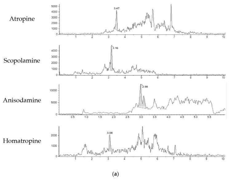
This diagnostic image displays four liquid chromatography-mass spectrometry (LC-MS) chromatograms for common tropane alkaloids: Atropine, Scopolamine, Anisodamine, and Homatropine. The content illustrates pharmacological analysis within toxicology and food safety domains. Each panel plots signal intensity (y-axis) against retention time (x-axis, in minutes) for a blank green tea sample spiked at the limit of quantification (LOQ). Key visual features include: 1) Atropine: A distinct peak at 3.47 minutes with subsequent complex matrix interference signals between 4-7 minutes. 2) Scopolamine: A primary peak at 3.16 minutes. 3) Anisodamine: Shows the highest relative signal intensity with a major peak at 2.98 minutes. 4) Homatropine: Displays a lower intensity main peak at 3.08 minutes relative to the other alkaloids. The chromatograms demonstrate the identification and quantification of these toxins, which are of clinical significance due to their potential for human poisoning from contaminated herbal infusions. The differing retention times and intensities illustrate the method's sensitivity and specificity for various tropane alkaloid species.
Food Poisoning (Foodborne Illness)
Definition
- Park's Textbook of Preventive and Social Medicine
Types of Food Poisoning
A. Non-Bacterial
B. Bacterial
- Textbook of Family Medicine 9e
Major Causes and Their Profiles
| Organism | Incubation | Key Foods | Mechanism | Hallmark Symptoms |
|---|---|---|---|---|
| Salmonella | 12-24 hours | Meat, poultry, eggs, milk | Live bacteria invade intestine | Chills, fever, nausea, vomiting, profuse watery diarrhea; lasts 2-3 days |
| Staphylococcus aureus | 1-8 hours | Salads, custards, milk products | Preformed heat-stable enterotoxin | Sudden vomiting, abdominal cramps, diarrhea; NO fever |
| Clostridium botulinum (Botulism) | 18-36 hours | Home-canned vegetables, smoked fish | Preformed toxin blocks parasympathetic nerves | Dysphagia, diplopia, ptosis, muscle weakness, paralysis - NOT typical GI; often fatal |
| Clostridium perfringens | 8-16 hours | Cooked meat, stews, gravies | Toxin produced in gut | Crampy abdominal pain and diarrhea |
| Bacillus cereus | 1-16 hours | Rice, fried foods | Preformed or gut-produced toxin | Vomiting type (1-6 h) or diarrhea type (8-16 h) |
| Campylobacter | 2-5 days | Poultry, unpasteurized milk | Invasive + toxin-producing | Bloody diarrhea, fever, abdominal pain |
| E. coli (EHEC/ETEC) | 1-8 days | Undercooked beef, raw vegetables | Toxin production or invasion | Watery to bloody diarrhea; risk of HUS with O157:H7 |
| Shigella | 1-4 days | Ready-to-eat foods, water | Invasive | Dysentery (small-volume bloody stools), fever |
| Norovirus | 12-48 hours | Shellfish, salads, person-to-person | Mucosal damage | Projectile vomiting, watery diarrhea, low-grade fever |
Timing as a Diagnostic Clue
- < 1 hour - Chemical agent (e.g., heavy metals, scombroid fish poisoning)
- 1-6 hours - Preformed toxin: S. aureus or B. cereus
- 8-16 hours - C. perfringens, B. cereus (diarrhea type)
- > 16 hours - Salmonella, Campylobacter, Shigella, norovirus, E. coli
Botulism - Special Warning
- Dysphagia (difficulty swallowing), diplopia (double vision), ptosis, blurred vision
- Progressive muscle weakness and paralysis
- No fever, consciousness retained
- Death can occur in 4-8 days from respiratory or cardiac failure
- The toxin is heat-labile - heating food to 100°C for a few minutes destroys it
- Park's Textbook of Preventive and Social Medicine
Symptoms Overview
- Nausea and vomiting
- Watery or bloody diarrhea
- Crampy abdominal pain
- Fever (not with Staph or Botulism)
- Malaise, headache
- Blurred/double vision, drooping eyelids
- Difficulty speaking or swallowing
- Muscle weakness, paralysis
When to See a Doctor (Red Flags)
- High fever (≥38.5°C / 101.3°F)
- Bloody diarrhea
- Severe dehydration (no urination, extreme dizziness)
- Neurological symptoms (vision changes, weakness, difficulty swallowing)
- Symptoms lasting more than 3 days
- Symptoms in elderly, pregnant women, infants, or immunocompromised individuals
- Hemolytic anemia or renal failure signs (possible E. coli HUS)
Treatment
Home Management (mild cases)
- Hydration is the cornerstone - replace fluids and electrolytes (oral rehydration salts)
- Rest; avoid solid food initially, then progress to bland foods (BRAT diet: bananas, rice, applesauce, toast)
- Anti-nausea medications (OTC): dimenhydrinate, ondansetron
- Antidiarrheal agents (loperamide): use with caution; avoid if bloody diarrhea or fever (could worsen some infections)
Medical Treatment
- IV fluids for severe dehydration
- Antibiotics for specific bacterial causes:
- Traveler's diarrhea / severe Shigella/Campylobacter: ciprofloxacin 500 mg twice daily x 5 days (first-line per Medscape emergency guidelines)
- TMP/SMX (Bactrim) as alternative
- Note: Antibiotics are NOT recommended for most E. coli O157:H7 infections (may increase HUS risk)
- Botulism: antitoxin + ICU supportive care (no antibiotics useful against toxin)
- Scombroid/histamine poisoning: antihistamines (diphenhydramine 25-50 mg IV) + H2 blockers
Prevention
- Cook food to safe internal temperatures (poultry: 75°C / 165°F)
- Refrigerate perishables within 2 hours (1 hour if ambient temperature >32°C)
- Wash hands thoroughly before and after handling food
- Avoid cross-contamination between raw meat and ready-to-eat foods
- Don't feed honey to infants under 12 months (botulism risk)
- Be cautious with home-canned or home-preserved foods
- Use safe water for drinking and food preparation