
Surgical anatomy of lateral wall of nose. Add a note on OMC in detail for 20 marks with diagram and flow charts from Scott Brown, Cummings, stell and marans, Zakir Hussain, dhingra, hazarika and related articles with recent advances according to rguhs university
Reading File
Finding Sources
Finding Sources
Searching PubMed
"ostiomeatal complex" AND "lateral wall" AND nose
Searching the Web
surgical anatomy lateral wall nose ostiomeatal complex RGUHS ENT 20 marks
Finding Sources
Finding Sources
Finding Sources
Searching PubMed
"ostiomeatal complex" AND sinusitis
Reading File
Reading File
Reading File
Reading File
Finding Sources
Loading Image
Reading File
Loading Image
Finding Sources
Reading File
Reading File
Loading Image
Reading File
Finding Sources
Searching PubMed
"lateral wall" nose anatomy turbinate FESS
Reading File
Reading File
Searching PubMed
anatomic variations ostiomeatal complex chronic rhinosinusitis
Reading File
Loading Image
Loading Image
Reading File
Finding Sources
Loading Image
Reading Web Page
https://www.theentresident.com/ent-notes-lectures/lateral-wa…
SURGICAL ANATOMY OF LATERAL WALL OF NOSE WITH NOTE ON OSTIOMEATAL COMPLEX (OMC)
(20 Marks - RGUHS ENT Examination)
INTRODUCTION
The lateral wall of the nose is the most complex and surgically relevant wall of the nasal cavity. It houses the turbinates (conchae), meatuses, and the ostiomeatal complex (OMC) - the final common pathway for drainage of the anterior group of paranasal sinuses. Messerklinger (1978) coined the term "ostiomeatal complex" and established its central role in the pathogenesis of sinusitis, forming the conceptual basis of modern Functional Endoscopic Sinus Surgery (FESS).
(Cummings Otolaryngology Head & Neck Surgery, 7th ed.; Scott-Brown's Otorhinolaryngology Head & Neck Surgery, Vol. 1)
PART I: SURGICAL ANATOMY OF THE LATERAL WALL OF NOSE
A. BONY FRAMEWORK
The lateral wall is formed by contributions from seven bones:
┌─────────────────────────────────────────────────┐
│     BONES FORMING THE LATERAL WALL OF NOSE     │
├────────────────────────┬────────────────────────┤
│ Bone                   │ Contribution            │
├────────────────────────┼────────────────────────┤
│ Nasal bone             │ Superior-anterior       │
│ Frontal process of     │ Anterosuperior          │
│   maxilla              │                         │
│ Lacrimal bone          │ Anterior middle         │
│ Ethmoid (labyrinth)    │ Middle portion          │
│ Inferior turbinate     │ Separate bone           │
│   (concha)             │ (only independent bone) │
│ Perpendicular plate    │ Posterior inferior      │
│   of palatine          │                         │
│ Medial pterygoid plate │ Posterior               │
│   of sphenoid          │                         │
└────────────────────────┴────────────────────────┘
B. TURBINATES (CONCHAE) AND MEATUSES
DIAGRAM 1 - Right Lateral Nasal Wall with Turbinates
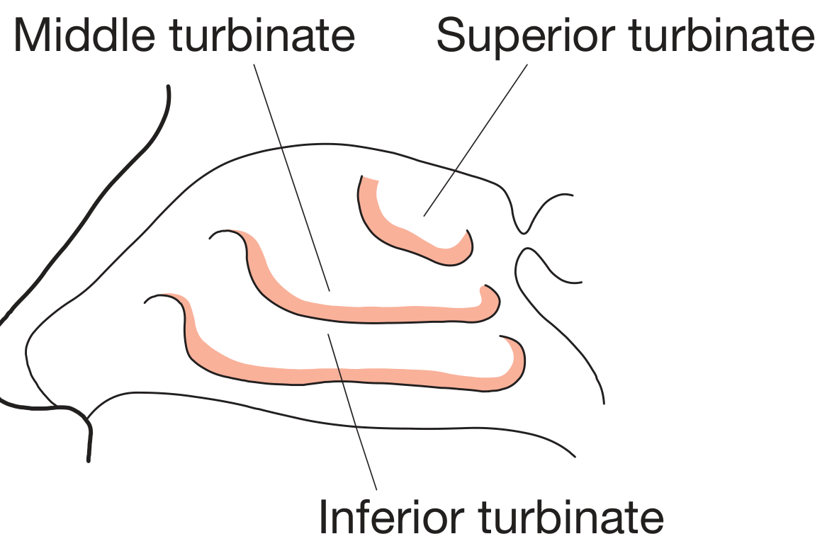
(Bailey & Love's Short Practice of Surgery, 28th ed., Fig. 51.34)
The lateral wall bears three turbinates - occasionally a fourth (supreme turbinate) is present. Each turbinate overhangs a meatus.
1. INFERIOR TURBINATE
- A separate, independent bone (not part of the ethmoid)
- Largest and most vascular turbinate
- Covered by specialized erectile mucosa with venous sinusoids
- Attached to the inferolateral nasal wall from just anterior to the Eustachian tube opening to the nasal valve anteriorly
- Blood supply: Lateral branch of the sphenopalatine artery (posteriorly); anterior ethmoid artery (anteriorly)
- Nerve supply: Trigeminal nerve (sensation); sympathetic fibers (vasoconstriction) and parasympathetic via Vidian nerve - sphenopalatine ganglion (vasodilation and secretion)
Inferior Meatus:
- Widest, longest, lowest meatus
- The nasolacrimal duct opens here - guarded by Hasner's valve (plica lacrimalis), 1-2 cm from the anterior end
- Site of puncture during Caldwell-Luc antral washout - trocar passed below the inferior turbinate through the thin medial wall of the maxillary sinus (Scott-Brown's, Vol. 1)
- Surgical significance: During crying, tears flow into the nose via this duct (explains "watery nose" with crying)
2. MIDDLE TURBINATE
- Part of the ethmoid bone
- Critical landmark in FESS - protects contents of the middle meatus
- Boomerang-shaped (Cummings)
- Has three distinct attachments (the basal lamella):
┌──────────────────────────────────────────────────────────────┐
│        THREE PARTS OF MIDDLE TURBINATE BASAL LAMELLA        │
├──────────────┬──────────────────────────────────────────────┤
│ Part         │ Orientation & Attachment                      │
├──────────────┼──────────────────────────────────────────────┤
│ 1. Anterior  │ Sagittal/vertical plane                       │
│   (vertical) │ Attaches to cribriform plate / crista         │
│              │ ethmoidalis of maxilla superiorly             │
├──────────────┼──────────────────────────────────────────────┤
│ 2. Middle    │ Coronal/oblique plane (45°)                   │
│   (oblique)  │ Attaches to lamina papyracea (key landmark)   │
│              │ = the TRUE basal lamella                      │
│              │ Divides ANTERIOR and POSTERIOR ethmoids       │
├──────────────┼──────────────────────────────────────────────┤
│ 3. Posterior │ Horizontal plane                              │
│  (horizontal)│ Attaches to medial wall of maxillary sinus   │
│              │ and lamina papyracea                          │
└──────────────┴──────────────────────────────────────────────┘
(Cummings, 7th ed., Chapter 44, Fig. 44.3)
Middle Meatus: Contains the key structures of the OMC (described in detail in Part II).
Pneumatization of middle turbinate = Concha Bullosa (most common anatomic variant - can obstruct the OMC).
3. SUPERIOR TURBINATE
- Smallest turbinate; part of the ethmoid
- Superior meatus: Receives the openings of the posterior ethmoid cells
- Sphenoethmoidal recess (between superior turbinate and nasal septum): Receives the sphenoid sinus ostium
C. STRUCTURES IN THE MIDDLE MEATUS (KEY CONTENTS)
DIAGRAM 2 - Lateral Nasal Wall with Turbinates Removed showing Sinus Ostia
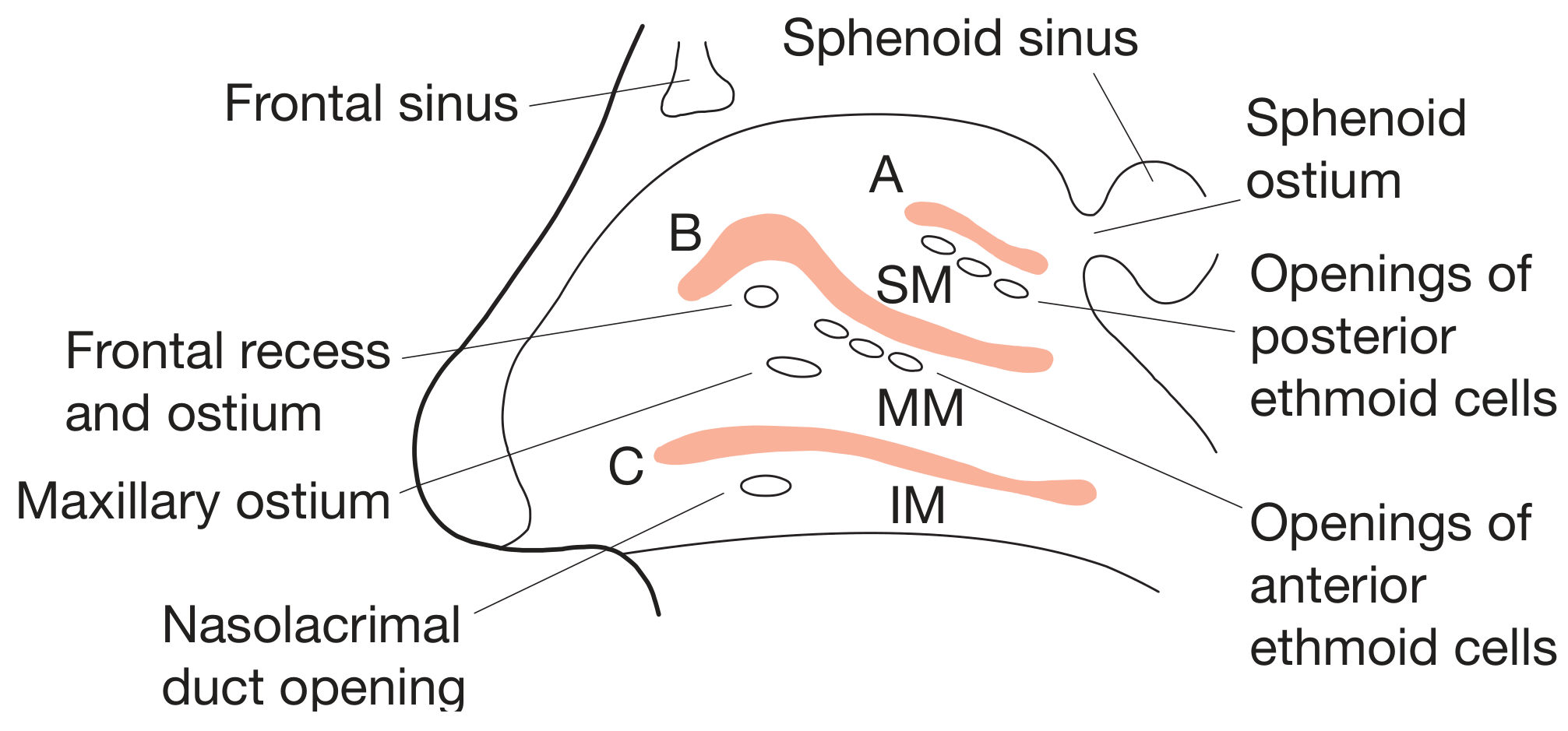
(Bailey & Love, Fig. 51.35 - SM = Superior Meatus, MM = Middle Meatus, IM = Inferior Meatus)
The middle meatus lies between the inferior turbinate below and the middle turbinate above. It contains:
- Uncinate process (sickle-shaped)
- Hiatus semilunaris
- Ethmoidal infundibulum
- Ethmoid bulla (bulla ethmoidalis)
- Agger nasi region
- Frontal recess
- Maxillary sinus ostium (natural)
(These form the OMC - detailed in Part II)
D. BLOOD SUPPLY OF LATERAL WALL
                BLOOD SUPPLY - LATERAL WALL OF NOSE
                ┌─────────────────────────────────┐
                │                                   │
     ┌──────────┴─────────┐         ┌──────────────┴──────────┐
     │  INTERNAL CAROTID  │         │   EXTERNAL CAROTID      │
     │  (via Ophthalmic A.)│         │   (via Maxillary A.)    │
     └──────────┬─────────┘         └──────────────┬──────────┘
                │                                   │
     ┌──────────┴──────────┐         ┌─────────────┴──────────────┐
     │ Anterior Ethmoidal  │         │ Sphenopalatine artery       │
     │ Artery (AEA)        │         │ → Posterolateral nasal      │
     │ Posterior Ethmoidal │         │   branches (to turbinates)  │
     │ Artery (PEA)        │         │ → Posterior septal branches │
     └─────────────────────┘         │ Greater Palatine artery     │
                                      │ (inferior)                  │
                                      └─────────────────────────────┘
- Kiesselbach's plexus (Little's area): Anastomosis on anterior nasal septum
- Woodruff's plexus: Posterior lateral wall, behind middle turbinate - site of posterior epistaxis
E. NERVE SUPPLY OF LATERAL WALL
         NERVE SUPPLY - LATERAL WALL
         ┌────────────────────────────────────┐
         │           TRIGEMINAL (CN V)         │
         └────────┬─────────────────┬──────────┘
                  │                  │
         ┌────────┴──────┐  ┌────────┴──────────┐
         │ Anterior 1/3  │  │  Posterior 2/3    │
         │               │  │                    │
         │ V1 - Anterior │  │ V2 - Posterolateral│
         │ Ethmoidal N.  │  │ nasal branches of  │
         │               │  │ sphenopalatine     │
         └───────────────┘  │ ganglion (via V2)  │
                            └────────────────────┘
         Autonomic supply via Vidian nerve (CN VII + sympathetic)
         → Sphenopalatine ganglion → nasal mucosa
F. LYMPHATIC DRAINAGE
- Anterior part: Submandibular nodes
- Posterior part: Upper deep cervical and retropharyngeal nodes
PART II: OSTIOMEATAL COMPLEX (OMC) - DETAILED NOTE
A. DEFINITION AND CONCEPT
The ostiomeatal complex (OMC) is not a single anatomical structure but a functional unit - a region on the lateral wall of the nasal cavity (deep to the middle turbinate) that represents the final common drainage pathway of the anterior group of paranasal sinuses (frontal, maxillary, and anterior ethmoid sinuses).
The term was popularized by Messerklinger (1978) and later expanded by Stammberger as the conceptual foundation of FESS.
(K.J. Lee's Essential Otolaryngology, 11th ed.; Cummings Otolaryngology, 7th ed., Chapter 44)
B. COMPONENTS OF THE OMC
DIAGRAM 3 - Coronal CT showing Normal Ostiomeatal Complex (Cummings, Fig. 8.62)
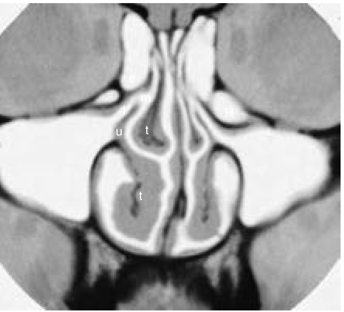
(Cummings Otolaryngology, Fig. 8.62 - e = ethmoid bulla, m = middle meatus, u = uncinate, t = turbinate, H = Haller cell, i = infundibulum)
DIAGRAM 4 - Endoscopic and Coronal View of OMC (Cummings, Fig. 44.1)
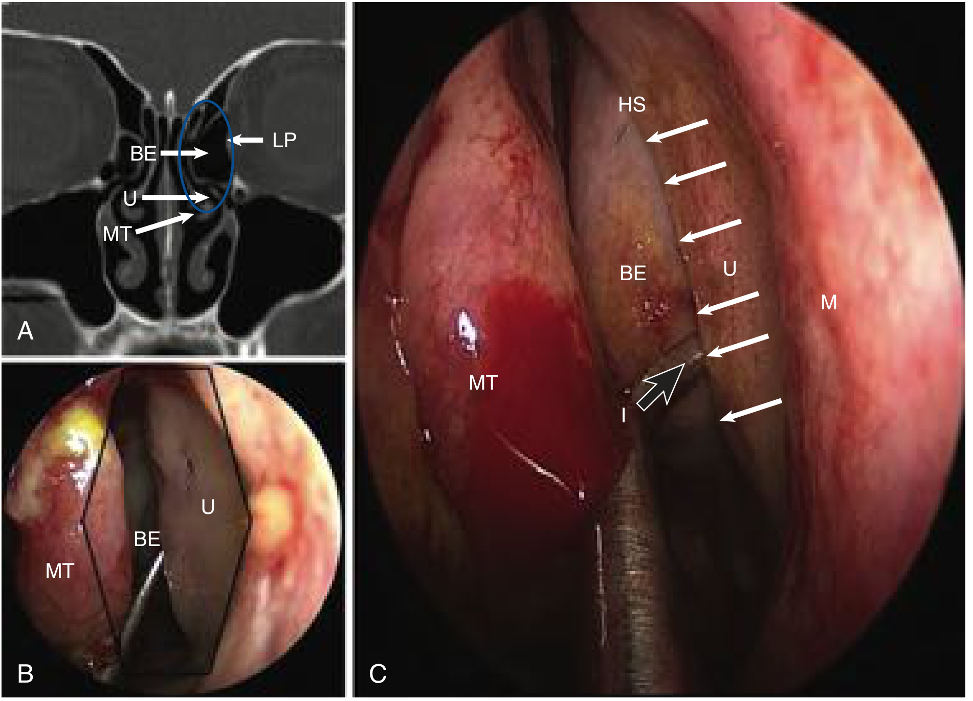
(Cummings, Fig. 44.1 - LP = lamina papyracea, MT = middle turbinate, BE = bulla ethmoidalis, U = uncinate, HS = hiatus semilunaris, I = infundibulum)
1. UNCINATE PROCESS
┌────────────────────────────────────────────────────────────────┐
│                    UNCINATE PROCESS                            │
├────────────────────────────────────────────────────────────────┤
│ Shape:    Sickle/hook-shaped; sagittally oriented              │
│ Size:     3-4 mm wide, 1.5-2 cm long                          │
│           (K.J. Lee's Essential Otolaryngology)               │
├────────────────────────────────────────────────────────────────┤
│ ATTACHMENTS:                                                    │
│ • Anterior/Superior: Frontal process of maxilla,              │
│   ethmoidal crest of maxilla, posterior lacrimal bone         │
│ • Posterior/Inferior: Ethmoidal process of inferior           │
│   turbinate, perpendicular plate of palatine bone             │
│ • SUPERIOR ATTACHMENT (crucial for frontal sinus drainage):   │
│   A: Lamina papyracea → creates recessus terminalis (blind)   │
│      → frontal sinus drains MEDIALLY beside middle turbinate  │
│   B: Ethmoid roof                                              │
│      → frontal sinus drains INTO infundibulum                 │
│   C: Middle turbinate                                          │
│      → frontal sinus drains INTO infundibulum                 │
├────────────────────────────────────────────────────────────────┤
│ SURGICAL SIGNIFICANCE:                                          │
│ • First structure removed in FESS (uncinectomy)                │
│ • Its posterior free edge forms medial wall of infundibulum   │
│ • The inferior part overlies the natural maxillary ostium     │
└────────────────────────────────────────────────────────────────┘
DIAGRAM 5 - Three variations of uncinate process superior attachment
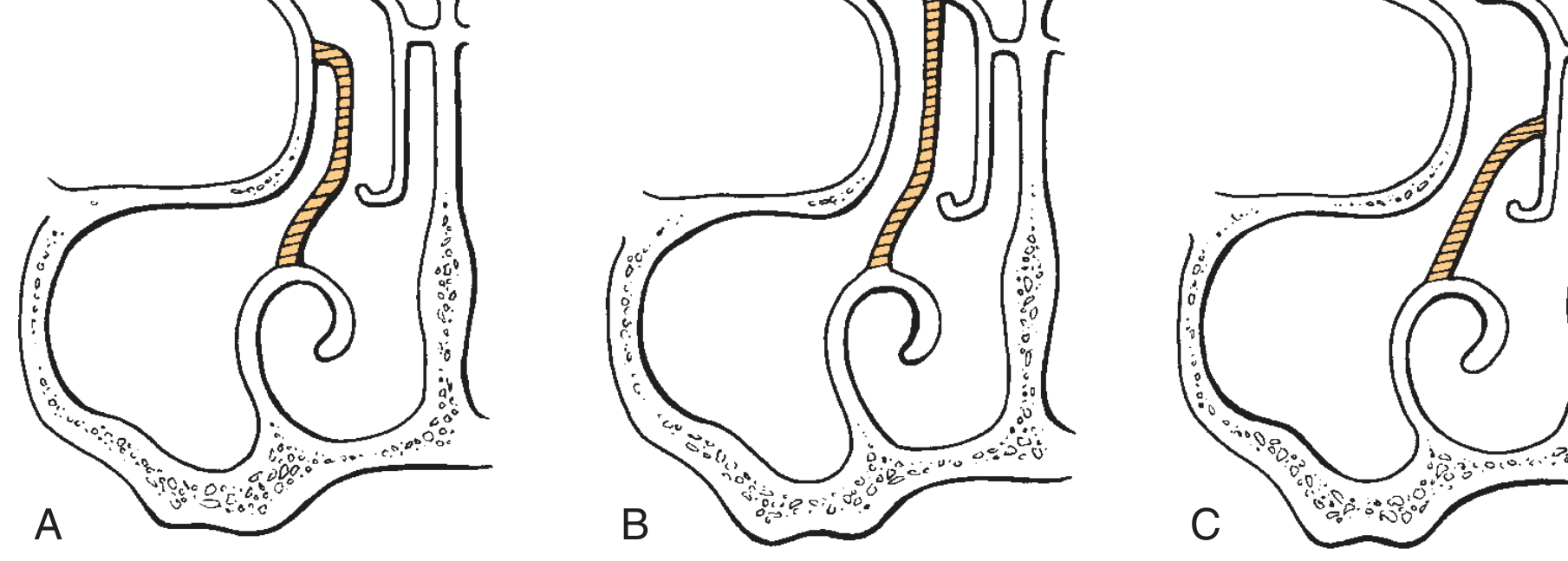
(Cummings, Fig. 44.2 - A = attachment to lamina papyracea, B = ethmoid roof, C = middle turbinate)
2. HIATUS SEMILUNARIS (INFERIOR)
- A two-dimensional, crescent/semilunar-shaped cleft (gap) between:
- Anteriorly/medially: Posterior free edge of the uncinate process
- Posteriorly/laterally: Anterior face of the ethmoid bulla
- Through this gap, the nasal cavity communicates with the ethmoidal infundibulum
- A "hiatus semilunaris superior" (hiatus secondarius) connects the middle meatus to the suprabullar/retrobullar recess (K.J. Lee's, p. 607)
3. ETHMOIDAL INFUNDIBULUM
┌──────────────────────────────────────────────────────┐
│              ETHMOIDAL INFUNDIBULUM                  │
├──────────────────────────────────────────────────────┤
│ Description: Funnel-shaped 3-dimensional space       │
│ that channels secretions → middle meatus             │
├──────────────────────────────────────────────────────┤
│ BOUNDARIES:                                           │
│ Medial:           Uncinate process                    │
│ Lateral:          Lamina papyracea (medial orbital   │
│                   wall)                               │
│ Posterior:        Anterior wall of ethmoid bulla     │
│ Anterior/Superior: Frontal process of maxilla        │
│ Superior/Lateral:  Lacrimal bone                     │
├──────────────────────────────────────────────────────┤
│ RECEIVES DRAINAGE FROM:                               │
│ • Maxillary sinus (via its natural ostium)           │
│ • Anterior ethmoid cells                             │
│ • Frontal sinus (if uncinate attaches to B or C)    │
└──────────────────────────────────────────────────────┘
(K.J. Lee's Essential Otolaryngology, p. 607; Cummings Chapter 44)
4. ETHMOID BULLA (BULLA ETHMOIDALIS)
- The largest and most constant anterior ethmoid air cell
- First cell encountered after removal of uncinate process during FESS
- Anterior wall: Forms the posterior boundary of the hiatus semilunaris
- Lateral wall: Is the medial wall of the orbit (lamina papyracea)
- Drains into suprabullar recess or retrobullar recess (collectively = Sinus Lateralis of Grunwald)
- When the bulla extends to skull base = bulla lamella
5. AGGER NASI AND FRONTAL RECESS
Agger Nasi:
- A bony mound at the attachment of the middle turbinate to the lateral wall
- When pneumatized = Agger Nasi Cell (ANC) - present in 98.5% of CT scans (Cummings)
- Most anterior of all ethmoid cells; key structure in frontal sinus surgery
Frontal Recess:
Boundaries:
• Lateral:    Lamina papyracea
• Medial:     Middle turbinate
• Anterior:   Posterior wall of Agger Nasi Cell (when present)
• Posterior:  Anterior face of ethmoid bulla (if it reaches skull base)
- Tapers superiorly into the frontal sinus ostium
(K.J. Lee's Essential Otolaryngology, p. 607)
C. BOUNDARIES OF THE OMC AS A UNIT
┌──────────────────────────────────────┐
│    BOUNDARIES OF THE OMC             │
├────────────────┬─────────────────────┤
│ Lateral        │ Lamina papyracea    │
│                │ (medial orbital     │
│                │ wall)               │
├────────────────┼─────────────────────┤
│ Medial         │ Middle turbinate    │
├────────────────┼─────────────────────┤
│ Anterior       │ Agger nasi region   │
├────────────────┼─────────────────────┤
│ Posterior      │ Basal lamella of    │
│                │ middle turbinate    │
├────────────────┼─────────────────────┤
│ Superior       │ Skull base /        │
│                │ fovea ethmoidalis   │
└────────────────┴─────────────────────┘
D. DRAINAGE PATHWAYS - FLOWCHART
DIAGRAM 6 - Anterior Ostiomeatal Channels (Cummings, Fig. 38.5)
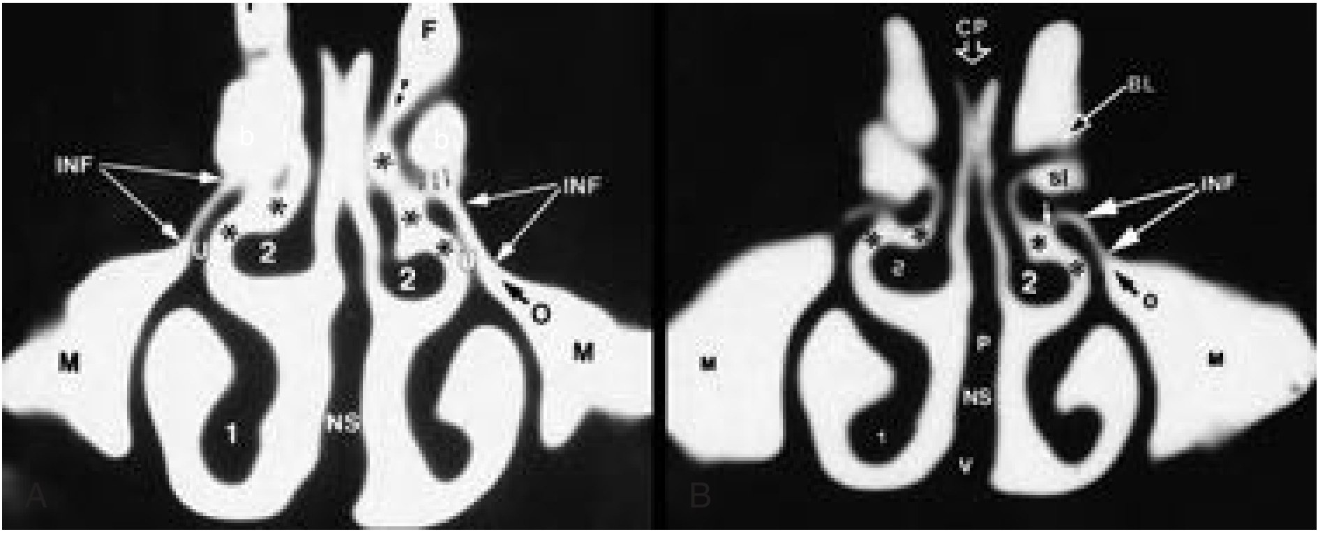
(Cummings, Fig. 38.5 - INF = infundibulum, U = uncinate, b = ethmoid bulla, O = maxillary sinus ostium, BL = basal lamella, CP = cribriform plate)
FLOWCHART 1 - Sinus Drainage via OMC
╔══════════════════════════════════════════════════════════════╗
║         MUCOCILIARY DRAINAGE OF PARANASAL SINUSES           ║
╠══════════════════════════════════════════════════════════════╣
║                                                              ║
║  ANTERIOR GROUP                    POSTERIOR GROUP          ║
║  (Via OMC / Middle Meatus)         (Via Superior Meatus)    ║
║  ┌─────────────┐                   ┌────────────────┐       ║
║  │Frontal Sinus│                   │Posterior Ethmoid│      ║
║  └──────┬──────┘                   │    Cells        │      ║
║         │ Frontal recess           └───────┬────────┘       ║
║         ↓                                  ↓                 ║
║  ┌──────────────────┐              ┌────────────────┐       ║
║  │  Anterior        │              │ Superior Meatus │       ║
║  │  Ethmoid Cells   │              └────────────────┘       ║
║  └──────┬───────────┘                                        ║
║         │                          ┌────────────────────┐   ║
║         ↓                          │  Sphenoid Sinus    │   ║
║  ┌──────────────────────┐          └────────┬───────────┘   ║
║  │  Ethmoidal           │                   │               ║
║  │  Infundibulum        │          ┌────────↓───────────┐   ║
║  └──────┬───────────────┘          │Sphenoethmoidal     │   ║
║         │ (via hiatus semilunaris) │Recess              │   ║
║         ↓                          └────────────────────┘   ║
║  ┌──────────────────┐                                        ║
║  │  Maxillary Sinus │                                        ║
║  │  → Natural ostium│                                        ║
║  └──────┬───────────┘                                        ║
║         │                                                    ║
║         ↓                                                    ║
║  ╔═════════════════╗                                         ║
║  ║  MIDDLE MEATUS  ║  ← FINAL COMMON PATHWAY                ║
║  ╚═════════════════╝                                         ║
╚══════════════════════════════════════════════════════════════╝
(Cummings Otolaryngology 7th ed., p. 784 - "anterior ostiomeatal unit" and "posterior ostiomeatal unit")
FLOWCHART 2 - How OMC Obstruction Leads to Sinusitis
┌─────────────────────────────────────────────────────────────────┐
│ TRIGGER: Viral URTI / Allergy / Anatomic Variant                │
└──────────────────────────┬──────────────────────────────────────┘
                           ↓
        ┌──────────────────────────────────┐
        │  Mucosal edema at OMC            │
        │  (uncinate, infundibulum,        │
        │   middle meatus)                 │
        └──────────────────┬───────────────┘
                           ↓
        ┌──────────────────────────────────┐
        │  Obstruction of sinus ostia      │
        │  (maxillary, frontal, ant.       │
        │   ethmoid)                       │
        └──────────────────┬───────────────┘
                           ↓
        ┌──────────────────────────────────┐
        │  ↓ O₂ tension in sinus           │
        │  ↓ mucociliary clearance         │
        │  Mucus accumulation              │
        └──────────────────┬───────────────┘
                           ↓
        ┌──────────────────────────────────┐
        │  Secondary bacterial infection   │
        │  (S. pneumoniae, H. influenzae,  │
        │   M. catarrhalis)                │
        └──────────────────┬───────────────┘
                           ↓
        ┌──────────────────────────────────┐
        │  ACUTE / CHRONIC RHINOSINUSITIS  │
        │  (involving multiple sinuses     │
        │   from one OMC lesion)           │
        └──────────────────────────────────┘
(Stammberger's concept of "key area"; Hazarika ENT, Dhingra Diseases of ENT)
E. ANATOMIC VARIANTS OF OMC (SURGICAL IMPORTANCE)
┌───────────────────────────────────────────────────────────────────┐
│              ANATOMIC VARIANTS OF OMC                             │
├────────────────────┬──────────────────────────────────────────────┤
│ Variant            │ Clinical Significance                         │
├────────────────────┼──────────────────────────────────────────────┤
│ Concha Bullosa     │ Most common; pneumatized middle turbinate;   │
│                    │ can obstruct OMC                             │
├────────────────────┼──────────────────────────────────────────────┤
│ Paradoxical middle │ Middle turbinate curves laterally instead of │
│ turbinate          │ medially; narrows middle meatus              │
├────────────────────┼──────────────────────────────────────────────┤
│ Haller cell        │ Now: Infraorbital ethmoidal cell (IOC)       │
│ (IOC)              │ Pneumatizes into orbital floor above         │
│                    │ maxillary ostium; can obstruct infundibulum  │
├────────────────────┼──────────────────────────────────────────────┤
│ Agger nasi cell    │ Large ANC can obstruct frontal recess        │
│ (ANC)              │                                              │
├────────────────────┼──────────────────────────────────────────────┤
│ Accessory maxillary│ Perforated posterior fontanelle (20-25%);   │
│ ostium             │ mucociliary recirculation                    │
├────────────────────┼──────────────────────────────────────────────┤
│ Onodi cell         │ Posterior-most ethmoid cell; intimate with  │
│ (sphenoethmoidal   │ optic nerve - risk during surgery           │
│ cell)              │                                              │
├────────────────────┼──────────────────────────────────────────────┤
│ Deviated nasal     │ Contralateral OMC narrowing                  │
│ septum             │                                              │
└────────────────────┴──────────────────────────────────────────────┘
(Cummings, Chapter 44; [Papadopoulou et al., 2022 systematic review, Int Arch Otorhinolaryngol, PMID: 35846811])
F. SURGICAL SIGNIFICANCE OF OMC - FESS
FLOWCHART 3 - Steps of Messerklinger's FESS technique (Anterior-to-Posterior)
Step 1: Middle turbinate medialized
        ↓
Step 2: UNCINECTOMY
        (Uncinate process removed - exposes infundibulum)
        ↓
Step 3: MAXILLARY ANTROSTOMY
        (Natural maxillary ostium identified and widened)
        ↓
Step 4: ETHMOID BULLA removed (anterior ethmoidectomy)
        (Exposes frontal recess and ethmoid roof)
        ↓
Step 5: FRONTAL SINUSOTOMY
        (Agger nasi cell opened; frontal recess cleared)
        ↓
Step 6: Posterior ethmoidectomy
        (Basal lamella entered; posterior cells cleared)
        ↓
Step 7: SPHENOIDOTOMY
        (Natural sphenoid ostium identified and widened)
(K.J. Lee's Essential Otolaryngology, 11th ed., Messerklinger technique; Cummings, Box 44.4)
Wigand's technique (Posterior-to-Anterior): Begins with sphenoidotomy, identifies skull base, and dissects anteriorly.
G. RADIOLOGICAL ASSESSMENT OF OMC
- Modality of choice: Coronal CT scan (non-contrast)
- Best demonstrates the OMC structures, infundibulum, ethmoid bulla, uncinate, Haller cells, and all anatomic variants
- The three "tight spots" to assess (Cummings, p. 784):
- Frontal recess - narrowest portion
- Infundibulum - connects maxillary sinus to middle meatus
- Sphenoethmoidal recess - sphenoid drainage
The Lund-Mackay CT scoring system evaluates OMC involvement in chronic rhinosinusitis.
PART III: RECENT ADVANCES (RGUHS Perspective)
1. Concept Revision - Haller Cell renamed Infraorbital Ethmoidal Cell (IOC)
The 2020 International Frontal Sinus Anatomy Classification (IFAC) and the European Position Paper on Rhinosinusitis (EPOS 2020) have recommended discontinuing the term "Haller cell" in favor of infraorbital ethmoidal cell (IOC), with a new classification of frontal ethmoidal cells (Types 1-4 Kuhn cells, now renamed as per IFAC 2020).
2. EPOS 2020 Guidelines
The European Position Paper on Rhinosinusitis and Nasal Polyps (EPOS 2020) remains the global standard for managing CRS, with CT evidence of OMC obstruction being a key indicator for surgical intervention.
3. CT vs. MRI
MRI is increasingly used to assess mucosal disease within the OMC (especially differentiating mucus from inflammatory tissue), while CT remains the surgical planning gold standard.
4. Endoscopic Anatomy - The "Key Area"
Stammberger's concept of the OMC as a "key area" has been validated: obstruction of this small region can result in concurrent maxillary, frontal, and anterior ethmoid sinusitis. Modern FESS aims to restore OMC drainage rather than obliterate sinuses (functional surgery over radical Caldwell-Luc).
5. Drug Delivery to OMC
Recent computational fluid dynamics studies show that gravity-dependent nasal drop positioning (head tilted back and to the affected side) delivers significantly more drug to the OMC and maxillary sinus (Seifelnasr et al., Drug Deliv Transl Res, 2024, PMID: 38044376).
6. Image-Guided FESS
Navigation systems (BrainLab, Medtronic Fusion) using preoperative CT/MRI allow real-time anatomical identification during surgery, reducing complications (CSF leak, orbital injury) in complex OMC anatomy.
7. Anatomic Variations and CRS
A 2024 CT-based Indian study (Nandyal & Benola, Indian J Otolaryngol, PMID: 38440475) confirmed that concha bullosa, deviated nasal septum, and IOC (Haller cells) are the most common anatomic variants predisposing to CRS by narrowing the OMC.
SUMMARY TABLE
┌──────────────────────────────────────────────────────────────────────┐
│           COMPONENTS OF OMC AND THEIR SURGICAL RELEVANCE            │
├────────────────────────┬─────────────────────────────────────────────┤
│ Structure              │ Key Points                                   │
├────────────────────────┼─────────────────────────────────────────────┤
│ Uncinate process       │ First to be removed in FESS; determines     │
│                        │ frontal sinus drainage pattern              │
├────────────────────────┼─────────────────────────────────────────────┤
│ Hiatus semilunaris     │ 2D gap between uncinate & ethmoid bulla;    │
│                        │ entry into infundibulum                     │
├────────────────────────┼─────────────────────────────────────────────┤
│ Ethmoidal infundibulum │ 3D funnel; common drainage channel          │
├────────────────────────┼─────────────────────────────────────────────┤
│ Ethmoid bulla          │ Largest anterior ethmoid cell; post.        │
│                        │ boundary of hiatus                          │
├────────────────────────┼─────────────────────────────────────────────┤
│ Frontal recess         │ Narrowest "tight spot"; most complex        │
│                        │ frontal drainage                            │
├────────────────────────┼─────────────────────────────────────────────┤
│ Agger nasi cell        │ Present in 98.5%; key for frontal surgery   │
├────────────────────────┼─────────────────────────────────────────────┤
│ Natural maxillary      │ Posteromedial sinus wall; always widen     │
│ ostium                 │ rather than create new opening (prevents    │
│                        │ mucociliary recirculation)                  │
└────────────────────────┴─────────────────────────────────────────────┘
REFERENCES
- Cummings Otolaryngology Head and Neck Surgery, 7th ed. - Flint PW et al. Chapter 38 (Imaging) and Chapter 44 (FESS). Elsevier, 2020. (Primary textbook - OMC anatomy, FESS steps, CT assessment)
- Scott-Brown's Otorhinolaryngology Head & Neck Surgery, 8th ed., Vol. 1 - Watkinson JC, Clarke RW (eds). CRC Press. (Turbinate anatomy, nerve supply, inferior turbinate details)
- K.J. Lee's Essential Otolaryngology, 11th ed. - Scholes MA, Ramakrishnan VR (eds). McGraw-Hill. (OMC components with detailed borders, ESS techniques)
- Bailey & Love's Short Practice of Surgery, 28th ed. - Williams NS et al. CRC Press. (Lateral wall anatomy, blood supply)
- Diseases of Ear, Nose and Throat, 7th ed. - Dhingra PL, Dhingra S. Elsevier. (Standard Indian ENT textbook - OMC concept)
- Hazarika P. - Textbook of Ear, Nose and Throat. (Indian context - standard RGUHS reference)
- Stell & Maran's Head and Neck Surgery, 5th ed. (OMC and FESS anatomy)
- Stammberger H. Functional Endoscopic Sinus Surgery. Mosby, 1991. (Original OMC concepts)
- Papadopoulou AM, Bakogiannis N, Skrapari I. "Anatomical Variations of the Sinonasal Area and Their Clinical Impact on Sinus Pathology: A Systematic Review." Int Arch Otorhinolaryngol, 2022 Jul. [PMID: 35846811]
- Nandyal CB, Benola A. "Evaluation of Anatomical Variations Associated with Chronic Rhinosinusitis by Computed Tomography of Paranasal Sinuses." Indian J Otolaryngol Head Neck Surg, 2024. [PMID: 38440475]
- Seifelnasr A, Zare F, Si XA. "Optimized gravity-driven intranasal drop administration delivers significant doses to the ostiomeatal complex and maxillary sinus." Drug Deliv Transl Res, 2024 Jul. [PMID: 38044376]
- EPOS 2020 Guidelines - Fokkens WJ et al. Rhinology, 2020;58(Suppl S29):1-464.
RGUHS Exam Key Points to Remember:
- OMC is NOT a single structure - it is a functional unit / region
- The uncinate process determines frontal sinus drainage pathway (3 variations of superior attachment)
- Coronal CT (non-contrast) is the imaging modality of choice for OMC evaluation
- Concha bullosa is the most common anatomic variant causing OMC obstruction
- Messerklinger's FESS goes anterior-to-posterior; Wigand's goes posterior-to-anterior
- Haller cell = IOC (infraorbital ethmoidal cell) per current nomenclature (IFAC 2020)
- The natural maxillary sinus ostium must always be identified and preserved/widened - never create a neo-ostium without identifying the natural one (risk of mucociliary recirculation)
- Agger nasi cell is present in 98.5% of patients and is the most anterior ethmoidal cell
This is a shared conversation. Sign in to Orris to start your own chat.