
Hi I'm a intern student i have completed my 1st year and here for my internship help me
What field is your internship in? · What kind of help are you looking for?
What I Can Help You With
- Drugs, dosages, mechanisms of action, side effects
- Disease diagnosis, pathophysiology, and management
- Anatomy, physiology, and lab values
- Understanding clinical cases and presentations
- Interpreting investigations (ECG, X-ray, labs, imaging)
- Procedures and patient management protocols
- Breaking down complex topics (pharmacology, pathology, surgery, medicine)
- Quick summaries of conditions you encounter on the ward
- Exam prep (MBBS finals, PG entrance, USMLE, etc.)
- Searching the latest clinical evidence and guidelines (PubMed)
- Understanding journal articles and studies
- "What's the management of diabetic ketoacidosis?"
- "Explain the mechanism of beta-blockers"
- "What does a raised troponin mean?"
Explain a specific medical topic I'm studying
Which medical topic would you like me to explain?
Which respiratory topic specifically?
Pulmonary Embolism (PE) - Complete Guide for Your Internship
1. What Is It?
- Murray & Nadel's Textbook of Respiratory Medicine: "PE is a life-threatening feature of venous thromboembolism and most frequently originates from deep venous thrombosis in the lower extremities."
2. Pathophysiology - Why Is It Dangerous?
Respiratory failure
- The blocked lung segment gets ventilated but NOT perfused - creating dead space (V/Q mismatch)
- Hypoxia results from shunting and atelectasis
- Surfactant loss can cause pulmonary infarction in peripheral emboli
Cardiovascular collapse
- Obstruction of the pulmonary vasculature increases right ventricular afterload suddenly
- The right ventricle (RV) is a thin-walled chamber - it is not built for high pressure
- RV dilation causes the interventricular septum to bow into the left ventricle (LV), reducing LV filling and cardiac output
- This leads to systemic hypotension and shock - the "obstructive shock" of massive PE
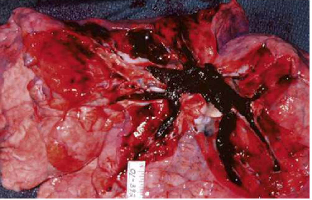
A large PE can obstruct the entire right ventricular outflow system, causing sudden cardiovascular collapse and cardiac arrest. About 25% of sudden cardiac deaths are thought to be due to PE. (Rosen's Emergency Medicine)
3. Risk Factors - Virchow's Triad
| Component | Examples |
|---|---|
| Hypercoagulability | Malignancy (lung, brain, pancreas, leukemia), thrombophilia (Factor V Leiden, antiphospholipid syndrome, protein C/S deficiency), oral contraceptives, pregnancy |
| Stasis | Prolonged immobility (long flights - "economy class syndrome"), bed rest, post-surgery, heart failure |
| Vessel wall injury | Surgery (especially hip/knee replacement), trauma, central lines |
- Murray & Nadel's: "Three predisposing factors - hypercoagulability, stasis, and venous injury or inflammation (Virchow's triad) - may each contribute to a clot that becomes clinically evident as DVT or PE."
4. Clinical Presentation
Symptoms
| Symptom | Frequency |
|---|---|
| Dyspnea | 75-80% (most common) |
| Chest pain (often pleuritic) | ~65% |
| Cough | ~50% |
| Hemoptysis | ~30% |
| Unilateral leg swelling | <30% |
| Syncope | <5% |
Signs
- Tachycardia (most common sign)
- Tachypnoea
- Low O2 saturations
- Hypotension (in massive PE only)
- Signs of DVT: unilateral leg swelling, redness, warmth
- Fever is usually low-grade; >38.6°C (101.5°F) suggests infection instead
5. Diagnosis
Step 1 - Estimate Pre-test Probability (PTP)
| Criterion | Points |
|---|---|
| Clinical signs/symptoms of DVT | 3 |
| PE more likely than alternative diagnosis | 3 |
| Heart rate >100 | 1.5 |
| Immobilization or surgery in past 4 weeks | 1.5 |
| Previous DVT/PE | 1.5 |
| Hemoptysis | 1 |
| Malignancy (on treatment or palliative) | 1 |
- Score >4: High probability - go straight to imaging
- Score ≤4: Low/moderate - test D-dimer first
Step 2 - PERC Rule (to avoid unnecessary testing)
- Age < 50
- Pulse < 100
- SaO2 > 94%
- No unilateral leg swelling
- No hemoptysis
- No recent trauma or surgery
- No prior PE/DVT
- No hormone use
Step 3 - D-Dimer
- Sensitivity 95-98%, specificity only 40-55%
- A negative D-dimer rules out PE in low/moderate probability patients
- A positive D-dimer is NOT diagnostic - requires imaging
- Age-adjusted D-dimer threshold: age × 10 ng/mL (for patients >50 years) - increases the number of patients who can be ruled out without imaging by 10-20%
Step 4 - Imaging
- CTPA (CT Pulmonary Angiography) - gold standard, most commonly used
- V/Q scan - when CTPA contraindicated (contrast allergy, pregnancy, renal failure)
- Echocardiography - useful in unstable patients to assess RV strain
- Chest X-ray - often normal; classic signs:
- Hampton's Hump (wedge-shaped pleural opacity from infarction)
- Westermark sign (oligaemia distal to embolus)
- Fleischner sign (enlarged pulmonary artery)
- ECG - most common finding is sinus tachycardia. The classic S1Q3T3 pattern (S in lead I, Q wave + T-wave inversion in lead III) suggests acute right heart strain - not sensitive but specific when present
Biomarkers
- Troponin - elevated in massive/submassive PE due to RV strain; indicates high risk for mortality
- BNP/NT-proBNP - elevated with RV dysfunction
- D-dimer - as above
6. Classification by Severity
| Type | Haemodynamics | Mortality | Key Feature |
|---|---|---|---|
| Massive (High-Risk) | Hypotension (SBP <90 mmHg) or shock | >15% | RV failure, haemodynamic instability |
| Submassive (Intermediate-Risk) | Normal BP but RV dysfunction on echo or raised troponin/BNP | 3-15% | RV strain without shock |
| Low-Risk | Normal BP, no RV dysfunction, normal biomarkers | <1% | Can manage outpatient |
7. Management
Immediate stabilisation
- O2 supplementation (target SaO2 >94%)
- IV access, cardiac monitoring
- Fluid resuscitation cautiously (excessive fluids worsen RV dilation)
- Vasopressors (noradrenaline) if haemodynamically unstable
Anticoagulation - the mainstay of treatment
- LMWH (Low Molecular Weight Heparin, e.g., enoxaparin) - drug of choice for most patients
- Unfractionated heparin (UFH) IV - preferred if thrombolysis planned or severe renal impairment
- DOACs (Direct Oral Anticoagulants) - e.g., rivaroxaban, apixaban - now first-line for most patients
- Warfarin - still used in certain cases (e.g., antiphospholipid syndrome, mechanical valves)
Thrombolysis (systemic or catheter-directed)
- Indicated in massive PE with haemodynamic instability (the patient is in shock or cardiac arrest)
- Drugs: alteplase (tPA), streptokinase
- Risk: major bleeding, including intracranial haemorrhage - so used only when risk of death outweighs bleeding risk
- Murray & Nadel's: "Primary difference between prophylaxis and treatment is the dosages of anticoagulants; treated patients receive a higher dosage."
Surgical embolectomy
- Reserved for massive PE when thrombolysis fails or is contraindicated
IVC filter
- When anticoagulation is absolutely contraindicated (e.g., active major bleed)
8. Special Scenarios to Know
| Scenario | Key Points |
|---|---|
| PE in pregnancy | 5x higher risk due to hypercoagulability + venous stasis; use LMWH (NOT warfarin in 1st trimester - teratogenic); modified V/Q or CTPA both used; D-dimer unreliable |
| Cancer-associated PE | Higher recurrence risk; LMWH or DOACs preferred; longer anticoagulation duration |
| Subsegmental PE | Small, peripheral - may not need anticoagulation if low DVT risk |
9. Complications
- Pulmonary hypertension - if PE recurs or goes untreated, chronic clot can scar the pulmonary arteries
- Chronic Thromboembolic Pulmonary Hypertension (CTEPH) - occurs in 1-4% after PE
- Right heart failure
- Post-PE syndrome - persistent dyspnea and reduced exercise tolerance after an index PE
Quick Memory Aid
- C - Cancer
- L - Long trips / immobility
- O - OCP / hormones / Obesity
- T - Trauma / surgery / Thrombophilia
- S - Surgery / Stasis / previous clot
- S - Sudden dyspnea
- A - Acute chest pain (pleuritic)
- D - DVT signs in leg
Syncope
Syncope - Complete Guide for Your Internship
1. Definition
- Abrupt onset
- Short duration
- Spontaneous and complete recovery
- Tintinalli's Emergency Medicine: "A brief loss of consciousness associated with inability to maintain postural tone that spontaneously resolves without medical intervention."
2. Pathophysiology
~10 seconds of complete disruption of blood flow to both cerebral cortices or to the brainstem reticular activating system, OR a 35-50% reduction in cerebral perfusion pressure
- Tintinalli's: "Cerebral perfusion and consciousness are restored by the supine position, autonomic autoregulatory response, or restoration of a perfusing cardiac rhythm."
3. Classification and Causes
A. Reflex-Mediated / Neural (Most Common - ~21%)
| Type | Trigger |
|---|---|
| Vasovagal | Pain, emotion, prolonged standing, heat, blood/needles - classic "fainting" |
| Situational | Coughing, micturition (urinating), defecation, swallowing |
| Carotid sinus syndrome | Tight collar, shaving, turning head - pressure on carotid sinus causes bradycardia |
- Trigger (e.g., pain, fear, prolonged standing)
- Prodrome: nausea, pallor, sweating, lightheadedness, tunnel vision, yawning
- Loss of consciousness (brief, usually <1 minute)
- Recovery: fatigue, nausea for hours after
B. Orthostatic Hypotension (~9%)
- Dehydration / blood loss / vomiting
- Medications: antihypertensives, diuretics, nitrates, alpha-blockers
- Autonomic neuropathy (diabetes, Parkinson's disease)
- Elderly (blunted baroreceptor response)
- Prolonged bed rest
C. Cardiac Syncope (~10%) - THE DANGEROUS ONE
- Bradycardias: Sick sinus syndrome, high-degree AV block, pacemaker malfunction
- Tachycardias: Ventricular tachycardia (VT), Ventricular fibrillation (VF), SVT
- Channelopathies: Long QT syndrome, Brugada syndrome, WPW syndrome
- Aortic stenosis (syncope on exertion = very ominous sign)
- Hypertrophic obstructive cardiomyopathy (HOCM)
- Pulmonary embolism
- Cardiac tamponade
- Pulmonary hypertension
- Atrial myxoma (tumour obstructing mitral valve)
D. Other Causes
- Neurologic (~4%): Seizures (not true syncope, but mimics it), transient ischaemic attack (TIA) of posterior circulation
- Metabolic: Hypoglycaemia, hypoxia, anaemia
- Medications: Beta-blockers, antihypertensives, antidepressants
- Unknown: 37-50% of cases remain unexplained even after workup
4. Key Clinical Features - History Is Everything
Before the event
- What were you doing? (exertion = cardiac; prolonged standing = vasovagal)
- Any prodrome? (nausea/sweating = vasovagal; NO prodrome = cardiac/arrhythmic)
- Any palpitations before? (arrhythmia)
- Any recent illness, vomiting, blood loss? (orthostatic)
- New medications?
During the event
- Witnessed? Duration?
- Limb jerking? (brief jerks can occur in syncope from hypoperfusion, but prolonged rhythmic jerking = seizure)
- Tongue biting? Incontinence? (seizure rather than syncope)
- Colour: Pale = vasovagal/cardiac; Blue = cyanotic/respiratory/arrhythmic
After the event
- How quickly did they recover? (<1 min = syncope; prolonged confusion = seizure - "postictal phase")
- Headache? Focal neurological deficit? (TIA/stroke)
- Chest pain? (ACS, PE, aortic dissection)
Red flag features suggesting cardiac cause
- Syncope during exertion (not after)
- Syncope when lying down or sitting (arrhythmia)
- No prodrome - sudden collapse
- Family history of sudden cardiac death or channelopathy
- Known structural heart disease
- Palpitations just before syncope
- Age >65 years
- Abnormal ECG
5. Examination
- Vital signs: BP in both arms (aortic dissection), HR, postural BP and HR (lying vs. standing 1 and 3 min)
- Cardiovascular: Murmurs (aortic stenosis, HOCM), signs of heart failure, carotid bruits
- Neurological: Any focal deficits? Tongue laceration?
- Skin: Pallor, cyanosis, diaphoresis
6. Investigations
Always do:
| Investigation | What it finds |
|---|---|
| 12-lead ECG | Arrhythmia, conduction defects, QT prolongation, Brugada pattern, LBBB, delta waves (WPW) |
| Blood glucose | Hypoglycaemia |
| Postural BP measurements | Orthostatic hypotension |
ECG - what to look for in syncope:
- Long QT (risk of Torsades de Pointes VT)
- Brugada pattern (right bundle branch block + saddle-shaped ST elevation in V1-V3)
- Delta waves (WPW - pre-excitation, risk of fast AF)
- S1Q3T3 (PE)
- Complete heart block / high-degree AV block
- Left bundle branch block (new LBBB = very high-risk)
- Right bundle branch block
If cardiac syncope suspected:
| Test | Purpose |
|---|---|
| Echocardiography | Structural disease (AS, HOCM, poor LV function, tamponade) |
| Troponin | ACS, PE |
| Holter monitor (24-48h) | Intermittent arrhythmia |
| Implantable loop recorder | Long-term arrhythmia monitoring (up to 3 years) - for unexplained recurrent syncope |
| Exercise stress test | Exertional syncope |
| Electrophysiology study (EPS) | Map arrhythmia circuits |
If orthostatic suspected:
- Tilt-table test (gold standard for vasovagal and orthostatic syncope)
If neurological cause suspected:
- CT/MRI brain - only if focal neuro signs or post-ictal state
- EEG - if seizure suspected
Important: Routine CT brain, EEG, or carotid Doppler are NOT recommended in isolated syncope with no neurological features.
7. Risk Stratification - Who Needs Admission?
Admit / High-risk if ANY of:
- Abnormal ECG
- History of structural heart disease or heart failure
- Age >65
- Syncope during exertion
- Syncope supine or without prodrome
- New-onset cardiac murmur
- Positive troponin
- SBP <90 mmHg on arrival
- Family history of sudden cardiac death
Can consider discharge / Low-risk if:
- Young patient
- Clear vasovagal trigger with prodrome
- Normal ECG
- No cardiac history
- Normal vital signs
8. Treatment - Based on Cause
| Cause | Treatment |
|---|---|
| Vasovagal | Reassurance + education (lie flat when prodrome felt), avoid triggers, increase fluid/salt intake, compression stockings. Beta-blockers do NOT help. |
| Orthostatic hypotension | Rehydrate, stop offending medications, fludrocortisone, midodrine, compression stockings |
| Bradycardia / AV block | Permanent pacemaker |
| Ventricular tachycardia | Antiarrhythmics (amiodarone), ICD (implantable cardioverter-defibrillator) |
| Aortic stenosis | Valve replacement (TAVR or surgical) |
| Long QT | Avoid QT-prolonging drugs, beta-blockers, ICD if high-risk |
| Carotid sinus syndrome | Pacemaker if cardioinhibitory type |
9. Syncope vs. Seizure vs. Cardiac Arrest - The Key Differential
| Feature | Syncope | Seizure | Cardiac Arrest |
|---|---|---|---|
| Prodrome | Nausea, sweating, pallor | Aura (smell, deja vu) | Usually none |
| Duration of LOC | Seconds to <1 min | 1-5 min | Prolonged unless treated |
| Movements | Brief myoclonic jerks | Prolonged tonic-clonic | Agonal |
| Tongue biting | Rare | Common (lateral) | No |
| Incontinence | Rare | Common | No |
| Postictal confusion | <30 seconds | Minutes to hours | Minutes to days |
| Colour | Pale | Cyanotic then flushed | Ashen/cyanotic |
| Recovery | Rapid, complete | Slow, confused | Requires CPR |
10. Mortality Risk by Cause (Framingham Heart Study)
| Cause | Risk |
|---|---|
| Cardiac syncope | Doubles the risk of death |
| Neurologic syncope | Increases risk of death by 50% |
| Unknown cause syncope | Increases risk of death by 30% |
| Vasovagal syncope | No increased risk of death |
Quick Memory Aid
- C - Cardiac history (structural or arrhythmia)
- R - R on T / abnormal ECG findings
- A - Age >65 or Arrhythmia-related
- S - Sudden (no prodrome), Supine
- H - Haemodynamic instability (hypotension, dyspnea)
Create a comparison poster: vasovagal vs cardiac syncope
The patient age is 19yrd old
Medical education comparison poster titled "Vasovagal vs Cardiac Syncope" for a 19-year-old patient case. Clean, professional clinical poster with two clearly divided columns. Left column labeled "Vasovagal Syncope" with a green/teal color scheme, right column labeled "Cardiac Syncope" with a red/orange color scheme. Each column contains sections for: Trigger, Prodrome, During Event, Recovery, ECG findings, Key Risk Factors, and Management. Include a middle section or footer with "Red Flags for Cardiac Cause" highlighted. Include relevant medical icons like heart, ECG waveform, brain. Add a note at bottom about 19-year-old presentation being typically vasovagal but must rule out channelopathies (Long QT, Brugada). Clear readable text, clinical infographic style suitable for medical interns.
400, message: Got more than 4194304 bytes when reading: b'data: {"candidates": [{"content": {"role": "model","parts": [{"inlineData": {"mimeType": "image/png"...'.
We encountered an error while processing your request. Please try again or reach out to us for help.
Vitals
Vital Signs - Complete Guide for Your Internship
What Are Vital Signs?
| # | Vital Sign | Abbreviation |
|---|---|---|
| 1 | Temperature | T |
| 2 | Heart Rate (Pulse) | HR / PR |
| 3 | Blood Pressure | BP |
| 4 | Respiratory Rate | RR |
| 5 | Oxygen Saturation | SpO2 |
1. Temperature
Normal
| Route | Normal Range |
|---|---|
| Oral | 36.5 - 37.5°C (97.7 - 99.5°F) |
| Axillary | 36.0 - 37.0°C (add 0.5°C to get core) |
| Rectal / Core | 37.0 - 38.0°C (most accurate) |
| Tympanic (ear) | 36.5 - 37.5°C |
Abnormal values and their names
| Term | Value | Common Causes |
|---|---|---|
| Hypothermia | <35°C | Cold exposure, sepsis (late/severe), hypothyroidism, overdose |
| Low-grade fever | 37.5 - 38.2°C | Inflammation, early infection, post-surgery |
| Fever (Pyrexia) | >38.3°C | Infection (bacterial, viral, fungal), malignancy, autoimmune |
| High-grade fever | >39°C | Serious infection, sepsis |
| Hyperpyrexia | >41°C | CNS injury, malignant hyperthermia, heat stroke - medical emergency |
Clinical pearls
- Fever pattern matters: intermittent (malaria), remittent (typhoid), continuous (lobar pneumonia)
- Elderly and immunocompromised patients may NOT mount a fever even in serious infection - a normal temp does NOT rule out sepsis in these groups
- In PE, fever is usually low-grade (<38.6°C); temperature above this suggests infection
- Fever + rigors = bacteraemia until proven otherwise
2. Heart Rate (Pulse)
Normal
| Age Group | Normal HR (beats/min) |
|---|---|
| Newborn (<1 yr) | 100 - 160 |
| 1-2 years | 90 - 150 |
| 2-5 years | 80 - 140 |
| 6-12 years | 70 - 120 |
| Adult (>12 yrs) | 60 - 100 |
| Trained athlete | 40 - 60 (normal for them) |
Abnormal values
| Term | Value | Common Causes |
|---|---|---|
| Bradycardia | <60 bpm | Athletes (normal), beta-blockers, hypothyroidism, AV block, sick sinus syndrome, raised ICP |
| Tachycardia | >100 bpm | Pain, fever, anxiety, dehydration, blood loss, PE, heart failure, hyperthyroidism, arrhythmia |
What to assess with pulse
- Rate: fast, normal, slow
- Rhythm: regular, irregularly irregular (AF), regularly irregular
- Volume: full/bounding (sepsis, CO2 retention) vs. weak/thready (shock, low cardiac output)
- Character: collapsing pulse (aortic regurgitation), slow rising (aortic stenosis)
Clinical pearls
- Shock index = HR / SBP - if >1.0, patient is in haemodynamic compromise
- A patient with PE who has HR > systolic BP is approaching cardiac arrest
- Pulsus paradoxus (exaggerated drop in BP >10 mmHg on inspiration) = cardiac tamponade / severe asthma
- Persistent tachycardia in an "apparently stable" patient is always concerning - find the cause
3. Blood Pressure
Normal (Adults)
| Category | Systolic (mmHg) | Diastolic (mmHg) |
|---|---|---|
| Optimal | <120 | <80 |
| Normal | 120 - 129 | 80 - 84 |
| High-normal | 130 - 139 | 85 - 89 |
| Hypertension Stage 1 | 140 - 159 | 90 - 99 |
| Hypertension Stage 2 | 160 - 179 | 100 - 109 |
| Hypertensive crisis | >180 | >120 |
| Hypotension | <90 systolic | - |
Key derived values
- Pulse pressure = Systolic - Diastolic (normal 40 mmHg)
- Wide (>60): aortic regurgitation, aortic stiffness, CO2 retention, sepsis
- Narrow (<25): aortic stenosis, cardiac tamponade, shock
- Mean Arterial Pressure (MAP) = Diastolic + (Pulse pressure / 3) - normal 70-100 mmHg; MAP >65 is the target in resuscitation
Abnormal states
| Term | Definition | Common Causes |
|---|---|---|
| Hypotension | SBP <90 mmHg | Blood loss (haemorrhage), sepsis, PE, cardiac failure, Addison's disease, drug overdose |
| Hypertension | SBP ≥140 or DBP ≥90 | Essential HT (>90%), secondary causes (renal, endocrine) |
| Hypertensive emergency | >180/120 + end-organ damage | Hypertensive encephalopathy, AKI, aortic dissection |
| Orthostatic hypotension | Drop ≥20 mmHg systolic on standing | Dehydration, medications, autonomic neuropathy |
Clinical pearls
- Always measure BP in both arms - >15 mmHg difference = aortic dissection or subclavian artery stenosis until proven otherwise
- Correct cuff size matters - too small a cuff = falsely high reading
- Auscultatory gap - Korotkoff sounds disappear and reappear in some hypertensive patients; can cause underestimation of BP
- In children: hypotension = SBP < 70 + (2 × age in years)
4. Respiratory Rate
Normal
| Age | Normal RR (breaths/min) |
|---|---|
| Newborn | 30 - 60 |
| 1-5 years | 22 - 34 |
| 6-12 years | 18 - 30 |
| Adult | 12 - 20 |
Abnormal values
| Term | Value | Common Causes |
|---|---|---|
| Bradypnoea | <12 breaths/min | Opiate overdose, CNS depression, hypothyroidism |
| Tachypnoea | >20 breaths/min | Infection/pneumonia, PE, heart failure, metabolic acidosis, anxiety, pain, sepsis |
| Apnoea | Cessation of breathing | Opiate overdose, cardiac arrest, sleep apnoea |
Breathing patterns to recognise
| Pattern | Description | Cause |
|---|---|---|
| Kussmaul | Deep, rapid, regular | Diabetic ketoacidosis (metabolic acidosis) |
| Cheyne-Stokes | Crescendo-decrescendo cycles with apnoea | Heart failure, brain injury, high altitude |
| Biot's | Irregular clusters with apnoea | Brainstem injury (pontine lesion) |
| Ataxic | Completely irregular | Medullary compression - pre-terminal |
Clinical pearls
- RR is the most sensitive early warning sign of clinical deterioration - often the first vital to change before everything else crashes
- RR is also the most commonly under-recorded vital sign on wards - many nurses write "18" on every patient without counting
- Count for a full 60 seconds at the bedside - do not estimate
- Tachypnoea + fever + productive cough = pneumonia until proven otherwise
- Tachypnoea + pleuritic chest pain = PE / pneumothorax
5. Oxygen Saturation (SpO2)
Normal
| Category | SpO2 |
|---|---|
| Normal (healthy adult) | 95 - 100% |
| Acceptable (COPD patients) | 88 - 92% (target - do NOT over-oxygenate) |
| Hypoxia | <94% |
| Severe hypoxia | <90% - requires urgent intervention |
Clinical pearls
- SpO2 measures peripheral oxygen saturation via pulse oximetry (infrared light through fingertip)
- Limitations - falsely normal in: carbon monoxide poisoning (carboxyhaemoglobin reads as oxyhaemoglobin), methaemoglobinaemia
- Falsely LOW with: nail varnish (esp. dark colours), poor perfusion/cold extremities, severe anaemia, motion artefact
- SpO2 is a late sign of respiratory failure - by the time it falls, the patient is already significantly hypoxic on the oxygen-haemoglobin dissociation curve
- In COPD patients: target SpO2 88-92% - high-flow O2 can suppress hypoxic drive and cause CO2 retention
Quick Abnormal Vitals Summary Table
| Vital Sign | Too Low | Too High |
|---|---|---|
| Temperature | Hypothermia <35°C | Fever >38.3°C / Hyperpyrexia >41°C |
| Heart Rate | Bradycardia <60 | Tachycardia >100 |
| Blood Pressure | Hypotension <90 systolic | Hypertension >140/90 |
| Respiratory Rate | Bradypnoea <12 | Tachypnoea >20 |
| SpO2 | Hypoxia <94% | N/A (100% can cause harm in some patients) |
The ABCDE Approach - How Vitals Fit In
A - Airway         → Is it patent?
B - Breathing      → RR, SpO2, work of breathing
C - Circulation    → HR, BP, capillary refill, skin
D - Disability     → GCS, blood glucose
E - Exposure       → Temperature, check whole body
- Always reassess vitals after any intervention
- Early Warning Scores (EWS/NEWS2) use all 6 parameters to flag deteriorating patients - learn your hospital's system