
Rigertz tumor ent
Reading File
The Rigertz tumor (also called inverted papilloma or Schneiderian papilloma) is a benign but locally aggressive sinonasal tumor. It arises from the Schneiderian membrane of the nasal cavity and paranasal sinuses, known for its tendency to invert into the underlying stroma, recur after surgery, and carry malignant transformation risk.
Searching sources...
Finding Sources
Finding Sources
Searching the Web
Rigertz tumor inverted papilloma ENT sinonasal classification features
Good, I found dedicated sections in Cummings and Scott-Brown's. Let me read the main content from both.
Reading File
Reading File
Reading File
Let me read more of the Cummings section for staging, imaging, and surgical management details.
Reading File
Let me also read the earlier part of the Cummings section for staging and surgical details.
Reading File
I now have comprehensive information. Let me fetch the endoscopic image shown in the text.
Loading Image
I now have all the information needed for a comprehensive answer.
Ringertz Tumor (Inverted Papilloma / Schneiderian Papilloma)
Nomenclature
The Ringertz tumor is the eponymous name for sinonasal inverted papilloma (IP), named after Ringertz who first described the characteristic feature of epithelium inverting into the underlying connective tissue stroma. It is also called:
- Schneiderian papilloma (most formal name - from the Schneiderian membrane)
- Inverted papilloma (most commonly used in clinical practice)
- Benign papilloma of the nasal cavity
- Cylindrical cell papilloma (when referring to the oncocytic variant)
Types of Schneiderian (Sinonasal) Papillomas
There are 3 types, classified by growth pattern:
| Feature | Inverted (Ringertz) | Exophytic (Fungiform) | Oncocytic (Cylindrical cell) |
|---|---|---|---|
| Frequency | Most common | Second | Rarest |
| Site | Lateral nasal wall, maxillary sinus | Nasal septum | Lateral nasal wall, maxillary sinus |
| Growth | Inverted into stroma | Outward (exophytic) | Mixed inverted + exophytic |
| M:F ratio | 2-3:1 | 10:1 | Equal |
| Peak decade | 5th-6th | 3rd-5th | 5th-6th |
| Malignant transformation | 5-15% | Exceptional | 4-17% |
| HPV association | Controversial (22-26%) | Yes (low-risk HPV 6, 11) | None demonstrated |
| Molecular marker | EGFR mutations | - | KRAS mutations |
Inverted Papilloma - Detailed Features
Epidemiology
- Represents 0.4-4.7% of all surgically removed nasal tumors
- Incidence: 0.74-2.3 new cases per 100,000 per year
- The 2nd most common benign sinonasal tumor after osteoma; the most common Schneiderian papilloma
- Peak: 5th and 6th decades, male predominance
Sites of Origin
- Most common: Lateral nasal wall and maxillary sinus (especially the medial wall/fontanelle region)
- Ethmoid region: 48%
- Lateral nasal wall and maxillary sinus: 28%
- Frontal sinus: only 2.5%
- 30% of cases involve multiple sites
- Bilateral involvement is exceptional
Histology
- Hyperplastic ribbons of basement membrane-enclosed epithelium growing downward (inverted) into the underlying stroma
- Epithelium is multilayered: squamous or ciliated columnar cells mixed with mucocytes and transmigrating neutrophils
- Distinct, intact basement membrane preserved
- Stroma may show inflammatory cells
Endoscopic Appearance
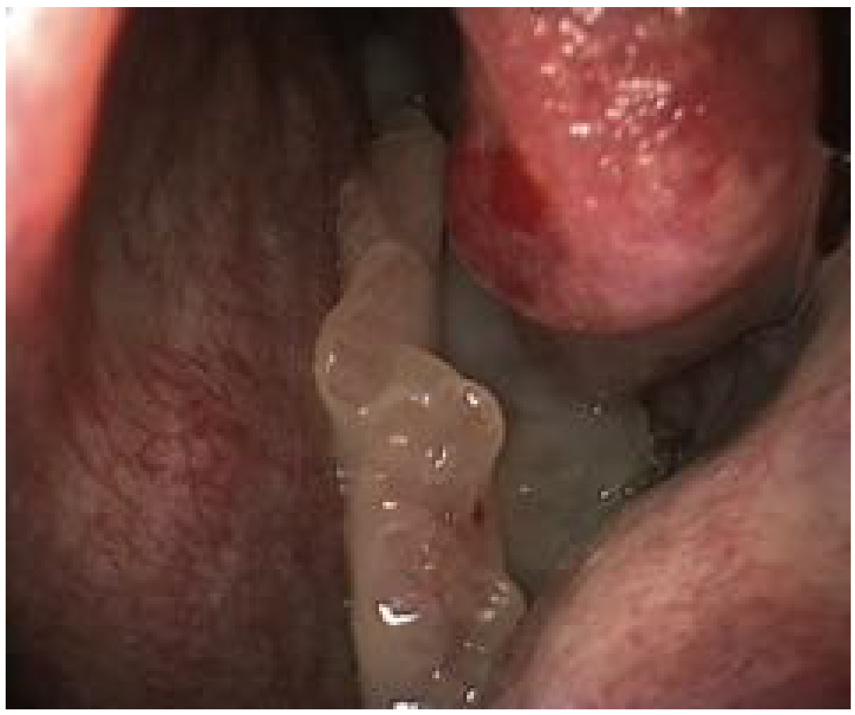
Fig. 50.1 - Typical endoscopic appearance: polypoid lesion with pale, papillary surface protruding from the middle meatus (Cummings Otolaryngology)
Etiology / Risk Factors
- Organic solvent exposure - significantly related, with dose-response relationship
- Smoking - risk factor for recurrence and malignant transformation (12x higher risk of malignancy in smokers; 26.4% vs 2.8% malignant transformation rate)
- HPV - role controversial; HPV DNA found in ~13-26% but transcriptional activity unclear; high-risk subtypes (16, 18) found more in cases with dysplasia/carcinoma
- EGFR mutations - characteristic molecular feature, preserved even in malignant transformation
Clinical Features
Symptoms:
- Unilateral nasal obstruction with watery rhinorrhea (most common presenting symptom)
- Unilateral rhinosinusitis (headache, facial pain) due to mechanical obstruction
- Epistaxis
- Anosmia (with extensive involvement)
Key clinical point: Any unilateral sinonasal mass should have inverted papilloma in the differential diagnosis.
Imaging
- CT - primary modality; shows a soft-tissue mass with bony remodeling; focal hyperostosis is a common finding and helps identify the site of tumor origin
- MRI - complementary; superior to CT in:
- Distinguishing mucus from papilloma (especially in the frontal sinus)
- Demonstrating the characteristic convoluted cerebriform pattern on T2/contrast-enhanced T1 - highly suggestive of IP
- Combined CT + MRI for staging and surgical planning
Malignant Transformation
- Rate: 5-15% (synchronous occurrence more common than metachronous)
- Smoking confers 12x higher risk
- Tumor types arising from IP:
- Squamous cell carcinoma (vast majority)
- Sinonasal undifferentiated carcinoma
- Mucoepidermoid carcinoma
- Verrucous carcinoma
Staging (Krouse Classification - most widely used)
| Stage | Description |
|---|---|
| T1 | Confined to nasal cavity, no extension to sinuses |
| T2 | Involves ostiomeatal complex, ethmoids, and/or medial part of maxillary sinus |
| T3 | Involves walls of maxillary sinus, frontal sinus, and/or sphenoid sinus |
| T4 | Involves extrasinus structures (orbit, intracranial, pterygoid plates) OR carcinoma |
Treatment
Surgical excision with negative margins is the treatment of choice. The key principle is removal of bone at the base to prevent recurrence.
- Endoscopic endonasal approach - preferred when possible (avoids facial scar from lateral rhinotomy)
- Open approach (lateral rhinotomy, medial maxillectomy) - for advanced tumors (T3/T4), frontal sinus disease, or when endoscopic access is limited
- For malignant transformation: surgery + adjuvant radiotherapy
Recurrence:
- Low with complete resection
- Increased with incomplete removal, smokers, and multi-site disease
- HPV low-risk types (especially HPV 6) promote recurrence
Key Summary Points
- Ringertz tumor = Inverted papilloma = most common Schneiderian papilloma
- Arises from the Schneiderian membrane (ectoderm-derived, unique to sinonasal tract)
- Triad: local aggressiveness + recurrence + malignant potential (10-15%)
- Most common site: lateral nasal wall and maxillary sinus
- Classic presentation: unilateral nasal obstruction in a middle-aged male
- CT shows soft-tissue mass with bony remodeling; MRI shows cerebriform pattern
- Treatment: endoscopic excision with bone removal at the base
- Smoking is the strongest modifiable risk factor for malignant transformation
Sources: Cummings Otolaryngology Head and Neck Surgery, Scott-Brown's Otorhinolaryngology Head & Neck Surgery, Sabiston Textbook of Surgery
This is a shared conversation. Sign in to Orris to start your own chat.