
Hi
Hi there! How can I help you today?
I need notes of General anatomy of ear with diagram Explain in simple language
Reading File
Finding Sources
Generating Image
Detailed labeled anatomical diagram of the human ear showing all three parts: outer ear (pinna/auricle, external auditory canal, cerumen glands), middle ear (tympanic membrane/eardrum, malleus, incus, stapes ossicles, Eustachian tube, oval window, round window), and inner ear (cochlea, semicircular canals, vestibule, auditory nerve/vestibulocochlear nerve). Clean educational style with clear labels and arrows, white background.
Reading File
Reading File
Finding Sources
Finding Sources
Reading File
Reading File
Finding Sources
Loading Image
Reading File
Loading Image
Here are your complete, easy-to-understand notes on the General Anatomy of the Ear, with diagrams from medical textbooks.
🦻 General Anatomy of the Ear
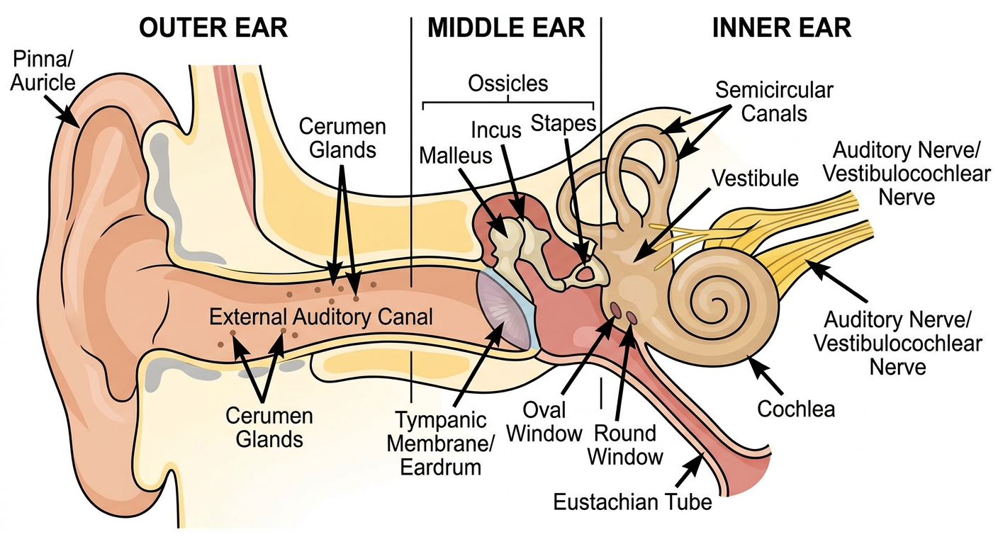
The ear is divided into 3 main parts:
- Outer Ear (External Ear)
- Middle Ear
- Inner Ear
OVERVIEW DIAGRAM
PART 1 - OUTER EAR (External Ear)
The outer ear is the part you can see and feel. Its job is to collect sound waves from the environment and send them inward.
1a. Auricle (Pinna)
The auricle (also called the pinna) is the visible flap of the ear on the side of your head.
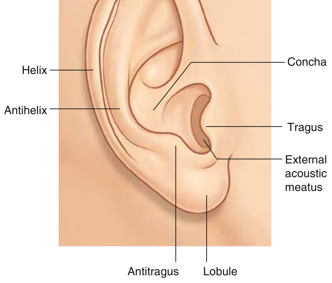
Gray's Anatomy for Students
Parts of the Auricle:
| Part | What it is (Simple Explanation) |
|---|---|
| Helix | The large outer curved rim of the ear |
| Antihelix | A smaller curved ridge just inside the helix |
| Tragus | Small bump in front of the ear canal opening |
| Antitragus | Small bump opposite the tragus, above the earlobe |
| Concha | The hollow bowl-shaped center of the auricle |
| Lobule | The soft fleshy earlobe - the ONLY part without cartilage |
| External Acoustic Meatus | The opening of the ear canal |
Key fact: The auricle is made of elastic cartilage covered by skin. The earlobe (lobule) has NO cartilage - that's why it's soft and flexible.
Function: Acts like a funnel - captures sound waves and directs them into the ear canal. It also helps with sound localization (figuring out where a sound is coming from).
1b. External Auditory Canal (External Acoustic Meatus)
This is the tube/passage that carries sound from the pinna to the eardrum.
- Length: about 2.5 cm long
- Lined with skin (stratified squamous epithelium)
- Contains hair follicles and special glands:
- Ceruminous glands - produce ear wax (cerumen)
- Sebaceous glands - produce oily secretions
- Outer 1/3 is supported by elastic cartilage
- Inner 2/3 is enclosed within the temporal bone
Ear wax (Cerumen): A protective, waxy substance with antimicrobial properties. It keeps the ear canal moist and traps dust and foreign particles.
PART 2 - MIDDLE EAR
The middle ear is an air-filled cavity inside the temporal bone. It sits between the eardrum and the inner ear.
Main job: Transfer sound vibrations from the eardrum to the inner ear, and amplify them.
2a. Tympanic Membrane (Eardrum)
- A thin, slightly transparent membrane at the end of the ear canal
- Acts like the skin of a drum - vibrates when sound hits it
- Made of 3 layers:
- Outer layer - skin (epidermis)
- Middle layer - fibrous connective tissue
- Inner layer - mucous membrane
2b. Ossicles (The 3 Tiny Bones)
These are the 3 smallest bones in the entire human body, forming a chain that carries vibrations from the eardrum to the inner ear.
| Bone | Nickname | Position |
|---|---|---|
| Malleus | Hammer | Attached to the eardrum |
| Incus | Anvil | In the middle of the chain |
| Stapes | Stirrup | Connects to the oval window of inner ear |
How it works: Eardrum vibrates → Malleus moves → Incus moves → Stapes pushes on oval window → Sound enters inner ear.
Important: The tympanic membrane is about 20x larger than the oval window. This size difference (plus the lever action of the ossicles) amplifies sound pressure, allowing vibrations to travel from air into the fluid-filled inner ear without losing energy.
2c. Eustachian Tube (Auditory Tube)
- A tube connecting the middle ear to the throat (nasopharynx)
- Normally collapsed, opens when you swallow or yawn
- Function: Equalizes air pressure on both sides of the eardrum
- This is why your ears "pop" on an airplane or when you yawn
- Can also be a pathway for infections to travel from the throat to the ear (causing otitis media - middle ear infection)
2d. Oval Window and Round Window
- Oval window: The opening where the stapes bone connects - transmits vibrations INTO the inner ear
- Round window: Acts as a pressure release valve for the inner ear fluid
PART 3 - INNER EAR (Labyrinth)
The inner ear is a complex system of fluid-filled spaces inside the temporal bone. It has two functions:
- Hearing (via the cochlea)
- Balance (via the vestibular system)
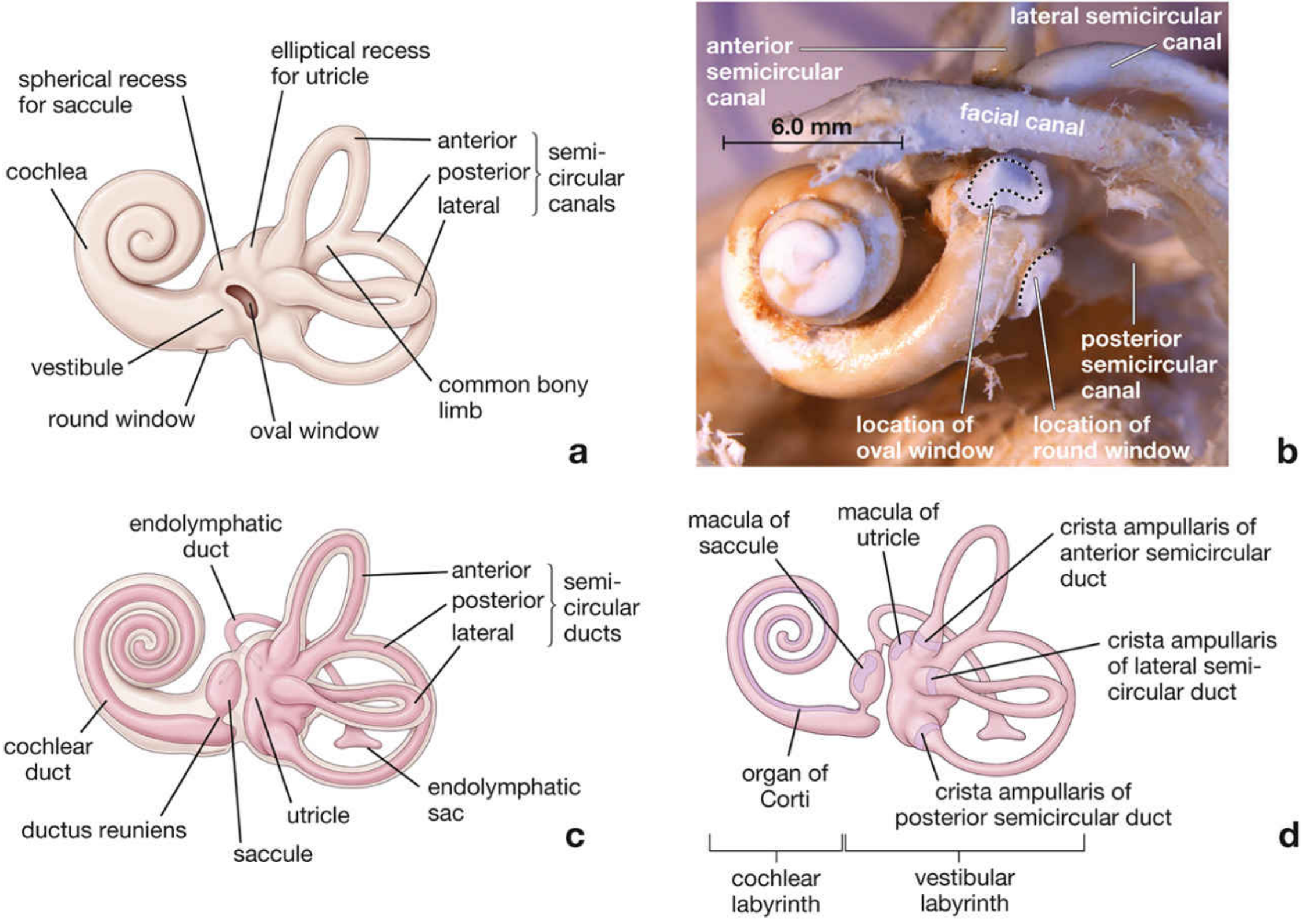
Histology: A Text and Atlas
3a. Cochlea (Hearing Organ)
- Looks like a snail shell - a coiled, fluid-filled tube
- Contains the Organ of Corti - the actual sensory receptor for hearing
- When the stapes pushes on the oval window, fluid waves travel through the cochlea and stimulate tiny hair cells in the Organ of Corti
- These hair cells convert mechanical vibrations into electrical nerve signals
3b. Vestibular System (Balance Organ)
Made up of:
| Structure | Function |
|---|---|
| Semicircular Canals (3 canals - anterior, posterior, lateral) | Detect rotational movement (turning your head) |
| Utricle | Detects horizontal linear movement and gravity |
| Saccule | Detects vertical linear movement and gravity |
The 3 semicircular canals are at right angles to each other - this allows detection of movement in all 3 planes of space.
3c. The Two Labyrinths
| Type | Description |
|---|---|
| Bony Labyrinth | The hard bony outer shell containing the structures |
| Membranous Labyrinth | Soft membrane tubes sitting inside the bony labyrinth |
- Fluid between the bony and membranous labyrinth = Perilymph (similar to cerebrospinal fluid)
- Fluid inside the membranous labyrinth = Endolymph
3d. Vestibulocochlear Nerve (Cranial Nerve VIII)
- The main nerve of the inner ear
- Has two branches:
- Cochlear nerve - carries hearing signals to the brain
- Vestibular nerve - carries balance signals to the brain
QUICK SUMMARY TABLE
| Part | Structures | Main Function |
|---|---|---|
| Outer Ear | Pinna, External auditory canal | Collects and channels sound |
| Middle Ear | Tympanic membrane, Malleus, Incus, Stapes, Eustachian tube | Amplifies and transmits vibrations |
| Inner Ear | Cochlea, Semicircular canals, Vestibule, CN VIII | Converts vibrations to nerve signals; Balance |
SOUND PATHWAY (Step by Step)
Sound waves
    ↓
Pinna (auricle) - collects sound
    ↓
External auditory canal
    ↓
Tympanic membrane (eardrum) - vibrates
    ↓
Ossicles: Malleus → Incus → Stapes
    ↓
Oval window
    ↓
Cochlea (fluid waves) → Organ of Corti (hair cells)
    ↓
Cochlear nerve (CN VIII)
    ↓
Brain (auditory cortex) - we HEAR!
Sources: Gray's Anatomy for Students; Junqueira's Basic Histology (17e); Medical Physiology (Boron & Boulpaep); Cummings Otolaryngology Head and Neck Surgery
Hi.
Hi! How can I help you?
This is a shared conversation. Sign in to Orris to start your own chat.