
How to differentiate between Polymyositis with aki and statin induced Rhabdomylisis with aki
Reading File
This is a complex clinical scenario. Let me search the medical library in parallel for all relevant topics.
Quick Answer: Differentiating polymyositis (PM) from statin-induced rhabdomyolysis (SIR) - both presenting with AKI - relies on: history of statin use (SIR), degree of CK elevation (extreme in rhabdomyolysis), pattern of muscle involvement, presence of autoantibodies (PM), muscle biopsy (inflammatory infiltrate in PM vs. necrosis without inflammation in SIR), and the time course of resolution after statin withdrawal.
Searching sources...
Finding Sources
Finding Sources
Searching PubMed
polymyositis rhabdomyolysis differentiation statin
Good hits. Now let me read the key sections on polymyositis and rhabdomyolysis in depth.
Reading File
Reading File
Finding Sources
Finding Sources
Reading File
Reading File
Reading File
Excellent content. Now let me get more on statin myopathy/IMNM (the immune-mediated necrotizing myopathy linked to statins), which is a critical differentiator, and the rhabdomyolysis diagnosis section.
Finding Sources
Reading File
Finding Sources
Reading File
Reading File
I now have excellent source material. Let me also fetch the muscle pathology image that was referenced, which shows PM biopsy vs other myopathies.
Loading Image
Finding Sources
I now have comprehensive information to build a thorough, well-structured answer. Let me compile it.
Differentiating Polymyositis (PM) vs. Statin-Induced Rhabdomyolysis (SIR) - Both with AKI
This is one of the more challenging clinical differentials in nephrology and rheumatology. The table below summarizes the key distinguishing features, followed by detailed discussion.
1. History and Onset
| Feature | Polymyositis | Statin-Induced Rhabdomyolysis |
|---|---|---|
| Drug history | No statin exposure needed | Statin use (current or recent) - essential |
| Onset | Subacute - weeks to months of progressive weakness | Acute to subacute; often precipitated by a trigger (new statin, dose increase, drug interaction, e.g., statin + cyclosporine + CYP3A4 inhibitor) |
| Duration before presentation | Months of insidious proximal weakness | Days to weeks of muscle pain/weakness |
| Preceding events | None specific | Strenuous exercise, infection, dehydration, or new interacting drug may precipitate it |
| Course after drug withdrawal | Does NOT improve with statin withdrawal alone (autoimmune process) | Typically improves after stopping statin (unless IMNM - see below) |
Key point: A subset of statin-exposed patients develop Immune-Mediated Necrotizing Myopathy (IMNM) - this is a true autoimmune condition triggered by statins that does NOT resolve on drug withdrawal and requires immunosuppression, closely mimicking polymyositis. - Goldman-Cecil Medicine, p. 2845
2. Clinical Features
| Feature | Polymyositis | Statin-Induced Rhabdomyolysis |
|---|---|---|
| Muscle pain | Often mild aching; severe pain actually argues against PM | Prominent myalgia, often severe; cramps, tenderness |
| Weakness pattern | Bilateral proximal (shoulder and hip girdle); insidious | Proximal weakness; may be more acute |
| Skin rash | Absent (if present → dermatomyositis) | Absent |
| Systemic features | Arthritis, ILD (interstitial lung disease), fever, Raynaud's phenomenon | Generally absent unless severe NMS or sepsis |
| Dark urine | Rare unless severe flare | Classic - red-brown/cola-colored urine (myoglobinuria) |
| Dysphagia/dyspnea | May occur (esophageal involvement, ILD) | Not typical |
| Malignancy association | Present (evaluate for occult cancer) | No |
3. Laboratory Features
| Parameter | Polymyositis | Statin-Induced Rhabdomyolysis |
|---|---|---|
| CK level | Elevated but often <10x ULN (though can reach very high in severe cases) | Massively elevated - often >10,000-100,000 U/L; AKI risk significant at >15,000-20,000 U/L |
| Degree of CK for AKI | AKI is less common in PM; if it occurs, usually due to severe active disease | AKI strongly associated with CK >15,000-20,000 U/L (pigment nephropathy) |
| Myoglobin (serum/urine) | Low/absent unless rhabdo-level flare | Elevated - urine dipstick heme-positive with few or no RBCs on microscopy (key diagnostic clue) |
| Urine sediment | May show mild proteinuria; no pigmented casts | Red-brown pigmented granular casts; heme-positive dipstick with absent RBCs on microscopy |
| FENa | Variable | Typically <1% early in disease course |
| LDH, AST, ALT | Elevated (muscle enzymes) | Elevated |
| Hyperkalemia | Less prominent | Prominent - from muscle necrosis |
| Hyperphosphatemia | Less prominent | Prominent |
| Hypocalcemia | Not typical | Occurs early due to Ca²⁺-phosphate complexing in necrotic tissue |
| Hyperuricemia | Not typical | Common |
| Metabolic acidosis | Mild | Often significant |
From the NKF Primer on Kidney Diseases: "The presence of myoglobin in the urine may make it appear reddish-brown. Urine dipstick shows significant heme protein positivity with few or no red blood cells on urine sediment - because the dipstick peroxidase reacts with both hemoglobin and myoglobin." - NKF Primer, p. 361
4. Autoantibodies (Critical Differentiator)
| Antibody | Significance |
|---|---|
| Anti-Jo-1 (anti-aminoacyl-tRNA synthetase) | Positive in polymyositis/antisynthetase syndrome - argues strongly FOR PM |
| Anti-Mi-2 | Dermatomyositis |
| Anti-MDA5 | Dermatomyositis with ILD |
| Anti-HMGR (anti-3-hydroxy-3-methylglutaryl-CoA reductase) | Positive in statin-associated IMNM - the statin drug target itself triggers autoimmunity |
| Anti-SRP (signal recognition particle) | IMNM subtype (may be paraneoplastic) |
| All antibodies negative | More consistent with simple toxic rhabdomyolysis |
Goldman-Cecil Medicine explains: anti-HMGR autoantibodies develop because statin-induced upregulation of HMGR in muscle is directly toxic and triggers an immune reaction against it. - Goldman-Cecil Medicine, p. 2841
5. Muscle Biopsy (Gold Standard)
This is the definitive differentiator:
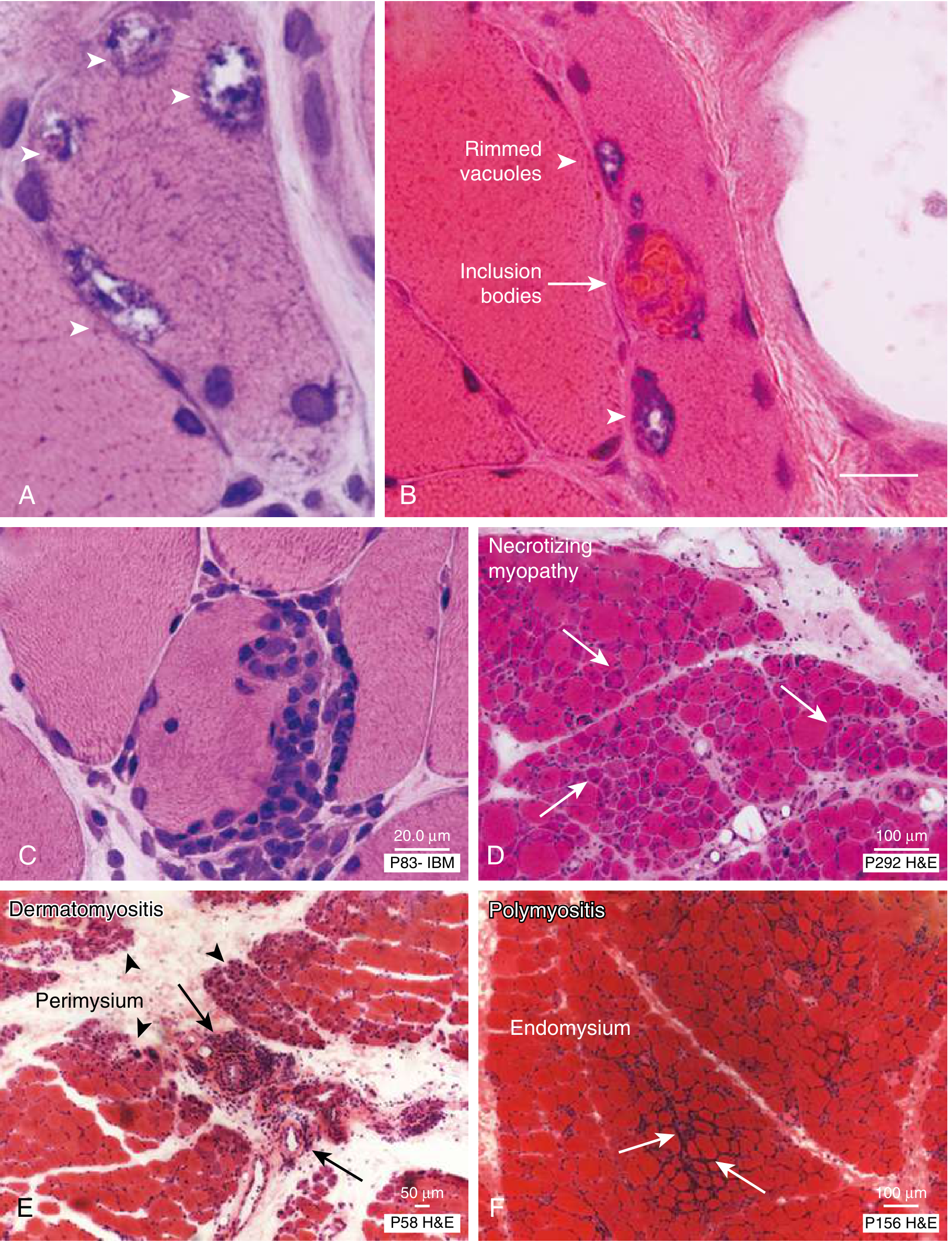
| Condition | Biopsy Finding |
|---|---|
| Polymyositis | Endomysial inflammation - CD8+ T cells invading non-necrotic muscle fibers (panel F above) |
| Statin toxic rhabdomyolysis | Widespread coagulative necrosis of muscle fibers, loss of architecture, no significant inflammatory infiltrate |
| Statin-associated IMNM (anti-HMGR) | Scattered necrotic and regenerating myofibers with macrophage invasion but minimal lymphocytic infiltrate (panel D above) - this is the key histological difference from PM |
| Dermatomyositis | Perimysial/perivascular inflammation with perifascicular atrophy (panel E) |
The core criteria for polymyositis diagnosis are: subacute proximal weakness + elevated CK + muscle biopsy showing endomysial inflammation without features of another diagnosis. - Goldman-Cecil Medicine, p. 2845
6. The Critical Overlap: Statin-Associated IMNM vs. Polymyositis
This is the hardest differentiation:
| Feature | Statin-IMNM | Polymyositis |
|---|---|---|
| Statin exposure | Yes (often for months/years) | Not required |
| Response to statin withdrawal | Does NOT improve (autoimmune) | Not applicable |
| Autoantibody | Anti-HMGR or anti-SRP positive | Anti-Jo-1, anti-PL-7, anti-PL-12 |
| Biopsy | Necrosis + regeneration, macrophages, minimal T-cell infiltrate | Endomysial CD8+ T-cell infiltrate |
| ILD | Less common | More common (antisynthetase) |
| Treatment | Requires immunosuppression (steroids ± steroid-sparing agents) | Same |
7. Response to Treatment
| Polymyositis | Statin-Toxic Rhabdomyolysis | Statin-IMNM | |
|---|---|---|---|
| Stop statin | No effect | CK falls, improves | Does NOT improve |
| Steroids | Responds | Not needed | Required |
| IV fluids | Supportive | First-line for AKI | Supportive |
| Expected CK trend | Slow decline with immunosuppression | Rapid decline within days of stopping statin | Persists until immunosuppression |
8. AKI Mechanism in Each
-
Statin-toxic rhabdomyolysis: Pigment nephropathy - myoglobin precipitates with Tamm-Horsfall protein in acidic urine forming obstructive tubular casts + direct myoglobin-mediated tubular toxicity (lipid peroxidation, oxidative injury) + renal vasoconstriction from volume depletion. AKI risk becomes significant at CK >15,000-20,000 U/L. - NKF Primer, p. 361
-
Polymyositis with AKI: Less commonly causes direct pigment nephropathy. AKI in PM more often relates to concurrent medications (NSAIDs), volume depletion, or systemic inflammation. If PM is severe enough to cause a rhabdomyolysis-level CK, pigment nephropathy can occur.
Summary Diagnostic Algorithm
Patient: Muscle weakness + elevated CK + AKI
            ↓
Is CK massively elevated (>10,000-15,000 U/L)?
  → YES: Rhabdomyolysis likely
            ↓
Is there dark urine + heme-positive dipstick with no RBCs?
  → YES: Myoglobinuria confirmed = rhabdomyolysis
            ↓
Is there current/recent statin use?
  → YES: Statin-induced rhabdomyolysis
  → Did CK normalize after stopping statin within days?
      YES → Toxic/direct mechanism (no biopsy needed if straightforward)
      NO after 4-6 weeks → Consider IMNM; send anti-HMGR, anti-SRP; biopsy
            ↓
Is CK moderately elevated + subacute proximal weakness over months?
Absence of myoglobinuria? Presence of ILD, arthritis, Raynaud's?
  → Consider Polymyositis
  → Send anti-Jo-1, anti-PL panel
  → Muscle biopsy → endomysial inflammation = PM confirmed
References
- Goldman-Cecil Medicine, International Edition - Chapter 248, pp. 2841-2845
- NKF Primer on Kidney Diseases, 8th Edition - Chapter 32, pp. 360-362
- Comprehensive Clinical Nephrology, 7th Edition - Chapter 77
Which muscle is preferred for biopsy site ?
Quick Answer: For inflammatory myopathies like polymyositis, the preferred biopsy site is a clinically or radiologically affected proximal muscle - most commonly the vastus lateralis (quadriceps) or deltoid. The key principle is to biopsy a muscle that is weak but not end-stage (avoid severely atrophied muscles and previously injected/biopsied sites).
Searching sources...
Finding Sources
Finding Sources
Finding Sources
Finding Sources
Searching the Web
preferred muscle biopsy site polymyositis inflammatory myopathy vastus lateralis deltoid
Reading File
Finding Sources
Reading File
I now have excellent material. Here is the comprehensive answer:
Preferred Muscle Biopsy Site in Inflammatory Myopathy (e.g., Polymyositis)
The Golden Rule
Biopsy a muscle that is clinically weak but not end-stage - specifically MRC grade 4 (weak but still functional). Muscles with full power (MRC 5) may not yet show pathological changes, while muscles that are too weak (MRC 1-2) show only non-specific end-stage fibrosis and fibrofatty replacement that masks the diagnostic histology. - Practical Neurology (Neuromuscular Notes)
Preferred Sites
| Preference | Muscle | Why Preferred |
|---|---|---|
| 1st choice | Vastus lateralis (quadriceps) | Large, accessible proximal muscle; consistently involved in PM/DM; easy open or needle biopsy; low complication risk |
| 2nd choice | Biceps brachii | Proximal upper limb; commonly affected in PM; good yield; used when quadriceps is too weak or already biopsied |
| 3rd choice | Deltoid | Accessible proximal muscle; reasonable yield; often used in clinical practice |
| Alternative | Triceps / Gastrocnemius | Used when the above are not suitable |
Clinical practice from FMUSP São Paulo: "Muscle biopsies were obtained from the vastus lateralis or brachial biceps, taking into consideration the clinical impairment (paretic, but not plegic limb)." - Brazilian Journal of Pathology and Laboratory Medicine
Key Principles for Site Selection
1. Choose the contralateral side from EMG
The EMG needle causes local inflammation and artifactual fiber injury that can mimic or obscure the true pathology. Always biopsy the contralateral limb to where EMG was performed.
"The EMG is also helpful in choosing a muscle for biopsy sampling, but care must be taken not to obtain tissue from precisely the same site as a recent EMG." - Adams and Victor's Principles of Neurology, p. 2808
2. Use MRI to guide selection
MRI (STIR/T2 sequences) identifies muscles with active inflammation (edema/increased signal) vs. those with fibrofatty replacement. Target muscles with MRI signal change, not those that are dark (fatty replaced) or entirely normal.
"MRI can refine the distribution of lesions and aid in targeting the muscle biopsy." - Adams and Victor's Principles of Neurology, p. 2812
3. Avoid previously biopsied or injected sites
Scar tissue and prior needle tracks distort the architecture and lead to false results.
4. Take multiple samples / multiple sections
Inflammatory infiltrates in PM are patchy and scattered - a single biopsy can be falsely negative if it misses the focal lesion.
"Because of the scattered distribution of inflammatory lesions, only part of the pathologic changes may be seen in any single specimen. More than one site of biopsy or multiple samples through one incision is advisable." - Adams and Victor's Principles of Neurology, p. 2817
5. Avoid end-stage atrophied muscle
A muscle that is severely wasted shows only fibrosis, fiber loss, and fatty replacement - non-specific changes that cannot confirm PM or any specific myopathy.
Summary
Clinical weakness in proximal muscle (MRC grade 4)?
         ↓
EMG done? → Biopsy CONTRALATERAL limb
         ↓
MRI STIR shows active edema? → Target that muscle
         ↓
Default sites:
  Lower limb → Vastus lateralis (1st choice)
  Upper limb → Biceps brachii (2nd choice)
         ↓
Open biopsy (preferred for larger sample) or
Needle/Bergstrom biopsy (less invasive, good for repeat)
         ↓
Take multiple samples / serial sections to avoid false negatives
Why Vastus Lateralis is the Most Common Choice
- Large muscle bulk - yields adequate tissue
- Proximal location - reliably involved in PM/DM
- Superficial - easily accessible
- Tolerates open biopsy well with minimal morbidity
- Allows multiple samples through a single incision
Sources:
- Adams and Victor's Principles of Neurology, 12th Ed., pp. 2808, 2812, 2817
- Practical Neurology - Neuromuscular Notes: When to Order a Muscle Biopsy
- Brazilian Journal of Pathology and Laboratory Medicine - DM/PM biopsy site study
This is a shared conversation. Sign in to Orris to start your own chat.