I now have sufficient material from the textbooks. Let me compile the comprehensive answers:---
Comprehensive Answers
QUESTION 1
(a) Draw and Label Different Parts of a Nephron
The nephron is the functional unit of the kidney, composed of the renal corpuscle (glomerulus + Bowman's capsule) and the renal tubules.
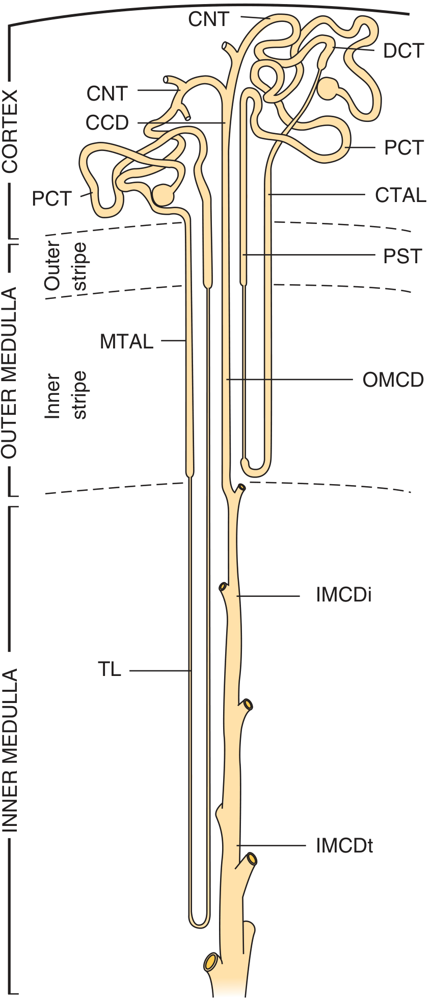
Diagram showing superficial and juxtamedullary nephron with labeled segments — Brenner and Rector's The Kidney
Labeled parts of the nephron:
RENAL CORPUSCLE
 ├── Glomerulus (capillary tuft)
 └── Bowman's capsule (parietal + visceral layer with podocytes)

RENAL TUBULE
 ├── Proximal Convoluted Tubule (PCT)
 ├── Proximal Straight Tubule (PST / pars recta)
 │
 ├── Loop of Henle
 │    ├── Descending thin limb
 │    ├── Thin ascending limb (long-loop nephrons)
 │    └── Thick Ascending Limb (TAL / pars recta of distal tubule)
 │
 ├── Distal Convoluted Tubule (DCT)
 └── Connecting Tubule (CNT) → leads to Collecting Duct system
Collecting Duct System (embryologically distinct from nephron, derived from ureteric bud):
- Initial Collecting Tubule (ICT)
- Cortical Collecting Duct (CCD)
- Outer Medullary Collecting Duct (OMCD)
- Inner Medullary Collecting Duct (IMCD)
Types of nephrons:
| Feature | Superficial/Cortical | Juxtamedullary |
|---|
| Glomerulus position | Outer cortex | Near cortico-medullary junction |
| Loop of Henle | Short (doesn't reach inner medulla) | Long (extends deep into inner medulla) |
| Role | Filtration | Urine concentration (countercurrent) |
— Brenner and Rector's The Kidney, p. 83–84
(b) Peculiarities of Renal Circulation and Their Significance
| Peculiarity | Significance |
|---|
| Two capillary beds in series — glomerular capillaries and peritubular capillaries, separated by the efferent arteriole | Allows independent regulation of filtration and reabsorption |
| High glomerular capillary pressure (~60 mmHg) — maintained by the efferent arteriole's high resistance | Drives ultrafiltration; provides the hydrostatic force for GFR |
| Low peritubular capillary pressure (~13 mmHg) — pressure drops across the efferent arteriole | Favors reabsorption of fluid from interstitium back into blood |
| Afferent and efferent arterioles — both have smooth muscle under neural/hormonal control | Allow precise control of glomerular pressure and GFR independently of systemic blood pressure (autoregulation) |
| Vasa recta — long straight vessels running parallel to loop of Henle in medulla, arranged in countercurrent fashion | Maintain the medullary osmotic gradient (hyperosmotic interstitium) essential for urine concentration; prevent washout of solutes |
| Cortical vs. medullary blood flow — cortex receives ~90% of renal blood flow; medulla receives ~10% | Cortex: high flow for filtration. Medulla: slow flow in vasa recta preserves concentration gradient |
| Portal-like arrangement — renal artery → afferent arteriole → glomerular capillary → efferent arteriole → peritubular capillary/vasa recta → renal vein | Allows the kidney to fine-tune both filtration AND reabsorption via the same blood supply |
| Myogenic autoregulation + tubuloglomerular feedback (TGF) — maintains RBF and GFR relatively constant over MAP 80–180 mmHg | Protects glomeruli from pressure swings; prevents massive salt/water loss during hypertension |
QUESTION 2
(a) Components of the Juxtaglomerular Apparatus (JGA) and Its Functions
The JGA is located at the vascular pole of the glomerulus, where the thick ascending limb (TAL) of the loop of Henle returns to touch its parent glomerulus.
Components:
-
Macula Densa — a plaque of specialized epithelial cells in the wall of the TAL. These cells have large, closely packed nuclei (hence the name), prominent lateral intercellular spaces, and are rich in neuronal nitric oxide synthase (nNOS) and cyclooxygenase-2 (COX-2). They act as sensors of luminal NaCl concentration.
-
Juxtaglomerular (JG) Granular Cells — modified smooth muscle cells (myoepithelial / epithelioid cells) in the wall of the afferent arteriole (and less commonly efferent arteriole). They contain membrane-bound electron-dense granules packed with renin (an aspartyl protease). These also express angiotensin II locally.
-
Extraglomerular Mesangium (Lacis / Polkissen) — a solid cluster of heavily branched cells and matrix located between the macula densa, afferent and efferent arterioles, and the glomerular tuft. Its cells are connected by gap junctions and by microfilaments. It serves as the structural relay between the tubular (macula densa) and vascular components.
-
Initial portion of the Efferent Arteriole (sometimes included).
— Brenner and Rector's The Kidney, p. 94; Comprehensive Clinical Nephrology, p. 31
Functions of the JGA:
| Function | Mechanism |
|---|
| Tubuloglomerular Feedback (TGF) | ↑ NaCl at macula densa → constriction of afferent arteriole → ↓ GFR (protects from excessive NaCl loss) |
| Renin secretion | JG granular cells release renin when: (a) perfusion pressure in afferent arteriole falls, (b) sympathetic stimulation (β₁), (c) macula densa senses ↓ NaCl delivery. Renin activates the RAAS → angiotensin II → aldosterone → Na⁺/water retention |
| Regulation of GFR and RBF | Through TGF and angiotensin II effects on afferent/efferent arterioles |
| Nitric oxide & prostaglandins from macula densa | Modulate vascular tone of afferent arteriole; counterbalance vasoconstrictive TGF signals |
(b) Autoregulation of GFR in Volume Depletion
Definition: Autoregulation is the intrinsic ability of the kidney to maintain a relatively constant GFR and renal blood flow (RBF) despite changes in arterial blood pressure (effective over MAP ~80–180 mmHg).
Two main mechanisms:
1. Myogenic Mechanism (intrinsic vascular mechanism):
- In volume depletion → ↓ arterial blood pressure → ↓ stretch in afferent arteriole wall
- Reduced stretch → relaxation of smooth muscle in afferent arteriole
- → afferent arteriole dilates → maintains glomerular capillary pressure → maintains GFR
2. Tubuloglomerular Feedback (TGF):
- In volume depletion → ↓ GFR → ↓ tubular flow → ↓ NaCl delivery to macula densa
- Macula densa senses ↓ NaCl → signals JG granular cells (via prostaglandins, adenosine, NO)
- → dilation of afferent arteriole (and/or constriction of efferent arteriole via angiotensin II)
- → ↑ glomerular capillary pressure → GFR restored toward normal
3. Angiotensin II (RAAS activation in volume depletion):
- ↓ volume → ↑ renin from JG cells → ↑ angiotensin II
- Angiotensin II preferentially constricts efferent arteriole more than afferent
- → maintains glomerular hydrostatic pressure → preserves GFR despite low perfusion
- This is critically important when GFR is pressure-dependent (e.g., heart failure, renal artery stenosis) — which is why ACE inhibitors can precipitate acute kidney injury in such states
Summary in volume depletion:
↓ BP / ↓ volume
    ↓
Myogenic: afferent arteriole dilates (↓ resistance)
TGF: ↓ NaCl at macula densa → afferent dilation + efferent constriction
RAAS: Ang II → efferent constriction → maintains glomerular pressure
    ↓
GFR maintained (autoregulated)
QUESTION 3
(a) Plasma Load and Tubular Transport Maximum (Tm)
Plasma Load (Filtered Load):
- The total amount of a substance that is filtered at the glomerulus per unit time.
- Formula: Plasma Load = GFR × Plasma concentration of substance
- Example: For glucose: Plasma load = 125 mL/min × 1 mg/mL = 125 mg/min
Tubular Transport Maximum (Tm):
- The maximum rate at which the renal tubules can reabsorb (or secrete) a substance per unit time.
- It reflects saturation of carrier proteins (transporters) on tubular epithelial cells.
- For glucose (TmG): ~375 mg/min (males), ~300 mg/min (females)
- When plasma load exceeds Tm → the excess cannot be reabsorbed → the substance appears in urine (e.g., glucosuria when blood glucose > ~180 mg/dL, the renal threshold)
- The renal threshold is the plasma concentration at which a substance first appears in urine; it corresponds approximately to the point when plasma load = Tm
- The "splay" in the titration curve of glucose is due to nephron heterogeneity — different nephrons have slightly different Tm values
| Term | Definition |
|---|
| Filtered load | GFR × Plasma [substance] |
| Tubular Tm | Maximum transport capacity of tubular carriers |
| Renal threshold | Plasma concentration at which substance first spills into urine |
(b) Reabsorption of Water (H₂O) from Renal Tubules
Water reabsorption is entirely passive and follows solute reabsorption osmotically. It occurs via aquaporins (AQPs) — water channels on tubular epithelial cells.
| Tubular Segment | % Water Reabsorbed | Mechanism | Regulation |
|---|
| Proximal Convoluted Tubule (PCT) | ~65–67% | Obligatory; follows Na⁺, glucose, amino acids, HCO₃⁻ reabsorption osmotically. AQP1 on apical and basolateral membranes. Isosmotic reabsorption. | Not hormonally regulated; always occurs |
| Descending thin limb of Loop of Henle | ~15% | Water moves out by osmosis into the increasingly hyperosmotic medullary interstitium. AQP1 present. | Driven by medullary gradient |
| Ascending thin limb & Thick Ascending Limb (TAL) | 0% | Impermeable to water. Active NaCl reabsorption here (NKCC2 cotransporter) — this is the "diluting segment." Creates free water and builds the medullary gradient. | — |
| Distal Convoluted Tubule (DCT) | ~5% | Limited water permeability. Some Na⁺-driven water reabsorption. AQP2 absent here. | — |
| Collecting Duct (cortical + medullary) | ~8–17% (variable) | ADH (vasopressin)-dependent. ADH binds V2 receptors → cAMP → inserts AQP2 into apical membrane of principal cells → water reabsorption down the osmotic gradient. AQP3 & AQP4 on basolateral side. | ADH (antidiuretic hormone / vasopressin) — key regulator |
Countercurrent Multiplication System (medullary gradient):
- The TAL actively pumps NaCl without water → creates a hyperosmotic medullary interstitium (up to ~1200 mOsm/kg at the papilla)
- Urea contributes ~50% of the inner medullary osmolality (recycled via inner medullary collecting duct)
- This gradient provides the osmotic driving force for water reabsorption in the collecting duct
ADH regulation:
- Secreted from posterior pituitary in response to ↑ plasma osmolality or ↓ blood volume
- In the absence of ADH → collecting duct is water-impermeable → dilute urine (diabetes insipidus)
- In the presence of ADH → AQP2 inserted → water reabsorbed → concentrated urine (up to 1200 mOsm/kg)
QUESTION 4
(a) Hormones Regulating Plasma Ca²⁺ and Their Role in Calcium Homeostasis
Normal plasma Ca²⁺: 8.5–10.5 mg/dL (total); ionized Ca²⁺ ~4.5–5.5 mg/dL
Three major hormones:
1. Parathyroid Hormone (PTH)
- Secreted by chief cells of parathyroid glands in response to ↓ ionized Ca²⁺ (sensed by CaSR)
- Chemical nature: Polypeptide, 84 amino acids
- Actions to raise Ca²⁺:
- Bone: Stimulates osteoclast activity → bone resorption → releases Ca²⁺ and PO₄³⁻ into blood
- Kidney: ↑ Ca²⁺ reabsorption in DCT; ↓ phosphate reabsorption (phosphaturia); activates 1α-hydroxylase → ↑ production of active vitamin D (calcitriol)
- Gut: Indirect effect via calcitriol → ↑ intestinal Ca²⁺ absorption
2. Calcitriol (1,25-dihydroxyvitamin D₃ / Active Vitamin D)
- Produced in proximal tubule of kidney (1α-hydroxylation of 25-OH-D, stimulated by PTH, hypophosphatemia)
- Actions to raise Ca²⁺:
- Gut: Principal action — stimulates synthesis of calcium-binding protein (calbindin) → ↑ intestinal absorption of Ca²⁺ and PO₄³⁻
- Bone: Enhances bone mineralization; also supports osteoclast activity (↑ Ca²⁺)
- Kidney: Mild ↑ Ca²⁺ and PO₄³⁻ reabsorption
3. Calcitonin
- Secreted by parafollicular C cells of thyroid gland in response to ↑ plasma Ca²⁺
- Chemical nature: Polypeptide, 32 amino acids
- Actions to lower Ca²⁺:
- Bone: Rapidly inhibits osteoclasts → ↓ bone resorption → lowers plasma Ca²⁺
- Kidney: ↑ urinary excretion of Ca²⁺ and PO₄³⁻
- Physiologic role in adults is minor (unlike PTH which is essential)
Summary table:
| Hormone | Source | Effect on Ca²⁺ | Main Site of Action |
|---|
| PTH | Parathyroid glands | ↑ | Bone, Kidney (→ Vitamin D activation), Gut (indirect) |
| Calcitriol (Vit D) | Kidney (skin + liver → kidney) | ↑ | Gut (primary), Bone, Kidney |
| Calcitonin | Thyroid C cells | ↓ | Bone, Kidney |
(b) Tetany and Features of Carpopedal Spasm
Tetany:
Tetany is a clinical syndrome of involuntary, sustained muscle contractions caused by hypocalcemia (or alkalosis, hypomagnesemia). Low ionized Ca²⁺ increases neuronal membrane permeability to Na⁺, lowering the action potential threshold → spontaneous repetitive firing of motor nerves and sensory nerves.
Causes of hypocalcemic tetany: hypoparathyroidism, vitamin D deficiency, malabsorption, alkalosis (↓ ionized Ca²⁺ without changing total Ca²⁺).
Features of Carpopedal Spasm (a manifestation of tetany):
Carpopedal spasm is the classic sign of tetany involving the hands and feet:
-
Hand (carpal spasm):
- "Obstetrician's hand" / "Main d'accoucheur" — the characteristic position
- Wrist flexion
- Metacarpophalangeal (MCP) joints flexed
- Interphalangeal (IP) joints extended
- Thumb adducted across the palm
- Fingers extended and adducted (pointed)
-
Foot (pedal spasm):
- Plantar flexion of the foot and toes (equinovarus posture)
- Inward turning of foot
Other signs of latent tetany:
- Chvostek's sign — tapping over the facial nerve (in front of ear) → ipsilateral facial muscle twitch
- Trousseau's sign — inflating BP cuff above systolic pressure for 3 minutes → carpopedal spasm (most reliable test for latent hypocalcemia)
- Erb's sign — increased neuronal excitability on galvanic stimulation
QUESTION 5 (OR)
(a) Ovulation — Definition and Mechanism
Definition:
Ovulation is the rupture of the mature (Graafian) follicle with release of the secondary oocyte (surrounded by the corona radiata) from the ovary into the peritoneal cavity and subsequently into the fallopian tube, occurring approximately on day 14 of a 28-day menstrual cycle.
Mechanism of Ovulation:
Step 1 — Preovulatory LH Surge:
- Rising estrogen from the mature follicle, after reaching a critical threshold (~200 pg/mL sustained for ~50 hours), switches from negative feedback to positive feedback on the hypothalamus-pituitary
- This triggers a massive LH surge (~6–10 fold increase) peaking ~16 hours before ovulation, accompanied by a smaller FSH surge (~2–3 fold)
- Without the LH surge, ovulation cannot occur
Step 2 — Follicular Changes:
LH acts on granulosa and theca cells → progesterone secretion begins → triggers:
- Proteolytic enzyme release from lysosomes of the follicular capsule (theca externa) → dissolution of the follicle wall → weakening and degeneration of the stigma (the small avascular area on the follicle surface)
- Neovascularization of follicle wall + secretion of prostaglandins (vasodilators) → plasma transudation into the follicle → rapid follicle swelling and increased intrafollicular pressure
Step 3 — Follicle Rupture:
- Combination of follicle swelling + stigma degeneration → rupture of the follicle at the stigma
- The secondary oocyte (in meiosis II) + zona pellucida + corona radiata are expelled
Step 4 — Corpus Luteum Formation:
- Remaining granulosa and theca interna cells undergo luteinization → form the corpus luteum
- Corpus luteum secretes progesterone (primarily) and estrogen for 14 days (if no pregnancy)
— Guyton and Hall Textbook of Medical Physiology
(b) Indicators of Ovulation
| Indicator | Change at Ovulation |
|---|
| Basal Body Temperature (BBT) | Rises 0.3–0.5°C at ovulation and remains elevated throughout the luteal phase (due to progesterone's thermogenic effect) |
| Mittelschmerz | Mid-cycle pelvic pain (due to follicle rupture and peritoneal irritation by follicular fluid) |
| Cervical mucus changes | Pre-ovulation: mucus becomes thin, clear, watery, stretchy (Spinnbarkeit) and shows ferning on microscopy; post-ovulation it becomes thick and viscous |
| LH surge (urine/blood) | Urinary LH surge detected by ovulation predictor kits ~24–36 hours before ovulation |
| Serum progesterone | ↑ in mid-luteal phase (day 21) confirms ovulation has occurred |
| Follicle tracking (USG) | Serial transvaginal ultrasound shows follicle growing to ~18–25 mm then disappearing after rupture |
| Endometrial biopsy | Secretory phase endometrium (days 15–28) confirms ovulation (progesterone effect) |
| Serum estrogen | Brief peak just before LH surge |
| Vaginal cytology | Cornification index (proportion of cornified cells) peaks at ovulation |
QUESTION 6
Clinical Scenario — 40-year-old Female with Weight Loss, Heat Intolerance, Sweating, Palpitations, Exophthalmos, Autoantibodies, RA history
(a) Most Likely Diagnosis
Graves' Disease (Autoimmune Hyperthyroidism)
Justification:
- Weight loss despite good appetite → hypercatabolic state (↑ thyroid hormones increase BMR)
- Heat intolerance + sweating → ↑ metabolic rate → excess heat production
- Palpitations, tachycardia (pulse 120/min) → thyroid hormone sensitizes heart to catecholamines (↑ β-adrenergic receptors)
- Exophthalmos (proptosis) — pathognomonic of Graves' disease (TSH receptor antibodies stimulate retro-orbital fibroblasts → glycosaminoglycan deposition + orbital fibrosis)
- Wide pulse pressure (150/50 mmHg) → ↑ stroke volume + ↓ peripheral resistance (vasodilation from heat)
- Autoantibody positive — TSI (Thyroid Stimulating Immunoglobulin) / TRAb (TSH receptor antibodies)
- History of Rheumatoid Arthritis — Graves' disease is also autoimmune; patients with one autoimmune condition have higher risk of others
(b) Reason for Heat Intolerance
Thyroid hormones (T₃ and T₄) increase the basal metabolic rate (BMR) by:
- ↑ Na⁺/K⁺-ATPase activity in virtually all tissues → increased ATP consumption → more heat generated
- ↑ mitochondrial oxidative phosphorylation — uncoupling effect → inefficient ATP production → excess heat release
- ↑ substrate cycling (e.g., glucose-fatty acid cycle) → futile metabolic cycles releasing heat
- ↑ cellular oxygen consumption — tissues burn more fuel
Result: body generates excess heat → patient feels uncomfortably warm (heat intolerance) → seeks cooler environments, prefers cold
(c) Reason for Sweating
The excessive heat generated by ↑ BMR (as above) must be dissipated to maintain core body temperature. The body's primary mechanism for heat dissipation is sweating (eccrine sweating):
- Hypothalamus detects rising core temperature → activates sympathetic cholinergic fibers to eccrine sweat glands → sweating
- Additionally, thyroid hormones increase adrenergic sensitivity → ↑ sympathetic nervous system activity → ↑ sweating even at lower core temperatures
- Peripheral vasodilation (cutaneous) also occurs to facilitate heat loss, contributing to the warm, moist skin characteristic of hyperthyroidism (in contrast to the cold, dry skin of hypothyroidism)
Note: The high pulse pressure (systolic 150, diastolic 50) reflects the high cardiac output state — ↑ stroke volume (from ↑ heart rate and contractility) with ↓ peripheral resistance (vasodilation for heat dissipation).
QUESTION 7
(a) Spermatogenesis — Definition and Location
Definition:
Spermatogenesis is the process by which diploid spermatogonia (2n) are transformed into haploid, mature spermatozoa (n) through a series of mitotic, meiotic, and morphological changes. One complete cycle takes approximately 64 days.
Location:
- Seminiferous tubules of the testes (along their full length)
- Specifically within the seminiferous epithelium, supported by Sertoli cells
- Leydig cells in the interstitial tissue provide testosterone (required for spermatogenesis)
- Post-testicular maturation occurs in the epididymis
— Costanzo Physiology, 7th Edition
(b) Steps of Spermatogenesis
Phase 1 — Mitotic Proliferation (Spermatocytogenesis):
- Type A spermatogonia (stem cells) divide mitotically → some replenish the stem cell pool, others become Type B spermatogonia
- Type B spermatogonia → mitosis → primary spermatocytes (diploid, 2n, 4C after DNA replication)
Phase 2 — Meiosis:
- Meiosis I (Reductive division): Primary spermatocyte (2n, 4C) → two secondary spermatocytes (n, 2C) — longest phase (~22 days)
- Meiosis II (Equatorial division): Secondary spermatocyte (n, 2C) → two spermatids (n, 1C) — haploid cells
Phase 3 — Spermiogenesis (morphological transformation):
Spermatids → mature spermatozoa through:
- Acrosome formation — Golgi apparatus forms the acrosome cap (contains hydrolytic enzymes: hyaluronidase, acrosin — needed to penetrate zona pellucida)
- Nuclear condensation — chromatin condenses; nucleus elongates and becomes the sperm head
- Flagellum formation — centrioles organize the axoneme (9+2 microtubule pattern) → midpiece + principal piece + end piece
- Mitochondrial sheath formation — mitochondria arrange helically around the midpiece (energy for motility)
- Loss of excess cytoplasm — residual body shed (phagocytosed by Sertoli cells)
Result: Mature spermatozoon with head (nucleus + acrosome), midpiece (mitochondrial sheath), and tail (flagellum)
— Costanzo Physiology, 7th Edition; Developing Human
(c) Hormones Regulating Spermatogenesis
| Hormone | Source | Role |
|---|
| GnRH (Gonadotropin-Releasing Hormone) | Hypothalamus (arcuate nucleus) — pulsatile secretion | Stimulates anterior pituitary to release FSH and LH |
| FSH (Follicle-Stimulating Hormone) | Anterior pituitary | Acts on Sertoli cells → ↑ androgen-binding protein (ABP) → concentrates testosterone in seminiferous tubules; supports spermatid development |
| LH (Luteinizing Hormone) | Anterior pituitary | Acts on Leydig cells → stimulates cholesterol desmolase → testosterone synthesis |
| Testosterone | Leydig cells | Essential for completion of meiosis and spermiogenesis; maintains seminiferous tubule function; paracrine effect within testis |
| Inhibin B | Sertoli cells | Negative feedback on FSH secretion (when spermatogenesis is proceeding normally) |
| Estrogen (local, from aromatization) | Sertoli cells (convert testosterone) | Required for normal spermatogenesis in small amounts |
Feedback loop:
- ↑ Testosterone → negative feedback on hypothalamus (↓ GnRH) and pituitary (↓ LH)
- ↑ Inhibin B → negative feedback on pituitary (↓ FSH specifically)
(d) Functions of Testosterone
During Fetal Development:
- Stimulates differentiation of Wolffian (mesonephric) ducts → internal male genitalia (epididymis, vas deferens, seminal vesicles)
- Converted to DHT by 5α-reductase → development of external male genitalia (penis, scrotum, prostate)
At Puberty (onset of masculinization):
- Growth and development of male reproductive organs
- Enlargement of the penis, scrotum, testes
- Growth of the prostate and seminal vesicles
- Development of male secondary sexual characteristics: facial, body, and pubic hair; voice deepening (laryngeal growth); male-pattern skin changes
- Pubertal growth spurt (anabolic effect), followed by epiphyseal closure
In the Adult Male:
- Spermatogenesis — essential for completion (acts on Sertoli cells; high local concentration required)
- Libido (sex drive) — acts on limbic system
- Anabolic effects:
- ↑ Protein synthesis → muscle hypertrophy
- ↑ Bone mineral density (↑ osteoblast activity)
- Stimulates erythropoiesis (↑ EPO + direct effect on bone marrow) → higher hematocrit in males
- Metabolic effects:
- Promotes nitrogen retention
- ↑ Basal metabolic rate
- Feedback: Negative feedback on hypothalamus and anterior pituitary to suppress GnRH, LH, FSH
QUESTION 8 — Pancreatic Hormones and Insulin
(a) Hormones of the Pancreas with Chemical Nature
| Hormone | Cell Type | Chemical Nature | % of Islet Cells |
|---|
| Insulin | Beta (β) cells | Polypeptide (51 amino acids; A-chain 21 aa + B-chain 30 aa, connected by disulfide bonds); derived from proinsulin | ~60% |
| Glucagon | Alpha (α) cells | Polypeptide (29 amino acids) | ~25% |
| Somatostatin | Delta (δ) cells | Polypeptide (14 amino acids; same as hypothalamic GHIH) | ~10% |
| Pancreatic Polypeptide (PP) | PP cells (F cells) | Polypeptide (36 amino acids) | Small number |
| Amylin (IAPP) | Beta (β) cells | Polypeptide (37 amino acids) — co-secreted with insulin | — |
— Guyton and Hall Textbook of Medical Physiology
(b) Mechanism of Action of Insulin
Receptor: Insulin binds to the insulin receptor — a transmembrane glycoprotein with intrinsic tyrosine kinase activity (receptor tyrosine kinase). It consists of 2 α subunits (extracellular, insulin-binding) and 2 β subunits (transmembrane + intracellular tyrosine kinase domain), linked by disulfide bonds (α₂β₂ tetramer).
Signal transduction cascade:
- Insulin binds α subunit → conformational change → autophosphorylation of β subunit (tyrosine residues) → activation of intrinsic tyrosine kinase
- Activated receptor phosphorylates Insulin Receptor Substrates (IRS-1, IRS-2) on tyrosine residues
- Phospho-IRS-1 recruits and activates PI3-kinase (PI3K) → generates PIP₃
- PIP₃ activates PDK1 (phosphoinositide-dependent kinase-1) → activates Akt (PKB)
- Akt activates downstream effectors:
- GLUT4 vesicle translocation to plasma membrane → ↑ glucose uptake in muscle and adipose
- Glycogen synthase kinase-3 (GSK-3) inactivation → ↑ glycogen synthesis
- mTOR activation → ↑ protein synthesis
- Inhibition of FOXO transcription factors → ↓ gluconeogenesis genes (PEPCK, G6Pase)
- MAP kinase pathway (Ras-Raf-MEK-ERK) also activated → mediates mitogenic/growth effects of insulin
Key result: GLUT4 transporter (the insulin-responsive glucose transporter) moves from intracellular vesicles to the cell membrane in muscle and adipose tissue → glucose entry increases up to 15-fold.
— Guyton and Hall Textbook of Medical Physiology
(c) Functions of Insulin
Carbohydrate Metabolism:
- ↑ Glucose uptake by muscle and adipose tissue (GLUT4 translocation)
- ↑ Glycogenesis (glycogen synthesis) in liver and muscle
- ↓ Glycogenolysis (inhibits glycogen phosphorylase)
- ↓ Gluconeogenesis in liver (inhibits PEPCK, G6Pase)
- Net effect: ↓ blood glucose
Fat (Lipid) Metabolism:
- ↑ Lipogenesis — converts excess glucose to fatty acids in liver; promotes triglyceride storage in adipose
- ↑ LPL (lipoprotein lipase) activity → ↑ uptake of circulating triglycerides by adipose
- ↓ Lipolysis (inhibits hormone-sensitive lipase) → ↓ free fatty acids in plasma
- ↓ Ketogenesis
Protein Metabolism:
- ↑ Amino acid uptake into cells
- ↑ Protein synthesis (ribosomal translation)
- ↓ Protein catabolism
- Net: anabolic; essential for growth (acts synergistically with GH)
Electrolyte Effects:
- Drives K⁺ into cells (↑ Na⁺/K⁺-ATPase) → used therapeutically in hyperkalemia
- ↑ Phosphate and Mg²⁺ cellular uptake
Other Effects:
- Inhibits glucagon secretion (paracrine)
- Hypothalamic effects: ↓ food intake (activates POMC neurons)
- Growth-promoting (mitogenic) effect via MAP kinase pathway
(d) Causes of Insulin Resistance
Insulin resistance = diminished biological response to a given concentration of insulin in target tissues (muscle, liver, adipose).
Physiological causes:
- Obesity (especially visceral/central adiposity) — most common; elevated FFAs and adipokines (TNF-α, IL-6) impair insulin signaling
- Pregnancy (especially 3rd trimester) — placental hormones (hPL, progesterone, cortisol)
- Puberty / adolescence — growth hormone excess
Hormonal / Endocrine causes:
- Excess counter-regulatory hormones:
- Growth hormone excess (acromegaly)
- Cortisol excess (Cushing syndrome / steroid therapy)
- Glucagon excess
- Catecholamine excess (pheochromocytoma)
- Thyroid hormone excess (hyperthyroidism)
- Polycystic Ovarian Syndrome (PCOS) — associated with hyperinsulinemia + insulin resistance
Genetic / Molecular causes:
- Mutations in insulin receptor gene (type A insulin resistance)
- Mutations in post-receptor signaling (IRS-1, PI3K, GLUT4)
- Lipodystrophy syndromes
Acquired / Metabolic causes:
- Elevated free fatty acids → inhibit PI3K pathway ("lipotoxicity")
- Inflammatory cytokines (TNF-α, IL-6) — activate serine kinases that phosphorylate IRS-1 at serine (inactivating) residues → impair insulin signaling
- Hyperinsulinemia itself → receptor downregulation
- Ectopic lipid deposition in liver and muscle
- Mitochondrial dysfunction
Drug-induced:
- Glucocorticoids
- Antipsychotics (especially atypicals: olanzapine, clozapine)
- Tacrolimus, cyclosporine
- Thiazide diuretics, niacin
Sources: Guyton and Hall Textbook of Medical Physiology; Costanzo Physiology, 7th Edition; Brenner and Rector's The Kidney; Comprehensive Clinical Nephrology, 7th EditionYou've run out of free credits. Sign up for a free account to continue using Orris.