
I have two terms which are orthodromic and antedromic. These are more related to ECG and cardiology. I want to know about them and everything around them and what does that mean. And I think it's related about some disease in cardiac system. I want to know that also. Give a detailed description about all of these terms.
Reading File
Finding Sources
Finding Sources
Finding Sources
Searching PubMed
"Wolff-Parkinson-White Syndrome"[MeSH Terms] AND "tachycardia"[MeSH Terms]
Searching the Web
orthodromic antidromic atrioventricular reentrant tachycardia ECG Wolff Parkinson White professional reference
Searching Images
ECG comparison orthodromic antidromic atrioventricular reentrant tachycardia Wolff Parkinson White
Reading File
Reading File
Reading File
Reading File
Loading Image
Reading Web Page
https://www.ncbi.nlm.nih.gov/sites/books/NBK539765
The short meaning
- Orthodromic means an electrical impulse travels in the usual, normal direction for that tissue.
- Antidromic means it travels in the opposite direction.
In cardiology, these words are most often used for types of a fast re-entry rhythm called atrioventricular re-entrant tachycardia (AVRT). This typically involves an extra electrical connection between atria and ventricles, called an accessory pathway.
The usual spelling is antidromic, not “antedromic.”
Normal cardiac electrical pathway
Normally, each heartbeat's electrical impulse follows this route:
SA node → atria → AV node → His bundle → bundle branches → ventricles
- The SA node is the heart’s normal pacemaker.
- The AV node is the normal electrical gateway from atria to ventricles. It slows the impulse briefly, allowing ventricular filling.
- The His-Purkinje system spreads the impulse rapidly through the ventricles, creating a relatively narrow QRS complex on ECG.
What is an accessory pathway?
An accessory pathway (AP) is an additional, abnormal electrical bridge that directly connects atrium and ventricle, bypassing the AV node.
It is usually congenital, meaning a person is born with it. The pathway may conduct:
- Forward from atrium to ventricle, called antegrade conduction
- Backward from ventricle to atrium, called retrograde conduction
- In some people, both directions
An accessory pathway creates the anatomical setting for a loop of electrical activation, called re-entry.
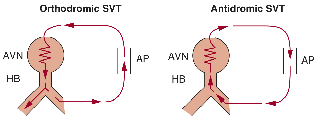
Accessory pathways can produce both orthodromic and antidromic AVRT and may be found on a screening ECG even in people with no symptoms. Fuster and Hurst's The Heart, 15th Edition, p. 1132.
1. Orthodromic AVRT
Orthodromic AVRT is the common form of AVRT.
Electrical circuit
Atria
  ↓ normal forward direction
AV node
  ↓
His-Purkinje system
  ↓
Ventricles
  ↑ backward through accessory pathway
Atria
So, in orthodromic AVRT:
| Part of circuit | Direction |
|---|---|
| AV node and His-Purkinje system | Forward, atrium to ventricle |
| Accessory pathway | Backward, ventricle to atrium |
The word orthodromic applies because the impulse reaches the ventricles through the normal AV node-His-Purkinje route.
ECG appearance
It usually produces:
- A regular, fast tachycardia
- Usually narrow QRS complexes: QRS duration <120 ms
- Rates commonly around 150 to 250 beats/min
- Retrograde P waves may be hidden within the QRS or appear shortly after it
- During the actual tachycardia, a delta wave is generally not visible because ventricular activation is using the normal conduction system.
This can resemble AV nodal re-entrant tachycardia (AVNRT), another common regular narrow-complex SVT. A 12-lead ECG during symptoms and, sometimes, an electrophysiology study distinguish them.
Why can the resting ECG be normal?
If the accessory pathway conducts only backward, it is called a concealed accessory pathway. It cannot pre-excite the ventricles during ordinary sinus rhythm, so the baseline ECG may look normal. Yet it can still participate in orthodromic AVRT.
2. Antidromic AVRT
Antidromic AVRT is less common.
Electrical circuit
Atria
  ↓ forward through accessory pathway
Ventricles
  ↑ backward through AV node and His-Purkinje system
Atria
So, in antidromic AVRT:
| Part of circuit | Direction |
|---|---|
| Accessory pathway | Forward, atrium to ventricle |
| AV node and His-Purkinje system | Backward, ventricle to atrium |
The term antidromic applies because the electrical impulse goes to the ventricles by an abnormal route rather than by the usual AV node-His-Purkinje route.
ECG appearance
It typically produces:
- A regular, rapid tachycardia
- Wide QRS complexes, often looking similar to ventricular tachycardia
- Often a very broad, unusual QRS morphology because the ventricles are activated cell-to-cell from the accessory pathway rather than quickly through the His-Purkinje network.
A wide-complex tachycardia should be treated seriously. It may be ventricular tachycardia, antidromic AVRT, or another rhythm with abnormal ventricular conduction. An ECG alone may not always settle the diagnosis promptly. In clinical practice, clinicians often manage an uncertain wide-complex tachycardia as ventricular tachycardia until proven otherwise.
Orthodromic vs antidromic: key comparison
| Feature | Orthodromic AVRT | Antidromic AVRT |
|---|---|---|
| Frequency | More common | Less common |
| Forward route to ventricles | AV node and His-Purkinje system | Accessory pathway |
| Return route to atria | Accessory pathway | AV node/His-Purkinje system, or occasionally another pathway |
| QRS during tachycardia | Usually narrow | Usually wide |
| ECG category | Regular narrow-complex SVT | Regular wide-complex tachycardia |
| Resting delta wave required? | No, can occur with a concealed pathway | Usually requires forward-conducting pathway |
| Main clinical look-alike | AVNRT | Ventricular tachycardia |
The disease most associated with these terms: WPW syndrome
Wolff-Parkinson-White pattern vs syndrome
This distinction matters:
WPW pattern
An ECG finding of pre-excitation, usually:
- Short PR interval
- Delta wave: slurred initial portion of QRS
- Often a wider-than-usual QRS complex
It indicates that an accessory pathway can conduct forward during sinus rhythm.
WPW syndrome
WPW pattern plus a clinically significant arrhythmia or arrhythmia-related symptoms, such as palpitations, documented AVRT, fainting, or sometimes pre-excited atrial fibrillation.
In other words:
Accessory pathway alone ≠ necessarily disease
WPW ECG pattern = pre-excitation visible on ECG
WPW syndrome = pre-excitation plus arrhythmia/symptoms
An accessory pathway may be present without symptoms, and some people first discover it from a routine ECG. The pathway itself is often not “heart muscle disease”; it is an extra electrical connection.
Why is pre-excitation present in WPW?
During sinus rhythm, if the accessory pathway conducts forward faster than the AV node, part of the ventricle is activated early.
That early ventricular activation causes:
- Short PR interval: ventricles begin depolarizing earlier than usual.
- Delta wave: the first part of ventricular activation spreads slowly from the pathway insertion site.
- Wider QRS: activation is a mixture of early abnormal activation and normal His-Purkinje activation.
The degree of pre-excitation can vary. A delta wave may be obvious, subtle, intermittent, or absent if the pathway is concealed.
Re-entry: why the tachycardia starts
A re-entry rhythm needs:
- Two possible pathways around a circuit
- Different conduction and recovery properties in those pathways
- Often a trigger, such as a premature atrial beat or premature ventricular beat
- One limb that can conduct while the other is temporarily refractory, allowing the impulse to circle back and keep repeating
Instead of one electrical impulse ending after activating the heart, it becomes trapped in a self-sustaining loop. Each loop creates another rapid heartbeat.
This is why AVRT often starts and stops suddenly. People may describe:
- “My heart suddenly took off”
- Abrupt episodes of racing heart
- Abrupt termination after seconds, minutes, or longer
Symptoms
Possible symptoms during AVRT include:
- Palpitations, often sudden-onset and regular
- Rapid pounding in the neck or chest
- Light-headedness or dizziness
- Shortness of breath
- Chest pressure or discomfort
- Anxiety, sweating, tremor
- Near-fainting or fainting
Some people have no symptoms at all.
Emergency symptoms include fainting, severe chest pain, severe breathlessness, confusion, signs of shock, or a persistent very fast rhythm. These warrant emergency evaluation.
The important dangerous rhythm: atrial fibrillation with WPW
The most important safety issue is not usually ordinary orthodromic AVRT. It is atrial fibrillation (AF) with pre-excitation.
In ordinary AF, the AV node blocks many of the chaotic atrial impulses, protecting the ventricles from extremely fast activation. But an accessory pathway may bypass that protective filter.
This can cause:
- An irregular wide-complex tachycardia
- Extremely fast ventricular rates, sometimes >250/min
- Rare progression to ventricular fibrillation and cardiac arrest in a susceptible person
Harrison's describes pre-excited AF as an irregular wide-complex or intermittently wide-complex tachycardia that can have rates above 250/min. Harrison’s Principles of Internal Medicine, 22e.
Treatment principles
Treatment depends on the rhythm, blood pressure and symptoms, the certainty of diagnosis, and the patient’s individual risk. This should be directed by clinicians who can assess the ECG.
If unstable
If there is low blood pressure, shock, severe chest pain, acute heart failure, altered consciousness, or ongoing severe instability:
- Urgent synchronized electrical cardioversion is generally indicated.
Stable regular narrow-complex tachycardia, likely orthodromic AVRT
Clinicians may use:
- Vagal manoeuvres, such as a modified Valsalva manoeuvre
- Adenosine if appropriate and with monitoring
- Other therapies or cardioversion if it does not terminate or the diagnosis is uncertain
For a clearly regular narrow-complex orthodromic AVRT, AV-node-directed treatment can interrupt the circuit because the AV node is part of the forward limb.
Antidromic AVRT or uncertain wide-complex tachycardia
This is a different situation. It needs urgent ECG-based clinical assessment. Antidromic AVRT may be hard to distinguish from ventricular tachycardia without an electrophysiology study. Procainamide is a commonly used option in stable pre-excited wide-complex tachycardias, while cardioversion is used if unstable. The NCBI AVRT review explains why uncertain wide-complex rhythms should not be casually treated as ordinary SVT.
Pre-excited AF or atrial flutter
Avoid AV-node blocking drugs unless a clinician has established the rhythm is not pre-excited AF/flutter. In particular, drugs such as adenosine, verapamil, diltiazem, beta-blockers, and digoxin can be harmful in pre-excited AF because blocking the AV node can favor conduction down the accessory pathway, potentially accelerating the ventricular rate. For stable pre-excited AF, procainamide or ibutilide may be used in monitored settings; electrical cardioversion is used for instability. See the clinical review of AVRT.
This warning is for medical decision-making, not self-treatment. Do not start, stop, or take rhythm medicines based on this explanation.
Long-term solution: catheter ablation
For symptomatic, recurrent, or higher-risk accessory-pathway arrhythmias, catheter ablation is often the definitive treatment.
An electrophysiologist:
- Maps the accessory pathway during an electrophysiology study.
- Delivers energy, most often radiofrequency or sometimes cryotherapy.
- Eliminates conduction through the pathway.
This can prevent recurrent AVRT and removes the substrate for pre-excited AF. The exact indication is individualized, including for people who have a WPW ECG pattern but no symptoms.
A recent systematic review evaluated exercise testing for identifying higher-risk accessory pathways, but this does not replace specialist assessment or invasive electrophysiology testing when indicated (2025 review).
Related terms you may encounter
| Term | Meaning |
|---|---|
| SVT | Supraventricular tachycardia, a broad category of fast rhythms arising at or above the ventricles |
| AVRT | Atrioventricular re-entrant tachycardia: a re-entry circuit using an accessory pathway |
| AVNRT | AV nodal re-entrant tachycardia: re-entry within or immediately around the AV node, with no accessory pathway required |
| Pre-excitation | Early ventricular activation through an accessory pathway |
| Antegrade conduction | Forward conduction from atrium to ventricle |
| Retrograde conduction | Backward conduction from ventricle to atrium |
| Concealed pathway | Usually conducts only retrogradely, so no delta wave during sinus rhythm |
| Delta wave | Slurred initial upstroke of the QRS in WPW pattern |
| Wide-complex tachycardia | Tachycardia with QRS ≥120 ms; VT must be considered |
| EP study | Electrophysiology study, an invasive procedure that maps cardiac electrical circuits |
A useful memory aid
- Ortho = ordinary direction through the AV node to ventricles -> usually narrow QRS.
- Anti = abnormal/opposite direction through the accessory pathway to ventricles -> usually wide QRS.
That is a helpful rule, but it is not enough to diagnose an ECG without clinical context.
If you have an ECG report mentioning pre-excitation, delta wave, WPW, AVRT, or a wide-complex tachycardia, it is appropriate to review it with a cardiologist, ideally a cardiac electrophysiologist.
This is a shared conversation. Sign in to Orris to start your own chat.