
Change takes place during development of graffian follicle
Reading File
Now let me search the medical library for content on Graafian follicle development.
Finding Sources
Finding Sources
Reading File
Reading File
Reading File
Reading File
Now let me fetch the Graafian follicle image from the histology textbook:
Loading Image
Finding Sources
I now have comprehensive information. Let me compile the full answer.
Changes During Development of the Graafian (Mature Vesicular) Follicle
The Graafian follicle represents the final, mature antral stage of folliculogenesis, just before ovulation. Its development involves a precise sequence of structural, cellular, and hormonal changes organized across four follicular stages.
1. Primordial Follicle (Starting point)
- A dormant primary oocyte (arrested in prophase I of meiosis) is surrounded by a single layer of flattened pregranulosa cells.
- These are maintained in a quiescent state throughout fetal life and childhood.
2. Primary Follicle
Changes:
- Pregranulosa cells transform from flattened to cuboidal under FSH, GFs (EGF, IGF-1), and calcium ion influence.
- Granulosa cells begin to proliferate (single layer initially).
- The zona pellucida - a glycoprotein shell - is deposited around the oocyte.
- Granulosa cell processes extend through the zona pellucida and form gap junctions with oocyte microvilli. These deliver cGMP into the oocyte, which inhibits PDE3A, maintaining high cAMP and keeping the oocyte arrested in meiosis I.
- Organelle redistribution occurs: Balbiani body breaks up, Golgi elements scatter, ribosomes, mitochondria, and rER all increase in number.
- Cortical granules (proteases released at fertilization) appear beneath the oolemma.
3. Secondary (Antral/Preantral) Follicle
Changes:
- Granulosa cells multiply to form 6-12 cell layers (stratum granulosum reaches ~600 cells).
- Theca cells differentiate from ovarian stroma surrounding the basement membrane:
- Theca interna: inner layer, develops LH receptors, acquires steroidogenic capacity (produces androgens).
- Theca externa: outer layer of smooth muscle cells and collagen bundles.
- A rich capillary network develops in the theca interna; the granulosa layer remains avascular (basal lamina restricts capillary penetration).
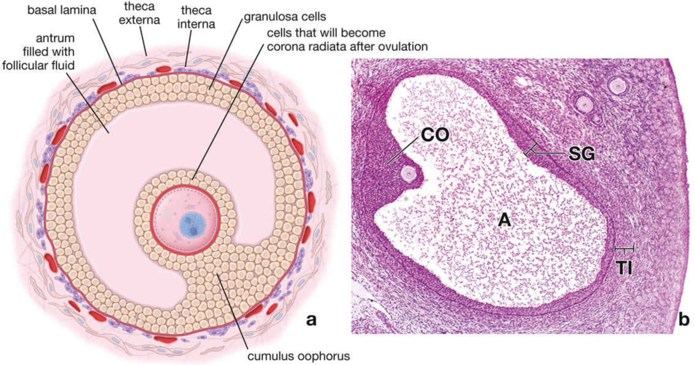
- Fluid-filled spaces (liquor folliculi - hyaluronan-rich) accumulate between granulosa cells, coalesce to form a crescent-shaped antrum - now the follicle is officially "antral/secondary."
- The oocyte reaches ~125 μm diameter and stops growing.
- Call-Exner bodies - PAS-positive extracellular material containing hyaluronan and proteoglycans - appear between granulosa cells.
4. Tertiary (Late Antral) Follicle
- Antrum enlarges further; FSH drives the transition from preantral to antral follicle.
- Granulosa cells differentiate into three subtypes:
- Mural granulosa cells: most metabolically active, highest LH receptor content, primary steroidogenic cells.
- Cumulus granulosa cells: shed with the oocyte at ovulation.
- Antral granulosa cells: remain in the follicle, become large luteal cells of the corpus luteum.
5. Mature Graafian Follicle
Changes:
- Follicle reaches 10-33 mm in diameter, extends through the full thickness of the ovarian cortex, and produces a visible bulge on the ovarian surface.
- The antrum nearly encircles the oocyte, except for a small mound - the cumulus oophorus - attaching the oocyte to the follicle wall.
- Cumulus cells immediately surrounding the oocyte form the corona radiata (single-cell layer at maturity); these cells remain with the oocyte after ovulation.
- Mitotic activity of granulosa cells decreases; stratum granulosum appears thinner as the antrum expands.
- Theca interna becomes prominent: cells accumulate lipid droplets and show ultrastructural features of steroid-secreting cells.
- Two-cell, two-gonadotropin steroidogenesis: Theca interna (LH-stimulated) produces androgens → converted by FSH-stimulated granulosa cells (via aromatase) into estradiol.
- LH surge (~36h before ovulation): triggers the oocyte to resume meiosis I, completing it to form a secondary oocyte and first polar body; arrests again in metaphase II.
- Antral fluid accumulates rapidly between 5-6 days before ovulation (dominant follicle selection).
Summary Table
| Stage | Key Change |
|---|---|
| Primordial | Flat pregranulosa cells, oocyte arrested in prophase I |
| Primary | Cuboidal granulosa cells, zona pellucida formed, gap junctions |
| Secondary | Multiple granulosa layers, theca differentiation, antrum forms, Call-Exner bodies |
| Tertiary | Antrum enlarges, granulosa subtypes differentiate, FSH-driven |
| Graafian | 10-33 mm; cumulus oophorus + corona radiata; LH surge resumes meiosis I |
Sources:
- Histology: A Text and Atlas with Correlated Cell and Molecular Biology (Kierszenbaum), Ch. 23
- Medical Physiology (Boron & Boulpaep), Ch. 55 - folliculogenesis stages
Cerebellar test from susan sulai
Reading File
The user is asking about cerebellar tests from "Susan Snell" (likely "Susan Standring" - Gray's Anatomy) or more probably "Susan O'Sullivan" - a Physical Therapy/Neurological examination textbook. Let me search the library.
Searching sources...
Finding Sources
Finding Sources
Finding Sources
Finding Sources
The library doesn't have a "Susan Sulai/Snell" book specifically. The user likely means Susan Standring's Gray's Anatomy or Susan O'Sullivan's Physical Rehabilitation - a well-known PT textbook that has a thorough section on cerebellar tests. Let me search more broadly and also read the Bradley & Daroff section which will have the clinical cerebellar tests.
Reading File
Finding Sources
Reading File
Reading File
Reading File
Finding Sources
I have enough information from the textbooks. The user is asking about cerebellar tests as described in "Susan Sulai" - this is most likely a reference to Susan Standring's Gray's Anatomy or, more commonly in medical education contexts (especially in South Asia), it refers to "Snell's Clinical Neuroanatomy" by Richard Snell - but the user said "Susan Sulai." This is likely a phonetic spelling of "Susan Standring" OR this could be a reference to "Suleiman" - but the most probable reference in clinical medicine exams (especially for MBBS) is that this is Susan O'Sullivan's Physical Rehabilitation textbook, OR simply a local/regional reference. In any case, the cerebellar tests are standard clinical signs and I have comprehensive content from authoritative neurology textbooks. Let me compile the complete answer.
Cerebellar Tests (Clinical Examination of Cerebellar Function)
Note: "Susan Sulai" likely refers to a local/regional teaching resource. The cerebellar tests described below are universal standard clinical signs, well-documented in Bradley and Daroff's Neurology in Clinical Practice and Localization in Clinical Neurology.
The neurological examination of cerebellar function covers 5 domains: eyes, speech, hands (upper limbs), legs (lower limbs), and gait. The mnemonic DANISH or VANISHD is commonly used to recall the signs.
1. GAIT TESTS
a. Ataxic (cerebellar) gait
- Patient walks with a wide base, irregular stride length, and tends to veer/stagger to the side of the lesion.
- Early: variable stride and veering; Late: broad-based gait as compensation.
b. Tandem walking (heel-to-toe)
- Walking heel-to-toe in a straight line - patient with cerebellar lesion cannot maintain balance.
c. Romberg test
- Cerebellar ataxia is present with eyes OPEN (positive Romberg is a sensory/dorsal column sign, NOT cerebellar).
- If the patient sways/falls with eyes open - think cerebellar.
2. STANCE TESTS
- Standing with feet together - truncal sway seen.
- Tandem stance (one foot in front of the other).
- Standing on one foot / hopping - to detect subtle cerebellar signs.
3. UPPER LIMB TESTS
a. Finger-Nose Test (Finger-to-Nose)
- Patient alternately touches their own nose and the examiner's index finger with the index finger.
- Dysmetria - overshooting or undershooting the target.
- Intention tremor - tremor that increases as the finger approaches the target (end-point tremor).
b. Finger Chase (Finger-Tracking) Test
- Examiner moves their finger; patient's index finger follows as precisely as possible.
- Cerebellar: over- or under-shoot (past-pointing).
c. Dysdiadochokinesia (Rapid Alternating Movements)
- Patient rapidly alternates pronation and supination of the hand ("pat the back of the hand as fast as possible").
- Cerebellar: slow, irregular, arrhythmic movements.
d. Rebound Phenomenon (Holmes' Rebound Test)
- Patient flexes the elbow against resistance; examiner suddenly releases.
- Normal: patient quickly checks the limb.
- Cerebellar: arm flies up and hits the face (loss of check reflex).
e. Pendular Reflexes
- Tendon jerk swings back and forth 3-4 times instead of the normal 1-2 (due to hypotonia).
4. LOWER LIMB TESTS
a. Heel-Shin (Heel-to-Knee-to-Shin) Test
- Patient lifts one leg, places the heel on the opposite knee, and slides it down the shin to the ankle.
- Cerebellar: heel falls off the shin, irregular movements.
- This is the lower limb equivalent of the finger-nose test.
5. SPEECH
Scanning dysarthria
- Slow, slurred, irregular speech with inappropriate pauses.
- Words broken into syllables with excess stress on each (explosive speech).
6. EYE SIGNS
| Sign | Description |
|---|---|
| Nystagmus | Jerky oscillation of eyes; most prominent (fast phase) toward side of lesion |
| Gaze-evoked nystagmus | End-gaze nystagmus in many ataxias |
| Saccadic pursuit | Breakdown of smooth pursuit into jerky saccades |
| Hypo/Hypermetric saccades | Undershooting or overshooting a target |
| Square-wave jerks | Saccadic intrusions in fixed gaze (e.g., Friedreich ataxia) |
7. HYPOTONIA
- Decreased muscle tone ipsilateral to cerebellar hemisphere lesion.
- Pendular knee jerk.
- Hypotonia is most prominent in proximal upper limb segments (relates to dentate nucleus injury).
8. SARA Scale (Bedside Clinical Scale)
The Scale for Assessment and Rating of Ataxia (SARA) covers:
- Gait
- Stance
- Sitting (without back support)
- Speech
- Finger chase
- Nose-finger test
- Fast alternating hand movements
- Heel-shin slide
Summary Mnemonic - "DANISH" (Signs of Cerebellar Disease)
| Letter | Sign |
|---|---|
| D | Dysdiadochokinesia |
| A | Ataxia (gait, limb) |
| N | Nystagmus |
| I | Intention tremor |
| S | Scanning dysarthria / Staccato speech |
| H | Hypotonia |
Sources:
- Bradley and Daroff's Neurology in Clinical Practice, Cerebellar Ataxia Chapter 23 - neurological examination domains and SARA scale
- Localization in Clinical Neurology, 8e - cardinal features of cerebellar dysfunction (hypotonia, dysmetria, dysdiadochokinesia, intention tremor, nystagmus)
This is a shared conversation. Sign in to Orris to start your own chat.