
advanced monitoring in oncoanesthesia
- Indications and risk stratification
- Invasive arterial pressure and blood sampling
- Central venous access and CVP
- Cardiac output and dynamic fluid-responsiveness monitoring
- Transesophageal echocardiography
- Pulmonary artery catheterization
- Neuromuscular, temperature, coagulation, and metabolic monitoring
- Tumor-specific contexts, such as major thoracoabdominal surgery, cytoreduction with HIPEC, mediastinal masses, and massive hemorrhage
- Postoperative critical-care monitoring
Which format would you like?
- Indications and risk stratification
- Invasive arterial pressure and blood sampling
- Central venous access and CVP
- Cardiac output and dynamic fluid-responsiveness monitoring
- Transesophageal echocardiography
- Pulmonary artery catheterization
- Neuromuscular, temperature, coagulation, and metabolic monitoring
- Tumor-specific contexts, such as major thoracoabdominal surgery, cytoreduction with HIPEC, mediastinal masses, and massive hemorrhage
- Postoperative critical-care monitoring
- Exam short note - definition, indications, modalities, and key points
- Practical intraoperative guide - monitor selection by surgical risk and physiology
- Tumor-specific monitoring - thoracic, mediastinal, head-neck, hepatic, cytoreductive surgery with HIPEC
- Comparison table - arterial line, CVP, cardiac output monitors, TEE, PAC, coagulation monitoring
prepare and give me a power point presentation on the topic advanced monitoring in oncoanesthesia
cancer surgery AND hemodynamic monitoring
advanced hemodynamic monitoring major cancer surgery perioperative guidelines
arterial pressure waveform monitoring anesthesia diagram
transesophageal echocardiography four chamber view diagram
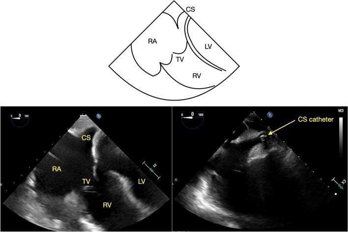
This diagnostic visual includes a side-by-side comparison of transesophageal echocardiography (TEE) images and an anatomical diagram depicting the mid-esophageal four-chamber (ME 4C) view. The educational focus is the visualization of the coronary sinus (CS) in its long axis. Key anatomical structures labeled include the right atrium (RA), right ventricle (RV), left ventricle (LV), and the tricuspid valve (TV). The left image demonstrates the normal ultrasound appearance of the CS as a clear, hypoechoic tubular structure positioned superior to the attachment of the tricuspid valve septal leaflet to the interventricular septum. The right image shows the clinical application of this view for monitoring catheterization, featuring a highly echogenic (bright) CS catheter positioned within the vessel. The accompanying line diagram provides a simplified schematic of these spatial relationships, emphasizing the CS's proximity to the RA and its orientation relative to the four cardiac chambers. This content is intended for advanced medical trainees learning TEE imaging protocols for electrophysiology or cardiac surgery.
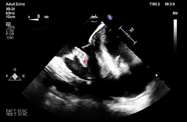
This diagnostic image is a transesophageal echocardiography (TEE) still, specifically a mid-esophageal four-chamber view of an adult heart. The primary finding is a discrete, echogenic mass (marked with a red asterisk) associated with the tricuspid valve, appearing to adhere to the posterior leaflet. The mass demonstrates a distinct texture and density compared to the surrounding cardiac chambers and septal walls. On-screen annotations include technical parameters such as a probe frequency of 53Hz, a 10cm imaging depth, and patient/probe temperatures (37.0°C and 37.8°C respectively). Mechanical and thermal indices (MI 0.9, TIS 0.2) are visible, along with a 0-degree sector sweep angle and a probe orientation diagram. This image is clinically significant for evaluating right-sided valvular pathology, specifically potential vegetations, thrombi, or primary cardiac tumors like fibroelastomas, within the context of intraoperative monitoring.
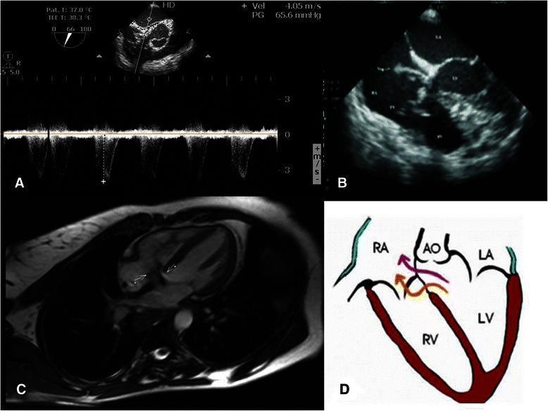
This composite figure illustrates the multi-modal diagnostic assessment of a C-type acquired Gerbode defect. Panel A displays a Transesophageal Echocardiography (TEE) with spectral Doppler, showing a high-velocity systolic shunt (4.05 m/s) with a pressure gradient of 65.6 mmHg, indicating a left-to-right shunt. Panel B is a Transthoracic Echocardiogram (TTE) in a four-chamber view, identifying an irregularly shaped, oscillating, and mobile echogenic mass (vegetation) attached above the septal leaflet of the tricuspid valve. Panel C presents a Cardiac Magnetic Resonance (CMR) image in a horizontal long-axis view, where white arrows highlight the pathological communication between the left ventricle (LV), right atrium (RA), and right ventricle (RV). Panel D provides a corresponding anatomical diagram mapping the shunt flow; it shows a complex C-type Gerbode defect consisting of both supravalvular (LV to RA) and infravalvular (LV to RV) components. This collection demonstrates how structural imaging and hemodynamics confirm the presence of infective endocarditis complicated by an acquired intracardiac shunt.
https://pubmed.ncbi.nlm.nih.gov/38392644/
~/
mkdir -p "/tmp/workspace/oncoanesthesia-monitoring/src" "/tmp/workspace/oncoanesthesia-monitoring/assets" "/tmp/workspace/oncoanesthesia-monitoring/output" && python /tmp/skills/shared/scripts/fetch_images.py "https://cdn.orris.care/cdss_images/pmc_clinical_VQA_6ef3b0188eeeae9f945185c84ff06700112356ca085528d6d71d168c70f656b6.jpg" > "/tmp/workspace/oncoanesthesia-monitoring/assets/image_fetch.json"
python - <<'PY' import base64, json, os from pathlib import Path p=Path('/tmp/workspace/oncoanesthesia-monitoring/assets/image_fetch.json') d=json.loads(p.read_text()) b64=d[0]['base64'].split(',',1)[1] Path('/tmp/workspace/oncoanesthesia-monitoring/assets/tee.jpg').write_bytes(base64.b64decode(b64)) PY cat > "/tmp/workspace/oncoanesthesia-monitoring/src/build_deck.py" <<'PY' from pptx import Presentation from pptx.util import Inches, Pt from pptx.enum.text import PP_ALIGN, MSO_ANCHOR from pptx.enum.shapes import MSO_SHAPE, MSO_CONNECTOR from pptx.dml.color import RGBColor from pptx.enum.dml import MSO_THEME_COLOR from pptx.enum.text import MSO_AUTO_SIZE from pathlib import Path OUT=Path('/tmp/workspace/oncoanesthesia-monitoring/output/Advanced_Monitoring_in_Oncoanesthesia.pptx') TEE='/tmp/workspace/oncoanesthesia-monitoring/assets/tee.jpg' prs=Presentation(); prs.slide_width=Inches(13.333); prs.slide_height=Inches(7.5) blank=prs.slide_layouts[6] NAVY=RGBColor(12,35,58); TEAL=RGBColor(0,139,139); CYAN=RGBColor(49,189,196); GOLD=RGBColor(238,171,56); RED=RGBColor(196,67,67); PALE=RGBColor(239,247,248); LIGHT=RGBColor(249,251,252); GREY=RGBColor(86,101,115); WHITE=RGBColor(255,255,255); GREEN=RGBColor(42,137,87) def bg(slide,color=WHITE): s=slide.background.fill; s.solid(); s.fore_color.rgb=color def rect(slide,x,y,w,h,fill, radius=False, line=None): sh=slide.shapes.add_shape(MSO_SHAPE.ROUNDED_RECTANGLE if radius else MSO_SHAPE.RECTANGLE, Inches(x), Inches(y), Inches(w), Inches(h)) sh.fill.solid(); sh.fill.fore_color.rgb=fill sh.line.color.rgb = line if line else fill if radius: sh.adjustments[0]=0.08 return sh def text(slide,x,y,w,h,txt,size=20,color=NAVY,bold=False,align=PP_ALIGN.LEFT, font='Aptos',val=MSO_ANCHOR.TOP): tb=slide.shapes.add_textbox(Inches(x), Inches(y), Inches(w), Inches(h)); tf=tb.text_frame; tf.clear(); tf.word_wrap=True tf.margin_left=Inches(.04);tf.margin_right=Inches(.04);tf.margin_top=Inches(.02);tf.margin_bottom=Inches(.02);tf.vertical_anchor=val for i,line in enumerate(txt.split('\n')): p=tf.paragraphs[0] if i==0 else tf.add_paragraph(); p.text=line; p.alignment=align; p.space_after=Pt(3) for r in p.runs: r.font.name=font; r.font.size=Pt(size); r.font.bold=bold; r.font.color.rgb=color return tb def bullets(slide,x,y,w,h,items,size=19,color=NAVY): tb=slide.shapes.add_textbox(Inches(x), Inches(y), Inches(w), Inches(h)); tf=tb.text_frame; tf.clear(); tf.word_wrap=True tf.margin_left=Inches(.12); tf.margin_right=0;tf.margin_top=Inches(.03) for i,item in enumerate(items): p=tf.paragraphs[0] if i==0 else tf.add_paragraph(); p.text=item; p.level=0; p.font.size=Pt(size); p.font.name='Aptos'; p.font.color.rgb=color; p.space_after=Pt(8); p.bullet=True return tb def title(slide,t,sub=None,n=None): rect(slide,0,0,13.333,.16,TEAL); text(slide,.55,.34,11.8,.5,t,28,NAVY,True) if sub:text(slide,.57,.9,11.7,.3,sub,12,GREY) if n is not None:text(slide,12.35,7.07,.45,.2,f'{n:02}',10,GREY,False,PP_ALIGN.RIGHT) text(slide,.55,7.07,5,.2,'ADVANCED MONITORING IN ONCOANESTHESIA',9,TEAL,True) def add_notes(slide, note): # notes text placeholder exists in python-pptx try: tf=slide.notes_slide.notes_text_frame tf.text=note except Exception: pass def card(slide,x,y,w,h,head,body,accent=TEAL, body_size=16): rect(slide,x,y,w,h,PALE,True); rect(slide,x,y,.09,h,accent) text(slide,x+.25,y+.22,w-.45,.3,head,17,accent,True) text(slide,x+.25,y+.67,w-.45,h-.8,body,body_size,NAVY) #1 title s=prs.slides.add_slide(blank); bg(s,NAVY); rect(s,0,0,13.333,.16,TEAL); rect(s,.72,1.15,.1,4.6,GOLD) text(s,1.05,1.2,10.8,1.35,'Advanced Monitoring\nin Oncoanesthesia',40,WHITE,True) text(s,1.08,3.05,9.2,.5,'A physiology-led approach to high-risk cancer surgery',21,RGBColor(203,225,229)) # visual motif for i,(lab,col) in enumerate([('PERFUSION',TEAL),('VOLUME',CYAN),('COAGULATION',GOLD),('TEMPERATURE',RED)]): rect(s,1.08+i*2.55,4.3,2.25,.63,col,True); text(s,1.08+i*2.55,4.48,2.25,.18,lab,12,WHITE,True,PP_ALIGN.CENTER) text(s,1.08,6.55,10,.3,'Educational presentation | For anesthesia trainees and perioperative teams',14,RGBColor(185,208,212)) add_notes(s,'Introduce the central premise: advanced monitoring is not routine technology for every patient. It is targeted monitoring that answers a clinical question and changes management.') #2 objectives s=prs.slides.add_slide(blank); bg(s); title(s,'Learning objectives','By the end, the audience should be able to select and interpret monitoring rationally.',2) for x,head,body,col in [(0.7,'Recognize risk','Identify procedures and patients that merit escalation.',TEAL),(3.85,'Match tool to question','Select arterial line, CO monitor, echo, coagulation and metabolic tests.',CYAN),(7.0,'Interpret in context','Use trends and dynamic indices rather than isolated numbers.',GOLD),(10.15,'Act on data','Link a signal to fluid, vasopressor, inotrope, transfusion or escalation.',RED)]: card(s,x,1.8,2.45,3.35,head,body,col,18) text(s,.85,5.85,11.4,.5,'Monitoring without a predefined response algorithm adds data, not safety.',25,NAVY,True,PP_ALIGN.CENTER) #3 why s=prs.slides.add_slide(blank); bg(s); title(s,'Why cancer surgery challenges conventional monitoring','The risk comes from the operation, patient physiology, and cancer-directed therapy.',3) card(s,.7,1.45,3.85,4.75,'Surgical drivers','• Prolonged major thoracoabdominal procedures\n• Major vascular exposure or resection\n• Large blood loss and rapid shifts\n• One-lung ventilation or high-risk positioning\n• Cytoreduction with HIPEC',TEAL,18) card(s,4.75,1.45,3.85,4.75,'Patient drivers','• Frailty, anemia, malnutrition\n• Coronary, pulmonary, renal disease\n• Chemotherapy-related cardiomyopathy\n• Prior radiation or difficult airway\n• Thromboembolic risk',CYAN,18) card(s,8.8,1.45,3.85,4.75,'Physiologic targets','• Oxygen delivery and perfusion pressure\n• Preload responsiveness, not CVP alone\n• Ventricular function and afterload\n• Hemostasis and temperature\n• Gas exchange and acid-base status',GOLD,18) #4 selection s=prs.slides.add_slide(blank);bg(s);title(s,'A tiered, question-driven monitoring strategy','Escalate when the expected information will change an immediate decision.',4) steps=[('1','Baseline','ASA standards: ECG, NIBP, SpO₂, capnography, inspired O₂, temperature, urine output.'),('2','Risk screen','Blood loss? Fluid shifts? Cardiorespiratory reserve? Need vasoactive support?'),('3','Add a modality','Choose the least invasive monitor that can answer the question.'),('4','Close the loop','Define thresholds, interventions, and reassessment before incision.')] for i,(num,h,b) in enumerate(steps): x=.72+i*3.15; rect(s,x,1.8,2.7,3.65,PALE,True); rect(s,x+.22,2.08,.56,.56,TEAL,True); text(s,x+.22,2.23,.56,.18,num,16,WHITE,True,PP_ALIGN.CENTER); text(s,x+.25,2.95,2.2,.32,h,19,NAVY,True); text(s,x+.25,3.48,2.18,1.25,b,15,GREY) if i<3: text(s,x+2.76,3.25,.3,.3,'→',25,TEAL,True,PP_ALIGN.CENTER) text(s,.8,6.05,11.6,.4,'Avoid treating a monitor number in isolation. Verify signal quality, trend, and clinical concordance.',19,RED,True,PP_ALIGN.CENTER) #5 arterial s=prs.slides.add_slide(blank);bg(s);title(s,'Arterial catheter: the foundation for high-risk cases','Continuous pressure data and rapid access to serial blood samples.',5) card(s,.7,1.35,3.7,4.85,'Useful when','• Anticipated rapid blood loss\n• Need for frequent ABG, Hb, electrolytes or lactate\n• Beat-to-beat BP needed for titration\n• Major thoracic, hepatic, vascular or HIPEC surgery\n• Significant cardiopulmonary disease',TEAL,17) card(s,4.82,1.35,3.7,4.85,'What it enables','• MAP trend and hypotension burden\n• Pulse contour-derived stroke-volume variables\n• Dynamic indices, when valid\n• ABG, PaCO₂, ionized Ca²⁺, glucose\n• Hemoglobin and lactate trending',CYAN,17) card(s,8.94,1.35,3.7,4.85,'Quality checks','• Level and zero at phlebostatic axis\n• Assess fast-flush waveform\n• Under/overdamping distorts systolic and pulse pressure\n• Correlate with cuff if unexpected\n• Protect limb perfusion and line sterility',GOLD,17) #6 flow s=prs.slides.add_slide(blank);bg(s);title(s,'Cardiac output and dynamic assessment','Use flow monitoring to distinguish hypovolemia, vasodilation, and pump failure.',6) card(s,.7,1.35,3.8,4.85,'Common technologies','Calibrated or uncalibrated pulse contour\nEsophageal Doppler\nBioreactance / bioimpedance\nPulmonary artery catheter in selected situations\nTEE when anatomy and function must be visualized',TEAL,17) card(s,4.78,1.35,3.8,4.85,'Parameters that matter','Cardiac index and stroke volume\nStroke-volume response to a test bolus\nSVV / PPV where valid\nSystemic vascular resistance estimate\nScvO₂ or mixed venous oxygen saturation in selected patients',CYAN,17) card(s,8.86,1.35,3.8,4.85,'Limits of dynamic indices','Best conditions: controlled ventilation, regular rhythm, adequate tidal volume, closed chest.\n\nUnreliable with atrial fibrillation, spontaneous breathing, low tidal volume, open chest, right-ventricular failure, or marked intra-abdominal hypertension.',RED,16) #7 algorithm s=prs.slides.add_slide(blank);bg(s);title(s,'Treat hypotension by mechanism, not reflex fluid','A practical bedside loop for a low MAP or falling stroke volume.',7) # flow chart nodes=[(0.7,'Low MAP / falling SV','Confirm artifact, depth, surgical event, rhythm.',TEAL),(3.55,'Preload responsive?','PLR or small test bolus, if dynamic index valid.',CYAN),(6.4,'If yes','Incremental fluid, then reassess SV/CO and lung tolerance.',GREEN),(9.25,'If no','Assess SVR and contractility: vasopressor for vasodilation; inotrope/echo for pump failure.',GOLD)] for x,h,b,c in nodes: rect(s,x,2.05,2.62,2.4,PALE,True);rect(s,x,2.05,2.62,.1,c);text(s,x+.18,2.35,2.25,.38,h,17,c,True,PP_ALIGN.CENTER);text(s,x+.18,2.95,2.25,.95,b,14,NAVY,False,PP_ALIGN.CENTER) if x<9: text(s,x+2.7,3.0,.5,.3,'→',27,TEAL,True,PP_ALIGN.CENTER) rect(s,1.15,5.3,11.05,.72,NAVY,True);text(s,1.35,5.51,10.65,.24,'A fluid challenge is a diagnostic test. Stop if stroke volume does not improve or signs of congestion appear.',18,WHITE,True,PP_ALIGN.CENTER) #8 TEE s=prs.slides.add_slide(blank);bg(s);title(s,'Focused intraoperative echocardiography and TEE','High-value when hemodynamic instability is unexplained or structural information changes care.',8) try: s.shapes.add_picture(TEE, Inches(.72), Inches(1.43), width=Inches(4.25), height=Inches(3.55)) except: pass text(s,.76,5.15,4.2,.42,'Example: mid-esophageal four-chamber view',13,GREY,False,PP_ALIGN.CENTER) card(s,5.35,1.42,3.4,4.7,'Questions answered','• Is LV systolic function depressed?\n• Is RV dilated or failing?\n• Is filling low, adequate, or excessive?\n• Is there tamponade, acute valvular pathology, or air?\n• Is there a regional wall-motion abnormality?',TEAL,16) card(s,9.0,1.42,3.6,4.7,'When to consider','• Major thoracic surgery or pulmonary hypertension\n• Mediastinal mass with dynamic obstruction risk\n• Significant chemotherapy-related cardiomyopathy\n• Refractory shock or suspected embolism\n• Complex hepatic / vascular resections',GOLD,16) text(s,.78,6.18,11.8,.35,'TEE requires trained operators and attention to contraindications, including relevant esophageal pathology.',16,RED,True,PP_ALIGN.CENTER) #9 oxygen/metabolic s=prs.slides.add_slide(blank);bg(s);title(s,'Perfusion, oxygen delivery, and metabolic surveillance','No single number proves adequate tissue oxygenation. Interpret serial trends.',9) for x,h,items,c in [(0.72,'Blood gas and labs',['pH / PaCO₂ / PaO₂','Hemoglobin','Ionized calcium','Potassium and glucose'],TEAL),(3.9,'Perfusion markers',['Serial lactate','Base deficit','Urine output in context','Peripheral temperature / capillary refill'],CYAN),(7.08,'Oxygen transport',['SpO₂ and arterial oxygenation','Cardiac output / cardiac index','ScvO₂ in selected high-risk cases','Hemoglobin and arterial content'],GOLD),(10.26,'Interpret wisely',['Lactate may rise from causes beyond hypoperfusion','Urine output is delayed and nonspecific','Trends beat single samples','Act on the clinical pattern'],RED)]: rect(s,x,1.55,2.35,4.45,PALE,True); text(s,x+.18,1.84,2,.35,h,18,c,True,PP_ALIGN.CENTER);bullets(s,x+.16,2.5,2.02,2.8,items,15,NAVY) #10 coagulation s=prs.slides.add_slide(blank);bg(s);title(s,'Hemostasis monitoring during major oncologic surgery','Combine a structured massive-bleeding protocol with point-of-care data where available.',10) card(s,.7,1.4,3.75,4.8,'Conventional tests','CBC / platelet count\nPT/INR, aPTT\nFibrinogen concentration\nABG-derived pH and ionized Ca²⁺\nTemperature\n\nUseful, but turnaround time may delay targeted treatment.',TEAL,17) card(s,4.8,1.4,3.75,4.8,'Viscoelastic testing','TEG / ROTEM assesses whole-blood clot initiation, clot strength and fibrinolysis.\n\nIt can support goal-directed component therapy in complex bleeding, particularly when rapid decisions are required.',CYAN,17) card(s,8.9,1.4,3.75,4.8,'Operational priorities','• Activate major hemorrhage pathway early\n• Maintain normothermia\n• Correct ionized hypocalcemia\n• Replace fibrinogen / platelets / factors by results and protocol\n• Reassess after each intervention',GOLD,17) #11 special s=prs.slides.add_slide(blank);bg(s);title(s,'Procedure-specific applications','Match the monitoring package to the predictable physiological insult.',11) rows=[('Thoracic / lung resection','A-line; ABG; careful ventilation; CO/TEE if poor reserve or RV risk.','Hypoxemia, one-lung ventilation, RV strain'),('Cytoreduction + HIPEC','A-line; serial ABG/electrolytes/lactate; temperature; CO for large shifts.','Hyperthermia, vasodilation, fluid loss, acidosis'),('Major hepatectomy','A-line; frequent labs; CO/TEE selectively; coagulation monitoring.','Blood loss, low-CVP strategy, ischemia/reperfusion'),('Mediastinal mass','A-line before induction when severe compression suspected; echo availability.','Airway or vascular collapse, positional compromise'),('Head and neck free flap','A-line in selected cases; temperature, Hb, perfusion and vasopressor plan.','Long duration, blood loss, flap perfusion')] for i,(a,b,c) in enumerate(rows): y=1.35+i*1.05; rect(s,.68,y,12.0,.82, LIGHT if i%2==0 else PALE,True);text(s,.9,y+.15,2.35,.25,a,15,TEAL,True);text(s,3.25,y+.12,5.55,.48,b,13,NAVY);text(s,9.1,y+.12,3.15,.48,c,13,RED) #12 evidence s=prs.slides.add_slide(blank);bg(s);title(s,'Evidence: use protocols, but avoid overclaiming','The benefit is most plausible when monitoring is linked to a response algorithm.',12) card(s,.75,1.35,5.7,4.6,'Major cancer surgery protocol study','In a before-after study of open abdominal cancer operations lasting >2 h, implementation of defined hemodynamic and depth-of-anesthesia targets was associated with less administered fluid, fewer selected complications, and a shorter median hospital stay.\n\nTargets included MAP >65 mmHg, SVV <12%, CI >2.5 L·min⁻¹·m⁻², ScvO₂ >70%, and BIS 40-60.',TEAL,17) card(s,6.85,1.35,5.7,4.6,'Interpretation and evidence limits','• Observational before-after design cannot prove causality.\n• A monitor does not improve outcome unless it changes care appropriately.\n• Indices and thresholds must be individualized.\n• Technology accuracy can degrade in arrhythmia, vasoplegia, altered compliance, and poor signal conditions.',GOLD,17) text(s,.85,6.32,11.6,.25,'Source: Lilot et al., Anaesthesia 2018, PMID 30074124. HPI study in 50 high-risk abdominal oncology patients: PMID 38392644.',11,GREY,False,PP_ALIGN.CENTER) #13 postop s=prs.slides.add_slide(blank);bg(s);title(s,'The monitoring plan extends beyond extubation','Handover should communicate physiology, trends, remaining risks, and response triggers.',13) for x,h,b,c in [(0.75,'ICU / HDU disposition','Need for vasoactive support, mechanical ventilation, serial labs, or ongoing resuscitation.',TEAL),(3.9,'Handover dataset','Blood loss, transfusion, net balance, vasopressors, CO trend, lactate, temperature, urine output.',CYAN),(7.05,'Surveillance','ECG and troponin when indicated, oxygenation, renal function, delirium, bleeding and thrombosis risk.',GOLD),(10.2,'Escalation triggers','Rising lactate, persistent hypotension, oliguria with instability, hypoxemia, recurrent bleeding, new arrhythmia.',RED)]: card(s,x,1.65,2.4,3.95,h,b,c,16) text(s,.8,6.17,11.7,.35,'Continuity of interpretation is as important as continuity of the monitor.',22,NAVY,True,PP_ALIGN.CENTER) #14 takeaway s=prs.slides.add_slide(blank);bg(s,NAVY);rect(s,0,0,13.333,.16,TEAL);text(s,.9,.72,11.5,.55,'Take-home messages',32,WHITE,True) items=[('1','Choose monitoring by anticipated physiology, not by the cancer label.'),('2','An arterial line and serial testing are often the first escalation in major cases.'),('3','Use cardiac output, dynamic tests, and echo to determine the cause of instability.'),('4','Make every monitor actionable through a pre-agreed response algorithm.'),('5','Trend perfusion, temperature, coagulation, and metabolic data throughout the perioperative pathway.')] for i,(n,b) in enumerate(items): y=1.6+i*.85;rect(s,.95,y,.44,.44,TEAL,True);text(s,.95,y+.09,.44,.16,n,13,WHITE,True,PP_ALIGN.CENTER);text(s,1.65,y+.04,10.9,.36,b,19,WHITE,False) text(s,.95,6.62,11.5,.25,'Advanced monitoring is a clinical decision-support system, not a substitute for clinical judgment.',14,RGBColor(178,214,218),False,PP_ALIGN.CENTER) #15 refs s=prs.slides.add_slide(blank);bg(s);title(s,'Selected references','For teaching and further reading.',15) refs=[ 'Miller’s Anesthesia, 10th ed. Sections on perioperative monitoring, hemodynamic monitoring, fluid therapy, and coagulation.', 'Morgan & Mikhail’s Clinical Anesthesiology, 7th ed. Perioperative monitoring and patient blood management.', 'Lilot M, et al. Outcome impact of hemodynamic and depth of anesthesia monitoring during major cancer surgery: a before-after study. Anaesthesia. 2018. PMID: 30074124.', 'Cylwik J, et al. Individualized perioperative hemodynamic management using Hypotension Prediction Index in oncological abdominal surgery. 2024. PMID: 38392644.', 'American Society of Anesthesiologists. Standards for Basic Anesthetic Monitoring. Accessed 2026.', 'Image on slide 8: indexed educational TEE four-chamber view, medical image library.' ] bullets(s,.9,1.42,11.55,4.95,refs,17,NAVY) text(s,.95,6.65,11.3,.22,'This deck is educational and should be adapted to local equipment, protocols, operator expertise, and individual patient risk.',12,RED,False,PP_ALIGN.CENTER) prs.save(OUT) print(OUT) PY python "/tmp/workspace/oncoanesthesia-monitoring/src/build_deck.py"
~/oncoanesthesia-monitoring/output
python - <<'PY' from pptx import Presentation from pathlib import Path p=Path('/tmp/workspace/oncoanesthesia-monitoring/output/Advanced_Monitoring_in_Oncoanesthesia.pptx') prs=Presentation(p) print('slides',len(prs.slides),'size',prs.slide_width,prs.slide_height) for i,s in enumerate(prs.slides,1): texts=[] for sh in s.shapes: if getattr(sh,'has_text_frame',False) and sh.text.strip(): texts.append(sh.text.replace('\n',' | ')[:80]) print(i, len(s.shapes), ' :: '.join(texts[:2])) print('file bytes',p.stat().st_size) PY
Advanced Monitoring in Oncoanesthesia
Presentation · PPTX
can u narrate it in the ppt like a story or with reference to a patiet
major abdominal cancer surgery perioperative hemodynamic monitoring goal directed therapy guidelines
major abdominal surgery AND goal-directed hemodynamic therapy
~/
cat > "/tmp/workspace/oncoanesthesia-monitoring/src/build_case_deck.py" <<'PY' from pptx import Presentation from pptx.util import Inches, Pt from pptx.enum.text import PP_ALIGN, MSO_ANCHOR from pptx.enum.shapes import MSO_SHAPE from pptx.dml.color import RGBColor from pathlib import Path OUT=Path('/tmp/workspace/oncoanesthesia-monitoring/output/Advanced_Monitoring_in_Oncoanesthesia_Case_Narrative.pptx') prs=Presentation();prs.slide_width=Inches(13.333);prs.slide_height=Inches(7.5);blank=prs.slide_layouts[6] N=RGBColor(12,35,58);T=RGBColor(0,139,139);C=RGBColor(49,189,196);G=RGBColor(238,171,56);R=RGBColor(196,67,67);P=RGBColor(239,247,248);W=RGBColor(255,255,255);GY=RGBColor(86,101,115);GR=RGBColor(42,137,87) def rect(s,x,y,w,h,f,round=True): sh=s.shapes.add_shape(MSO_SHAPE.ROUNDED_RECTANGLE if round else MSO_SHAPE.RECTANGLE,Inches(x),Inches(y),Inches(w),Inches(h));sh.fill.solid();sh.fill.fore_color.rgb=f;sh.line.color.rgb=f return sh def txt(s,x,y,w,h,a,z=18,col=N,b=False,al=PP_ALIGN.LEFT): tb=s.shapes.add_textbox(Inches(x),Inches(y),Inches(w),Inches(h));tf=tb.text_frame;tf.clear();tf.word_wrap=True;tf.margin_left=Inches(.05);tf.margin_right=Inches(.05);tf.margin_top=Inches(.02);tf.margin_bottom=0 for i,l in enumerate(a.split('\n')): p=tf.paragraphs[0] if i==0 else tf.add_paragraph();p.text=l;p.alignment=al;p.space_after=Pt(4) for run in p.runs: run.font.name='Aptos';run.font.size=Pt(z);run.font.bold=b;run.font.color.rgb=col return tb def bullets(s,x,y,w,h,items,z=17): tb=s.shapes.add_textbox(Inches(x),Inches(y),Inches(w),Inches(h));tf=tb.text_frame;tf.clear();tf.word_wrap=True;tf.margin_left=Inches(.13) for i,it in enumerate(items): p=tf.paragraphs[0] if i==0 else tf.add_paragraph();p.text=it;p.bullet=True;p.space_after=Pt(7);p.font.name='Aptos';p.font.size=Pt(z);p.font.color.rgb=N return tb def base(s,title,stage,n): s.background.fill.solid();s.background.fill.fore_color.rgb=W;rect(s,0,0,13.333,.14,T,False);txt(s,.55,.3,10.8,.45,title,27,N,True);txt(s,.57,.82,10.4,.25,stage,12,GY);txt(s,.55,7.06,6,.18,'CASE NARRATIVE | ADVANCED MONITORING IN ONCOANESTHESIA',9,T,True);txt(s,12.35,7.06,.4,.2,f'{n:02}',10,GY,False,PP_ALIGN.RIGHT) def card(s,x,y,w,h,head,body,col=T): rect(s,x,y,w,h,P);rect(s,x,y,.08,h,col,False);txt(s,x+.22,y+.18,w-.4,.3,head,17,col,True);txt(s,x+.22,y+.62,w-.42,h-.75,body,16,N) def note(s,n): try:s.notes_slide.notes_text_frame.text=n except:pass # 1 s=prs.slides.add_slide(blank);s.background.fill.solid();s.background.fill.fore_color.rgb=N;rect(s,0,0,13.333,.14,T,False);txt(s,.85,1.0,11,1.1,'A patient’s journey through\nadvanced monitoring in oncoanesthesia',35,W,True);txt(s,.9,2.85,9.5,.4,'A case-based presentation: use the monitor to answer the next clinical question.',20,RGBColor(195,223,226));rect(s,.9,4.25,11.4,1.0,T);txt(s,1.15,4.49,10.9,.25,'“What does this patient need right now: volume, vascular tone, contractility, blood, or time?”',19,W,True,PP_ALIGN.CENTER);txt(s,.9,6.55,10,.25,'Fictional case for educational purposes',13,RGBColor(178,214,218));note(s,'Open by inviting the audience to follow one patient rather than memorize a list of monitors. The case is fictional but the decisions are typical of high-risk oncologic surgery.') #2 s=prs.slides.add_slide(blank);base(s,'Meet Mrs Rao','Scene 1: the preoperative visit',2);card(s,.75,1.35,5.8,4.9,'The patient','Mrs Rao, 68 years old\n\n• Metastatic colorectal cancer with liver metastases\n• Planned open right hepatectomy, expected duration 6-8 h\n• Hypertension, diabetes, mild chronic kidney disease\n• Previous anthracycline exposure for breast cancer\n• Hb 10.1 g/dL; echocardiogram: LVEF 48%',T);card(s,6.85,1.35,5.7,4.9,'The question','She is stable while sitting in clinic. But will intermittent cuff pressures and routine monitoring be enough during hepatic transection, major blood loss, low-CVP management and potential vasodilation?\n\nThe answer is no: we need continuous, actionable information.',G);note(s,'Narrate that the operation itself changes risk. Her reserve is limited, blood loss may be rapid, and low central venous pressure can reduce surgical bleeding but conflicts with the need to preserve organ perfusion. We plan escalation before the first incision.') #3 s=prs.slides.add_slide(blank);base(s,'Before entering theatre','Scene 2: turn risk into a monitoring plan',3);card(s,.75,1.38,3.72,4.75,'Anticipated insults','• Major hemorrhage\n• Large fluid shifts\n• Low-CVP surgical phase\n• Vasodilation after induction\n• Limited cardiac reserve\n• Hypothermia and coagulopathy',R);card(s,4.8,1.38,3.72,4.75,'Questions we must answer','• Is MAP adequate?\n• Is she fluid responsive?\n• Is low flow or low SVR the cause?\n• Is ventricular function deteriorating?\n• Is bleeding becoming coagulopathic?',C);card(s,8.85,1.38,3.72,4.75,'Monitoring package','• ASA standard monitoring\n• Radial arterial line before induction\n• Two large-bore IVs + central access if needed\n• Serial ABG, Hb, lactate, ionized Ca²⁺\n• CO trend / dynamic assessment\n• TEG/ROTEM available',T);note(s,'Emphasize that a central line is not placed simply to display CVP. Low CVP may be part of a surgical strategy, but CVP alone is a poor guide to fluid responsiveness. Each planned device needs a purpose.') #4 s=prs.slides.add_slide(blank);base(s,'The first monitor goes in','Scene 3: before induction',4);card(s,.75,1.32,3.7,4.9,'Arterial line','Placed awake under local anesthesia before induction.\n\nWhy?\nBeat-to-beat pressure during induction, blood sampling, and access to waveform-derived trends.',T);card(s,4.82,1.32,3.7,4.9,'Baseline data','MAP 86 mmHg\nHR 78/min, sinus rhythm\nHb 10.1 g/dL\nLactate 1.2 mmol/L\nNormal pH and ionized calcium\n\nThese baseline values make later trends meaningful.',C);card(s,8.9,1.32,3.7,4.9,'Signal discipline','Level and zero at the phlebostatic axis.\n\nPerform a fast-flush test. An overdamped trace can under-read systolic pressure; artifact must not become a treatment target.',G);note(s,'The arterial catheter is the foundation here. Explain waveform quality, not just placement. The ability to trend ABG, hemoglobin, calcium and lactate makes it particularly useful in liver surgery.') #5 s=prs.slides.add_slide(blank);base(s,'Induction: the first turning point','Scene 4: MAP falls from 86 to 58 mmHg',5);txt(s,.8,1.3,11.7,.38,'The surgeon has not started. Blood loss is zero. The monitor tells us “hypotension” but not yet “why.”',20,R,True,PP_ALIGN.CENTER);card(s,.75,2.1,3.7,3.7,'Do not reflexively give fluid','Check: line artifact, anesthetic depth, ventilation, rhythm, and recent drugs.\n\nHer low MAP after induction is often vasodilation, but the patient’s low EF means we should not assume.',R);card(s,4.82,2.1,3.7,3.7,'Use a flow question','What happened to stroke volume / cardiac output?\n\nA stable stroke volume with falling MAP points toward falling vascular tone. A falling stroke volume needs a different pathway.',C);card(s,8.9,2.1,3.7,3.7,'Action in this case','MAP low, SV and CO approximately unchanged, no evidence of hypovolemia.\n\nTreat vasodilation with titrated vasopressor, reassess MAP and flow.',GR);note(s,'This is the first story lesson: one number cannot make the diagnosis. In this fictional case, the likely mechanism is vasodilation after induction. A small dose of vasopressor restores MAP without loading an already vulnerable ventricle with unnecessary fluid.') #6 s=prs.slides.add_slide(blank);base(s,'Hepatic transection begins','Scene 5: low CVP, rising surgical risk',6);card(s,.75,1.35,5.8,4.8,'The surgical request','The surgeon requests a low venous pressure to reduce hepatic venous bleeding.\n\nThis is a balancing act: less venous congestion can reduce blood loss, but excessive restriction may jeopardize systemic and renal perfusion.\n\nCVP is a context variable, not a standalone volume target.',G);card(s,6.85,1.35,5.7,4.8,'What we trend together','• MAP and its time below acceptable threshold\n• Stroke volume / CO trend\n• Dynamic index only if valid\n• Urine output as delayed contextual data\n• Serial lactate, ABG, Hb and ionized Ca²⁺\n• Surgical field and measured blood loss',T);note(s,'Explain the conflict in hepatic surgery. Literature describes low-CVP strategies, but they must be reconciled with organ perfusion. This is exactly why a combination of monitoring is more useful than a single CVP target.') #7 s=prs.slides.add_slide(blank);base(s,'The second turning point','Scene 6: a falling stroke volume during transection',7);txt(s,.75,1.28,11.8,.35,'Thirty minutes later: MAP 62 mmHg, stroke volume down 20%, rising PPV under controlled ventilation and sinus rhythm.',19,R,True,PP_ALIGN.CENTER);card(s,.75,2.0,3.7,3.9,'Step 1: validate','Are the conditions for PPV/SVV valid?\n\nControlled ventilation, regular rhythm, no spontaneous breathing, adequate tidal volume and no major RV failure.',C);card(s,4.82,2.0,3.7,3.9,'Step 2: test','A small, incremental fluid challenge is a diagnostic test.\n\nIf stroke volume rises meaningfully, the patient is likely preload responsive. Observe surgical field and lung tolerance.',T);card(s,8.9,2.0,3.7,3.9,'Step 3: reassess','In Mrs Rao, stroke volume improves after 200 mL balanced crystalloid, MAP recovers, and bleeding remains acceptable.\n\nStop rather than continuing a blind infusion.',GR);note(s,'Frame the fluid bolus as a test, not a ritual. Explain that dynamic indices fail in common circumstances such as atrial fibrillation, spontaneous breathing, low tidal volumes, open chest, and RV failure. Here the assumptions are met, making a small test reasonable.') #8 s=prs.slides.add_slide(blank);base(s,'Bleeding accelerates','Scene 7: information must arrive faster',8);card(s,.75,1.35,3.7,4.85,'New clinical data','Surgical blood loss is now 1.2 L.\n\nMAP becomes labile.\nHemoglobin: 7.4 g/dL\nIonized calcium: falling\nTemperature: 35.4°C\nLactate: rising from baseline.',R);card(s,4.82,1.35,3.7,4.85,'What the arterial line enables','Rapid, repeated ABGs and laboratory results guide correction of: \n\n• Oxygen-carrying capacity\n• Ionized hypocalcemia\n• Acidemia\n• Ventilation / oxygenation\n• Perfusion trend',C);card(s,8.9,1.35,3.7,4.85,'Response','Activate the local major hemorrhage protocol.\n\nCoordinate surgical control, warmed blood products, calcium replacement, active warming, and repeat measurement.\n\nDo not wait for a single late “perfect” laboratory result.',T);note(s,'This is the transition from elective monitoring to active resuscitation. No monitor replaces surgical hemostasis. The arterial line provides rapid data to correct the physiologic consequences of hemorrhage alongside blood-product and temperature management.') #9 s=prs.slides.add_slide(blank);base(s,'Coagulation becomes the next question','Scene 8: targeted rather than empirical replacement',9);card(s,.75,1.35,5.8,4.85,'Viscoelastic testing','TEG/ROTEM adds a dynamic whole-blood picture of clot formation, clot strength, and fibrinolysis.\n\nTogether with platelet count, fibrinogen, conventional tests, temperature and clinical bleeding, it can support targeted component therapy.',T);card(s,6.85,1.35,5.7,4.85,'Case response','The trace and laboratory profile suggest reduced clot strength with hypofibrinogenemia.\n\nUse the local protocol to select fibrinogen replacement and reassess. Continue to correct hypothermia and hypocalcemia, which worsen hemostasis.',G);note(s,'Avoid presenting TEG or ROTEM as a magic test. It is most useful within a protocol and must be interpreted with the clinical bleeding pattern and local transfusion thresholds. Stress that treatment belongs to institutional policy.') #10 s=prs.slides.add_slide(blank);base(s,'Shock persists after blood replacement','Scene 9: when echo changes the diagnosis',10);txt(s,.8,1.25,11.7,.35,'Blood pressure remains unstable despite surgical control and appropriate replacement. Is this still simply hypovolemia?',20,R,True,PP_ALIGN.CENTER);card(s,.75,2.0,3.7,3.9,'Why add focused echo / TEE?','The question has changed.\n\nWe now need to see ventricular function, filling, right-heart strain, regional wall motion, or possible tamponade rather than infer physiology from pressure alone.',T);card(s,4.82,2.0,3.7,3.9,'Fictional finding','Focused TEE shows a small, vigorously contracting LV with no new regional wall-motion abnormality and no major RV dilation.\n\nThis supports underfilling rather than primary pump failure.',C);card(s,8.9,2.0,3.7,3.9,'Management consequence','Continue carefully titrated resuscitation while monitoring stroke-volume response and lung tolerance.\n\nIf echo instead showed ventricular failure or RV strain, fluid alone could worsen the patient.',G);note(s,'This is the pivotal role of TEE: it answers the anatomy and function question in real time. Mention that it requires trained personnel and must be used with attention to esophageal contraindications. The case finding is illustrative, not a protocol.') #11 s=prs.slides.add_slide(blank);base(s,'End of surgery: numbers become a handover','Scene 10: prevent the next complication',11);card(s,.75,1.35,3.7,4.8,'What has improved','MAP stable with low-dose vasopressor\nStroke volume near baseline\nNormothermia restored\nIonized calcium corrected\nHemostasis improved\nLactate has stopped rising',GR);card(s,4.82,1.35,3.7,4.8,'What remains uncertain','Renal and hepatic perfusion after low-CVP period\nDelayed bleeding\nMyocardial injury risk\nPulmonary edema after resuscitation\nDelirium and respiratory risk',G);card(s,8.9,1.35,3.7,4.8,'ICU handover','Communicate not only totals but trends:\n\n• Blood loss and product replacement\n• Vasoactive trajectory\n• CO/SV and lactate trajectory\n• Current respiratory status\n• Trigger points for escalation',T);note(s,'Advanced monitoring does not stop at skin closure. A high-quality handover transmits the physiologic story, especially which values are improving, which are unresolved, and exactly what should trigger intervention overnight.') #12 s=prs.slides.add_slide(blank);base(s,'What Mrs Rao’s case teaches us','The story is the framework; the monitors are supporting characters.',12);items=[('Risk predicts need','Major blood loss, low-CVP surgery, long duration and limited cardiac reserve justified escalation.'),('Pressure is not flow','MAP alone could not distinguish vasodilation, hypovolemia, hemorrhage, or pump failure.'),('Tests need conditions','Dynamic indices and fluid challenges are useful only when their assumptions are met.'),('Echo resolves uncertainty','TEE or focused echo can redirect treatment when shock does not fit the expected pattern.'),('Monitoring must close the loop','Every data stream needs a pre-agreed response and reassessment plan.')] for i,(h,b) in enumerate(items): y=1.28+i*.92;rect(s,.8,y,11.7,.68,P);rect(s,.8,y,.12,.68,T,False);txt(s,1.12,y+.1,2.2,.2,h,16,T,True);txt(s,3.35,y+.1,8.7,.25,b,16,N) note(s,'Close by returning to the opening question. The goal was never to attach more devices. It was to ask the right question at the right time and to alter management based on reliable trends.') #13 s=prs.slides.add_slide(blank);base(s,'Selected references','Adapt monitoring and treatment to local protocols and expertise.',13);refs=['Miller’s Anesthesia, 10th ed. Perioperative monitoring, hemodynamic monitoring, fluid therapy and coagulation sections.','Morgan & Mikhail’s Clinical Anesthesiology, 7th ed. Hepatic surgery and perioperative monitoring.','Lilot M, et al. Outcome impact of hemodynamic and depth of anesthesia monitoring during major cancer surgery. Anaesthesia. 2018. PMID: 30074124.','Alves MRD, et al. Goal-directed therapy guided by the FloTrac sensor in major surgery: systematic review and meta-analysis. 2024. PMID: 38775544.','Ripollés-Melchor J, et al. Intraoperative goal-directed hemodynamic therapy to optimize stroke volume: meta-analysis. 2024. PMID: 39243815.'] bullets(s,.9,1.4,11.5,4.8,refs,16);txt(s,.9,6.5,11.5,.3,'The patient and clinical sequence are fictional, created to illustrate monitoring principles.',13,R,False,PP_ALIGN.CENTER);note(s,'Offer these references for further reading. Reinforce that local major hemorrhage, transfusion, and hemodynamic protocols supersede examples in this teaching case.') prs.save(OUT);print(OUT) PY python "/tmp/workspace/oncoanesthesia-monitoring/src/build_case_deck.py"
python - <<'PY' from pptx import Presentation from pathlib import Path p=Path('/tmp/workspace/oncoanesthesia-monitoring/output/Advanced_Monitoring_in_Oncoanesthesia_Case_Narrative.pptx') prs=Presentation(p) print('slides:',len(prs.slides)) print('bytes:',p.stat().st_size) for i,slide in enumerate(prs.slides,1): words=[] for sh in slide.shapes: if getattr(sh,'has_text_frame',False) and sh.text.strip(): words.append(sh.text.replace('\n',' | ')[:60]) print(i, 'shapes',len(slide.shapes),'title',words[0] if words else '[none]') PY
- Preoperative risk assessment
- Choosing monitoring before induction
- Hypotension after induction
- Low-CVP phase during liver transection
- Fluid responsiveness and stroke-volume changes
- Major hemorrhage and metabolic monitoring
- TEG/ROTEM-guided hemostasis
- TEE for persistent instability
- ICU handover and key learning points
Advanced Monitoring in Oncoanesthesia Case Narrative
Presentation · PPTX
teach me in detail about advanced monitoring in onco anaesthesia
cancer surgery AND hemodynamic monitoring
perioperative hemodynamic monitoring goal directed therapy major cancer surgery guidelines 2025
diagram arterial line pressure waveform pulse pressure variation anesthesia
Topic map
-
Principles and indications
- Standard versus advanced monitoring
- Which cancer patients and operations merit escalation
-
Invasive arterial pressure monitoring
- Indications, setup, waveform quality, arterial blood gas sampling
- Limitations and complications
-
Central venous access and CVP
- Appropriate purposes for a central line
- Why CVP is not a reliable standalone guide to fluid responsiveness
-
Cardiac output and goal-directed hemodynamic therapy
- Stroke volume, cardiac output/index, SVR
- Pulse-contour systems, esophageal Doppler, bioreactance, pulmonary artery catheter
- Fluid challenge, passive leg raise, PPV, and SVV
-
Focused cardiac ultrasound and TEE
- Assessment of LV/RV function, filling, tamponade, valvular disease, acute pulmonary embolic physiology
- Indications, limitations, and esophageal contraindications
-
Perfusion and metabolic monitoring
- Serial ABG, hemoglobin, lactate, base deficit, ionized calcium, temperature, glucose, urine output
- Using trends to judge adequacy of resuscitation
-
Coagulation and blood-loss monitoring
- TEG/ROTEM, fibrinogen, platelet count, conventional tests
- Targeted therapy during major hemorrhage
-
Depth of anesthesia, cerebral oximetry, and neuromuscular monitoring
- Selected use in frail or high-risk patients
- Avoiding excessive anesthetic depth and residual paralysis
-
Tumor- and procedure-specific plans
- Hepatectomy, cytoreduction plus HIPEC, thoracic surgery, mediastinal masses, head-neck free flap surgery, and major pelvic surgery
-
Practical algorithms and postoperative handover
- A structured response to hypotension, falling stroke volume, hypoxemia, or bleeding
- ICU/HDU monitoring and trend-based handover
High-level framework
- Hemodynamic monitoring and fluid responsiveness
- Arterial line, central line, and CVP
- TEE and focused ultrasound
- TEG/ROTEM and massive hemorrhage
- Tumor-specific monitoring plans
- Complete practical case: major hepatectomy