
Hs in medical terms
Hs medical terms meaning ACLS cardiology mnemonic
ACLS Hs and Ts reversible causes cardiac arrest mnemonic
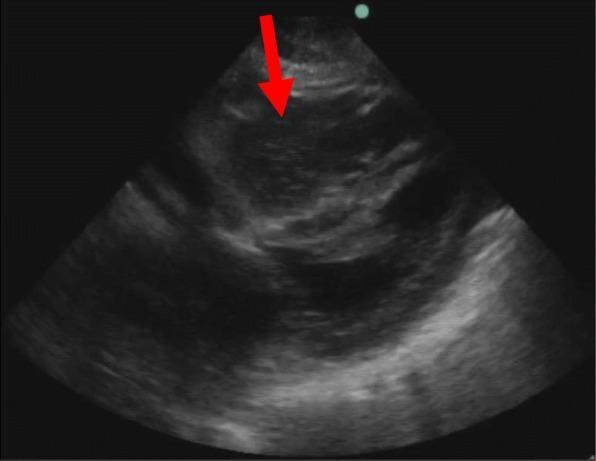
This diagnostic image is a point-of-care ultrasound (POCUS) echocardiogram, likely from a subxiphoid or parasternal view, demonstrating significant pathology in a patient during cardiac arrest. The image reveals a prominent, dilated right ventricle containing a heterogeneous, echogenic mass (indicated by a red arrow). This visual finding is characteristic of a large coagulum or blood clot formation, often associated with massive pulmonary embolism as an underlying cause of cardiac arrest. In comparison, the left ventricle appears markedly underfilled and compressed, exhibiting reduced chamber size relative to the right side. The overall appearance highlights the loss of normal cardiac chamber proportions and the presence of intracavitary thrombus, which are critical diagnostic markers in emergency resuscitation settings. This visual serves as an educational example of identifying reversible causes of cardiac arrest, such as obstructive shock, using real-time ultrasonography.
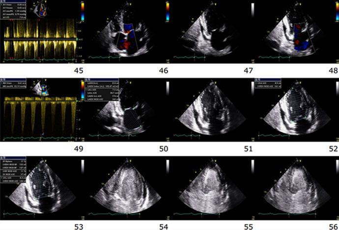
A composite of 12 transthoracic echocardiographic frames and Doppler waveforms documenting the physiological progression toward cardiac standstill during ACLS. The series primarily utilizes apical four-chamber views to demonstrate hemodynamic collapse. Early frames (45-48) show biventricular hypokinesis with color Doppler indicating mitral and tricuspid regurgitation, accompanied by a trace circumferential pericardial effusion. As the sequence progresses through frames 49-53, there is a visible reduction in cardiac wall motion and atrioventricular valve excursion. Starting in frame 54 and intensifying through frame 56, there is an abrupt appearance and rapid progression of dense spontaneous echo contrast (SEC) within the left ventricle, indicative of blood stasis and incipient thrombus formation. The final frames capture the 'dying heart' phenotype, characterized by cessation of mechanical activity, tethered atrioventricular valves with a fixed mid-annular gap, and increasing intracavitary echogenicity across all chambers. This educational series illustrates the echocardiographic correlates of pulseless electrical activity and the transition to cardiac arrest.
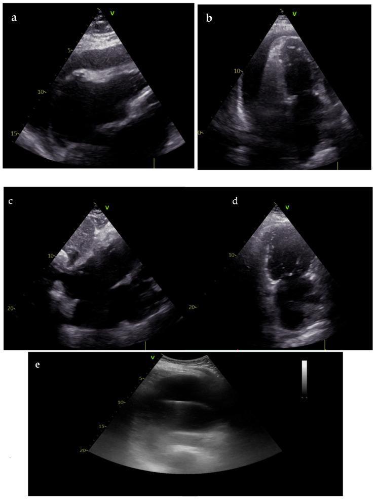
This composite of transthoracic echocardiogram (TTE) images illustrates the diagnosis, procedural management, and resolution of cardiac tamponade. Panels (a) and (b) show parasternal long-axis and apical views during cardiac arrest, revealing a large, circumferential anechoic pericardial effusion. Key diagnostic features include visible compression of the right heart chambers (atrial and ventricular collapse), indicative of high intrapericardial pressure causing tamponade. Panel (c) demonstrates an ultrasound-guided pericardiocentesis, showing the insertion of the drainage needle into the pericardial space. Panels (d) and (e) represent the post-resuscitation phase, displaying near-complete resolution of the pericardial effusion and restoration of normal cardiac chamber dimensions and filling. This series highlights the critical role of Point-of-Care Ultrasound (POCUS) in identifying reversible causes of pulseless electrical activity (PEA) and facilitating life-saving interventions such as emergency pericardiocentesis.
The "Hs" in Medical Terms
The 6 Hs of Cardiac Arrest
| H | Full Name | What It Means |
|---|---|---|
| H1 | Hypovolemia | Low blood volume - most common cause of PEA; treat with IV fluids/blood |
| H2 | Hypoxia | Low oxygen - ensure adequate airway and ventilation |
| H3 | Hydrogen ion (Acidosis) | Metabolic or respiratory acidosis lowering pH; treat with bicarbonate if severe |
| H4 | Hypo-/Hyperkalemia | Abnormal potassium levels causing arrhythmias |
| H5 | Hypothermia | Core body temperature below 35°C; "not dead until warm and dead" |
| H6 | Hypoglycemia | Low blood sugar (included in some ACLS versions; especially in peds/Washington Manual) |
Some older versions list only 5 Hs (omitting hypoglycemia), while current AHA guidelines include 5-6 depending on the institution.
The Paired "Ts" (for completeness)
- Tension pneumothorax
- Tamponade (cardiac)
- Toxins (drug overdose, poisoning)
- Thrombosis - pulmonary (PE)
- Thrombosis - coronary (MI)
Clinical Importance
Other Uses of "Hs" in Medicine
- H&P - History and Physical
- H/O - History of
- hs (lowercase) - hora somni (Latin: "at bedtime") - a prescription abbreviation
- hsCRP - high-sensitivity C-reactive protein
- hsTnT/hsTnI - high-sensitivity troponin (cardiac biomarkers)
Sources: Roberts and Hedges' Clinical Procedures in Emergency Medicine, p. 314; Fuster and Hurst's The Heart, 15th Ed.; The Washington Manual of Medical Therapeutics; ACLS Online - Hs and Ts