
Seminomatous vs non seminomatous tumours
Reading File
Seminomatous tumors are testicular germ cell tumors (GCTs) arising from germinal epithelium - pure seminomas are the most common single histologic type (~50% of GCTs), while non-seminomatous GCTs (NSGCTs) include embryonal carcinoma, yolk sac tumor, choriocarcinoma, and teratoma, or mixed combinations.
Searching sources...
Finding Sources
Finding Sources
Finding Sources
Finding Sources
Good - Bailey and Love's has a dedicated section on testicular tumours. Let me read the relevant blocks.
Reading File
Reading File
Finding Sources
Finding Sources
Finding Sources
Excellent! Now I have the right files. Let me read the key sections from Harrison's and Bailey & Love's, and Smith & Tanagho's.
Reading File
Reading File
Reading File
I now have rich content. Let me read more of the Bailey & Love section on NSGCT types and management, and Harrison's on pathology/staging:
Reading File
Reading File
I now have comprehensive content from multiple authoritative textbooks. Let me also fetch the seminoma gross pathology image from Bailey & Love's and read the staging/management section.
Loading Image
Reading File
I now have everything I need. Let me also read the NSGCT types and management from Bailey & Love:
Reading File
Reading File
Now I have all the content needed for a thorough comparison. Let me also fetch the teratoma image:
Reading File
I now have everything I need to deliver a comprehensive, well-structured comparison. Let me now read the Harrison's staging/management section:
Reading File
I now have all the content needed to write a comprehensive, textbook-quality comparison. Here is the full response:
Seminomatous vs Non-Seminomatous Germ Cell Tumours (GCTs)
Testicular GCTs account for 95% of all testicular neoplasms. The fundamental clinical distinction is between seminomas and non-seminomatous germ cell tumours (NSGCTs) - this split drives every management decision. Importantly, for a tumour to be classified as a pure seminoma, it must be 100% seminoma histologically; any mixed GCT is managed as an NSGCT. - Harrison's Principles of Internal Medicine 22E, p. 748
All adult GCTs share a pathognomonic genetic marker: isochromosome of the short arm of chromosome 12 (i12p). - Bailey and Love's Short Practice of Surgery 28th Ed, p. 1591
Gross Pathology
Seminoma - a homogeneous, pinkish-cream cut surface that appears to compress neighbouring tissue:
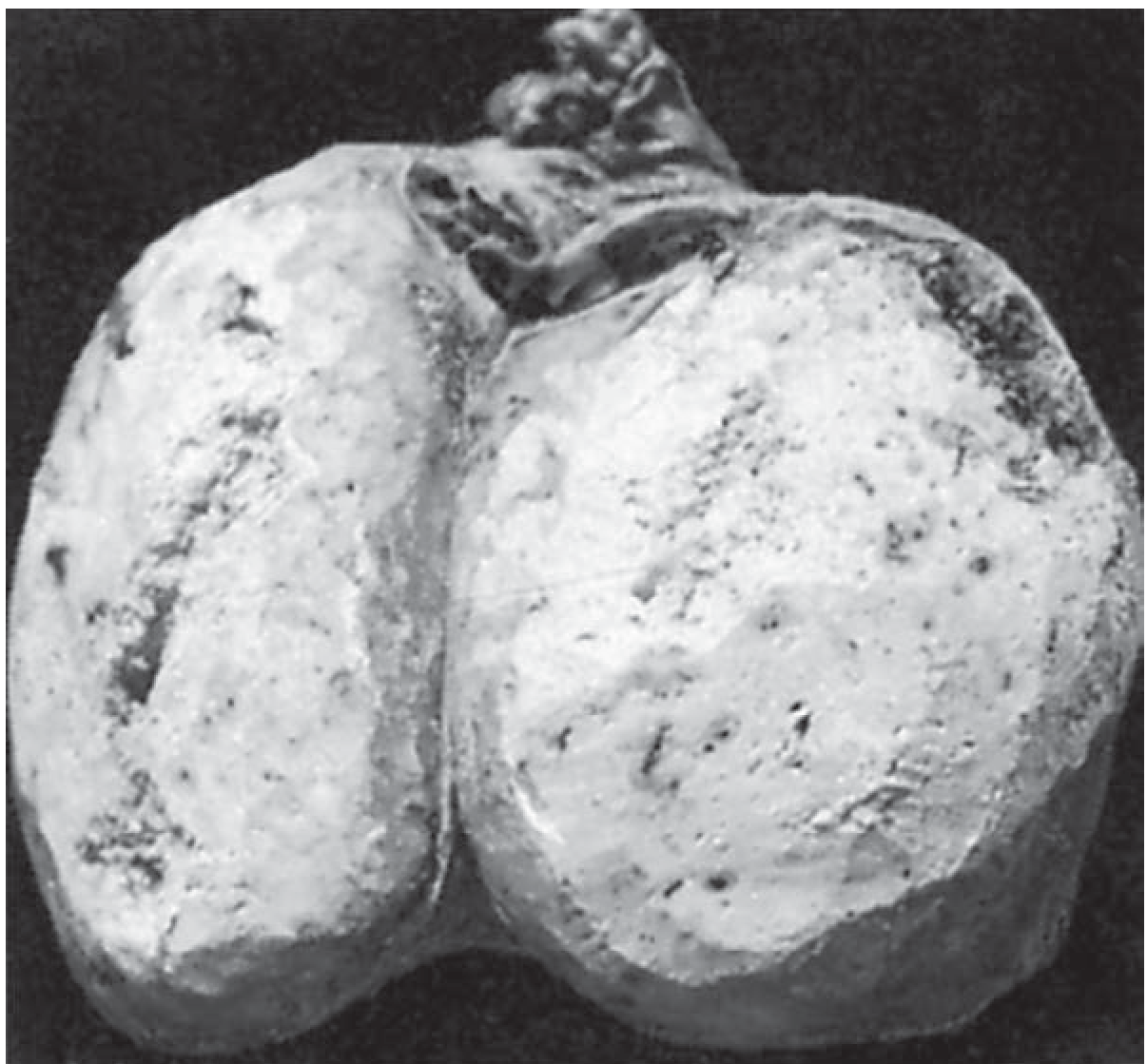
1. Classification
| Feature | Seminoma | NSGCT |
|---|---|---|
| Proportion of GCTs | ~50% | ~50% |
| Peak age | 4th decade | 3rd decade |
| Histologic rule | Must be 100% seminoma | Any mixed GCT = NSGCT |
| Subtypes | Classic seminoma; spermatocytic seminoma (separate entity, >50 yrs, rarely metastasises) | Embryonal carcinoma, Yolk sac tumour, Choriocarcinoma, Teratoma - often mixed |
Sources: Harrison's 22E, p. 748; Bailey & Love's 28th Ed, p. 1590
2. Histology
Seminoma:
- Oval cells with clear cytoplasm and large, rounded nuclei with prominent acidophilic nucleoli
- Sheets of cells resembling spermatocytes, separated by fine fibrous stroma
- Active lymphocytic infiltration = good host response, better prognosis
- May contain syncytiotrophoblastic cells (which can secrete hCG, but AFP is never elevated)
NSGCT subtypes:
- Embryonal carcinoma - most undifferentiated subtype; highly malignant; can differentiate into other subtypes; may invade cord structures; secretes AFP, hCG, both, or neither
- Yolk sac tumour - loose stroma; secretes AFP; commonest childhood testicular malignancy (rare pure form in adults); part of mixed tumour in adults
- Choriocarcinoma - highly malignant; early haematogenous + lymphatic spread; secretes hCG (often at very high levels); extremely rare as pure form
- Teratoma - derived from ≥2 germinal layers (ectoderm, mesoderm, endoderm); may be mature, immature, or malignant; all can metastasise; importantly, teratomas are chemotherapy resistant and must be treated surgically
Sources: Harrison's 22E, p. 748; Bailey & Love's 28th Ed, p. 1591
3. Tumour Markers
This is one of the most clinically tested differences:
| Marker | Seminoma | NSGCT subtypes |
|---|---|---|
| AFP | Never elevated (AFP↑ in a "seminoma" = treat as NSGCT) | Elevated in ~60-70% of NSGCTs; yolk sac (always), embryonal carcinoma (70%), teratoma (38%), choriocarcinoma (0%) |
| hCG (β-hCG) | Elevated in only ~7% (via syncytiotrophoblast cells) | Elevated variably: choriocarcinoma (100%), embryonal (60%), teratoma (25%) |
| LDH | Elevated in high tumour burden | Less specific; elevated in recurrent NSGCT |
AFP is never secreted by seminoma. A seminoma patient with elevated AFP must be managed as NSGCT. - Harrison's 22E, p. 749; Smith & Tanagho's General Urology 19th Ed, p. 394
4. Pattern of Spread
- Both initially spread via lymphatics to para-aortic nodes (lymphatic drainage of the testes follows the gonadal vessels, not inguinal nodes - inguinal nodes are involved only if scrotal skin is breached)
- Seminoma - predominantly lymphatic spread; haematogenous spread is uncommon
- Choriocarcinoma (NSGCT) - early haematogenous spread (lungs most common distant site); most aggressive of all subtypes
- Subsequent spread: retrocrural → mediastinal → supraclavicular nodes, then haematogenous to lungs, liver, bone, brain
5. Sensitivity to Treatment
| Seminoma | NSGCT | |
|---|---|---|
| Radiotherapy | Exquisitely sensitive | Relatively resistant |
| Chemotherapy (platinum-based) | Highly sensitive | All subtypes sensitive except teratoma |
| Teratoma specifically | N/A | Chemotherapy resistant - must be resected |
6. Staging
Both use TNM staging (AJCC):
- Stage I - confined to testis
- Stage II - retroperitoneal lymph node involvement
- Stage III - beyond retroperitoneum and/or distant metastases
The International Germ Cell Cancer Collaborative Group (IGCCCG) risk classification differs between seminoma and NSGCT:
| Risk | Seminoma | NSGCT |
|---|---|---|
| Good | Any primary site; normal AFP, any hCG, any LDH; no non-pulmonary visceral mets | Testicular/retroperitoneal primary; no non-pulmonary visceral mets; markers at S1 level |
| Intermediate | Any primary; nonpulmonary visceral mets present | Testicular/retroperitoneal primary; nonpulmonary visceral mets absent; intermediate marker level |
| Poor | No poor-risk category for seminoma | Mediastinal primary OR nonpulmonary visceral mets OR high markers |
Importantly, seminoma has no "poor risk" IGCCCG category. - Harrison's 22E, p. 749
7. Stage-Based Management
Stage I Seminoma:
- ~70% of seminomas present at Stage I
- ~15% have occult retroperitoneal micrometastases
- Options: active surveillance (preferred), adjuvant paraaortic radiotherapy, or carboplatin monotherapy (1-2 cycles)
- Active surveillance is the standard as the vast majority are cured by orchiectomy alone
Stage I NSGCT:
- Options: active surveillance, nerve-sparing retroperitoneal lymph node dissection (RPLND), or adjuvant BEP chemotherapy x1 cycle
- RPLND is both diagnostic and therapeutic
Advanced disease (Stage II/III) - both types:
- BEP chemotherapy (bleomycin, etoposide, cisplatin) is the backbone
- Post-chemotherapy residual masses in NSGCT require surgical resection (teratoma is chemo-resistant; viable cancer may be present)
- Post-chemotherapy residual masses in seminoma: PET scan is used (FDG-avid = residual disease; PET-negative masses can be observed as they may be necrosis/fibrosis)
Source: Harrison's Principles of Internal Medicine 22E, pp. 749-751; Bailey & Love's 28th Ed
Quick Reference Summary
| Feature | Seminoma | NSGCT |
|---|---|---|
| Age peak | 4th decade | 3rd decade |
| AFP | Never elevated | Often elevated |
| hCG | Rarely (~7%) | Variably elevated |
| Radiotherapy | Sensitive | Resistant |
| Chemo (platinum) | Sensitive | Sensitive (except teratoma) |
| Teratoma | N/A | Chemo-resistant; needs surgery |
| IGCCCG poor risk | Does NOT exist | Exists |
| Spread | Mainly lymphatic | Lymphatic + early haematogenous (especially choriocarcinoma) |
| Prognosis | Generally excellent (~95% cure) | Excellent if good-risk (~95% cure), worse if poor-risk |
| Post-chemo residual | PET to assess; observe if negative | Resect (may be teratoma or viable cancer) |
The ~95% overall cure rate makes testicular GCT a "model for a curable neoplasm" - one of oncology's landmark achievements. - Harrison's Principles of Internal Medicine 22E, p. 748
Syphilitic aneurysm
A syphilitic (luetic) aneurysm is a late complication of tertiary syphilis, typically affecting the ascending aorta and aortic arch. It results from obliterative endarteritis of the vasa vasorum, causing ischemic damage to the aortic media, with loss of elastic tissue and smooth muscle, leading to aortic dilatation.
Searching sources...
Finding Sources
Finding Sources
Excellent sources found. Let me read the key sections in parallel:
Reading File
Reading File
Reading File
Excellent content from three authoritative sources. Let me read the dedicated Syphilitic Aortitis section from Fuster's Heart and the Harrison's infective aortitis section:
Reading File
Reading File
Now let me get the dedicated Fuster/Hurst section on syphilitic aortitis and also check for pathology images:
Reading File
Finding Sources
Now let me read Robbins Pathology for the microscopic pathology details:
Reading File
Reading File
I now have comprehensive, multi-textbook content. Let me compile the definitive answer.
Syphilitic Aneurysm
A syphilitic (luetic) aneurysm is a late cardiovascular complication of tertiary syphilis, typically manifesting 10 to 30 years after the primary Treponema pallidum infection. It predominantly affects the ascending aorta and aortic arch.
Epidemiology & Historical Context
Syphilis was once the most common cause of ascending aortic aneurysms. The advent of penicillin has made it a rarity in developed nations, but syphilitic aneurysms remain a significant cause of morbidity and mortality in other parts of the world. The emergence of HIV in the 1980s caused a rise in syphilis incidence - because syphilitic aortitis presents 10-30 years after primary infection, a future increase in associated aneurysms is possible.
Cardiovascular syphilis develops in approximately 10% of patients with untreated tertiary syphilis and is the primary cause of death in a similar proportion. At autopsy, evidence of aortitis is found in about half of patients who have had untreated syphilis for more than 10 years. - Fuster and Hurst's The Heart, 15th Ed
Pathogenesis - Step by Step
The mechanism is unique and clinically important:
- Spirochetemic seeding: During the spirochetemic phase of primary/secondary syphilis, T. pallidum organisms lodge in the adventitia of the vasa vasorum (the small vessels supplying the aortic wall)
- Inflammatory response: A perivascular lymphocytic and plasma cell infiltrate develops around the vasa vasorum
- Obliterative endarteritis: The vasa vasorum undergo obliterative endarteritis, compromising blood supply to the aortic media
- Medial ischemia and destruction: Ischemic injury causes destruction of collagen and elastic fibers - loss of the structural skeleton of the aortic wall. There is patchy medial necrosis and elastic fiber fragmentation
- Aneurysm formation: The weakened wall dilates, scar tissue forms, and calcification occurs
- Characteristic linear calcification: These changes account for the linear calcification of the ascending aorta visible on chest X-ray - a hallmark radiographic sign
"The initial lesion is an obliterative endarteritis of the vasa vasorum, especially in the adventitia... Destruction of collagen and elastic tissues leads to dilation of the aorta, scar formation, and calcification." - Harrison's Principles of Internal Medicine 22E
The process is most severe in the ascending aorta and arch because the density of vasa vasorum is greatest there. - Fuster and Hurst's The Heart 15th Ed
Morphology
| Feature | Detail |
|---|---|
| Shape | Typically saccular (not fusiform) |
| Location | ~90% in ascending aorta or aortic arch |
| Gross appearance | "Tree bark" appearance of the intima - wrinkled with irregular transverse corrugations, often with superimposed atherosclerotic plaques |
| Dissection | Does NOT cause dissection - the medial scars interrupt the wall transversally, preventing the longitudinal cleavage needed for dissection |
| Calcification | Linear calcification of ascending aorta on CXR |
Cardiovascular Complications
Syphilitic aortitis produces three main cardiovascular manifestations, in order of frequency:
- Aortic regurgitation (most common - 20-30%): Results from aortic root dilatation, not cusp disease (valve cusps are normal but the annulus dilates). The decrescendo murmur is often loudest along the right sternal margin (rather than the typical left sternal border), due to the ascending aortic involvement
- Coronary ostial stenosis (25-30%): Syphilitic inflammation narrows the ostia of the coronary arteries, causing angina pectoris. Historically more common than coronary atherosclerosis as a cause of angina. MI is rare
- Aneurysm formation (least frequent - 5-10% of those with syphilitic aortitis): Typically saccular; may compress or erode adjacent mediastinal structures (e.g., trachea, bronchus, esophagus, superior vena cava, recurrent laryngeal nerve). An aneurysm may present as a pulsating mass bulging through the anterior chest wall
Sources: Fuster and Hurst's The Heart 15th Ed; Goldman-Cecil Medicine; Harrison's 22E
Clinical Presentation
- Usually asymptomatic for years; often found incidentally on CXR
- Symptom onset: 15-30 years after initial infection
- Symptoms may arise from:
- Aortic regurgitation (dyspnoea, wide pulse pressure, diastolic murmur loudest at right sternal border)
- Coronary ostial stenosis (angina)
- Compression of adjacent structures: dysphagia (esophagus), stridor/dyspnoea (trachea/bronchus), hoarseness (recurrent laryngeal nerve), SVC syndrome
- Rupture (major complication - high mortality)
- Between 10-20% of patients with cardiovascular syphilis have coexistent neurosyphilis - always screen
Diagnosis
| Test | Finding |
|---|---|
| Chest X-ray | Linear calcification of the ascending aorta; widened mediastinum; saccular aneurysm silhouette |
| Serology | RPR / VDRL (non-treponemal, screening), FTA-ABS (treponemal, confirmatory) |
| Echocardiography | Aortic root dilatation, aortic regurgitation, aneurysm |
| CT/MRI | Confirms aneurysm size, extent, and complications |
| Intraoperative | "Tree bark" appearance; uniform involvement of ascending aorta surface; inflammation in all three layers; sparing of the aortic root (contrast with atherosclerotic disease) |
Treatment
- Antibiotics: Penicillin G remains first-line for the underlying syphilitic infection. Whether this retards progression once aortitis is established is not clearly proven
- Surgery: Surgical excision and repair is required for symptomatic aneurysms. Without surgery, symptomatic syphilitic aortic aneurysms carry a high mortality rate. Early antibiotic treatment of primary syphilis is the most important preventive measure
- The prognosis for uncomplicated syphilitic aortitis is comparable to the general population, but becomes poor once aneurysms become symptomatic
Key Distinguishing Points (vs. Other Aortic Aneurysms)
| Feature | Syphilitic | Atherosclerotic | Marfan/Cystic Medial Degen. |
|---|---|---|---|
| Location | Ascending aorta/arch | Descending/abdominal | Ascending (aortic root) |
| Shape | Saccular | Fusiform | Fusiform |
| Dissection | Rarely/never | Possible | Common |
| Mechanism | Obliterative endarteritis of vasa vasorum | Plaque-induced medial thinning | Fibrillin-1/TGF-β defect |
| CXR sign | Linear ascending aortic calcification | Egg-shell calcification | - |
| Histology | Obliterative endarteritis, medial fibrosis, scarring | Atherosclerotic plaque, thinned media | Mucoid degeneration of media |
Sources: Harrison's Principles of Internal Medicine 22E; Fuster and Hurst's The Heart 15th Ed; Goldman-Cecil Medicine; Robbins & Kumar Basic Pathology; Schwartz's Principles of Surgery 11th Ed
DMD vs Beckers MD
Reading File
Duchenne MD is caused by frameshift/nonsense mutations in the dystrophin gene leading to complete absence of dystrophin, while Becker MD results from in-frame mutations producing a truncated but partially functional dystrophin - hence Becker is milder and later-onset.
Searching sources...
Finding Sources
Finding Sources
Excellent sources. Let me read the key sections in parallel from Adams & Victor's Neurology, Robbins Pathology, Goldman-Cecil, and Harriet Lane:
Reading File
Reading File
Reading File
Good content. Let me now read more of the Goldman-Cecil section on Becker MD and treatment, and get the Adams & Victor detailed clinical features:
Reading File
Reading File
Now let me read the Becker MD section from Goldman-Cecil and the Harriet Lane clinical features:
Reading File
Reading File
Finding Sources
Finding Sources
Finding Sources
Finding Sources
Reading File
Now let me get the Adams & Victor clinical features section for DMD/BMD:
Reading File
Reading File
Excellent detailed content. Let me read the Becker section and the cardiac/respiratory complications in one more pass:
Reading File
I now have comprehensive, multi-textbook content. Let me compile the full comparison.
Duchenne Muscular Dystrophy (DMD) vs Becker Muscular Dystrophy (BMD)
Both are X-linked dystrophinopathies - allelic disorders caused by mutations in the same gene (the DMD gene on the X chromosome), differing in the type of mutation and the resulting amount and function of the dystrophin protein. They represent a spectrum of severity rather than two entirely distinct diseases.
The "Reading Frame Rule" - The Core Concept
This is the fundamental molecular distinction:
| DMD | BMD | |
|---|---|---|
| Mutation type | Frameshift or nonsense mutation - disrupts the reading frame | In-frame deletion/duplication - preserves the reading frame |
| Resulting protein | Dystrophin is completely absent | Dystrophin is present but truncated/abnormal (partially functional) |
| Consequence | Severe, early-onset disease | Milder, later-onset disease |
"Whereas dystrophin is absent in patients with the Duchenne phenotype, it is present but structurally abnormal in the Becker type. Moreover, phenotypes that fall between the classic Duchenne and Becker forms exist and are characterized by a lower-than-normal amount of dystrophin." - Adams and Victor's Principles of Neurology 12th Ed
Most mutations are deletions (~65%), with duplications and point mutations making up the rest. The same deletion can cause DMD in one patient and BMD in another depending on whether it disrupts the reading frame. - Henry's Clinical Diagnosis and Management
Pathogenesis
The mechanism of muscle damage is identical in both:
- Normal function: Dystrophin anchors the sarcomere F-actin cytoskeleton to the sarcolemma (cell membrane) via the dystrophin-associated protein (DAP) and dystroglycan complex, which in turn connects to the extracellular matrix via merosin/laminin
- Loss of dystrophin disrupts this mechanical link
- Sarcolemmal breaks and tears occur during muscle contraction
- Extracellular calcium leaks into the fiber
- Calcium activates proteases, increases protein degradation, causes myofiber necrosis
- CK and other muscle enzymes leak into the serum
- Repeated cycles of necrosis → fibrosis → fat replacement → muscle wasting
"Loss of dystrophin leads to a parallel loss of DAPs and to disruption of the dystroglycan-protein complex. This change renders the sarcolemma susceptible to breaks and tears during muscle contraction." - Adams and Victor's Principles of Neurology 12th Ed
Dystrophin is also expressed in cardiac and smooth muscle, and in the brain (a slightly different isoform in neurons, astrocytes, Purkinje cells) - accounting for cardiac involvement and cognitive effects.
Genetics
| Feature | DMD | BMD |
|---|---|---|
| Inheritance | X-linked recessive | X-linked recessive |
| Gene | DMD gene (Xp21.2) - largest known human gene, >2 Mb, 79 exons | Same DMD gene |
| Incidence | ~1 in 3,300-5,000 male births | ~1 in 18,500 male births (3-6 per 100,000) |
| De novo mutations | ~30% of cases | ~15% of cases |
| Affected sex | Almost exclusively males | Almost exclusively males |
| Female carriers | Usually asymptomatic; 2.5-10% develop myalgias, proximal weakness, cardiomyopathy; rarely full Duchenne phenotype (XO karyotype/skewed X-inactivation) | Same |
Clinical Features
Age of Onset
| DMD | BMD | |
|---|---|---|
| Onset | Typically recognised by age 2-5 years (almost always before age 6) | Mean onset age 12 years; range 5-45 years |
| Wheelchair dependence | By age 12 (early second decade) | Many walk well into adult life |
Motor Features
DMD:
- Delayed motor milestones; falls frequently, difficulty running/climbing stairs
- Proximal muscle weakness: iliopsoas, quadriceps, gluteals first → pretibial muscles (foot drop, toe-walking) → pectoral girdle (serrati, pectorals, latissimus, biceps, brachioradialis)
- Waddling gait, excessive lumbar lordosis, protuberant abdomen
- Gower's sign: To rise from the floor, the child assumes a four-point position and "walks" hands up the thighs to extend the trunk - classic sign of proximal lower limb weakness (first described by Gowers in 1879)
- Pseudohypertrophy of calves (also deltoids, lateral vasti) - firm, rubbery feel; represents fat and fibrous tissue replacing degenerated muscle fibres. In early stages there may be true hypertrophy, which gives way to pseudohypertrophy
- Progressively loses the ability to raise head fully off the bed (absent in DMD)
- Tendon reflexes diminish and disappear as muscle fibers are lost; ankle jerk is last to go
- Contractures: Heel-cord (Achilles tendon) contractures, then elbow and knee flexion contractures; scoliosis follows loss of ambulation
BMD:
- Weakness and pseudohypertrophy in the same distribution as DMD
- Onset much later and course much slower
- Retains ability to raise head off the bed (distinguishes from DMD)
- May serve in the military with disease undetected
- If maternal uncles with the disease are still walking, diagnosis is relatively easy
Cognitive / Neuropsychiatric Features
| DMD | BMD | |
|---|---|---|
| Intellectual disability | Average IQ 85; ~25% have IQ <70; range 40-130. Also associated with ADHD, autism, learning disorders | Usually normal mentation |
| Mechanism | Deficiency of brain-specific dystrophin isoforms in neurons, astrocytes, Purkinje cells | Less prominent |
Cardiac Involvement
| DMD | BMD | |
|---|---|---|
| Cardiomyopathy | Dilated cardiomyopathy - nearly universal by adolescence; arrhythmias common. ECG: prominent R waves in right precordial leads, deep Q waves in left precordial and limb leads (basal left ventricular wall fibrosis) | Cardiomyopathy occurs but far less frequent and later; heart failure may be the initial presenting manifestation in some BMD patients |
| Cause of cardiac death | Heart failure, arrhythmias | Heart failure, arrhythmias (later course) |
Respiratory Involvement
| DMD | BMD | |
|---|---|---|
| Respiratory failure | Progressive; primary cause of death (late 20s-early 30s without ventilation) | May occur but in later course |
| Management | Noninvasive ventilation prolongs life into 3rd-4th decade | Screening still required |
Investigations
| Investigation | DMD | BMD |
|---|---|---|
| Serum CK | 25-200x normal (markedly elevated) | Elevated, but typically less than in DMD |
| EMG | Myopathic: fibrillations, positive waves, brief low-amplitude polyphasic motor unit potentials | Similar myopathic pattern |
| Genetic testing | Confirms frameshift/nonsense mutation; positive in ~90-95% | Confirms in-frame mutation |
| Muscle biopsy | Absent dystrophin on immunostaining (except "revertant fibres"); chronic myopathy pattern (variation in fibre size, necrosis, fibrosis, fat replacement) | Decreased/abnormal molecular weight dystrophin on immunostaining/Western blot; less severe changes |
| Cardiac | Echo/ECG: dilated cardiomyopathy, ECG changes | Echo/ECG monitoring required |
Treatment
Both conditions require multidisciplinary management:
DMD-specific treatments:
| Treatment | Details |
|---|---|
| Corticosteroids | Prednisone 0.75 mg/kg/day OR deflazacort 0.9 mg/kg/day - prolong ambulation, improve muscle strength; less weight gain with deflazacort; may slow scoliosis progression |
| Exon-skipping therapies (antisense oligonucleotides) | Induce exon skipping to convert frameshift → in-frame mutation (effectively converting DMD toward BMD): Eteplirsen (exon 51 skip), Golodirsen + Viltolarsen (exon 53 skip), Casimersen (exon 45 skip) - all conditionally FDA-approved |
| Gene therapy | AAV9 carrying mini-dystrophin under muscle-specific promoter - phase Ib trials promising; CRISPR-Cas9 gene editing in research stage |
| Cardiac | ACE inhibitors (slow myocardial fibrosis), beta-blockers |
| Respiratory | Noninvasive ventilation (BiPAP/CPAP) |
| Orthopedic | Physical therapy, Achilles tendon release, spinal fusion for scoliosis |
BMD:
- Largely supportive; corticosteroids rarely used
- Cardiac monitoring and management (heart transplantation in severe cardiomyopathy)
- Respiratory function monitoring
Prognosis
| DMD | BMD | |
|---|---|---|
| Ambulation lost | By ~age 12 | Retained well into adult life |
| Survival without ventilation | Late adolescence; <25% survive beyond age 25 | Variable - many have normal lifespan |
| Survival with ventilation | Often into 3rd-4th decade | Generally longer |
| Primary cause of death | Respiratory failure + pneumonia; cardiac (heart failure/arrhythmia) | Heart failure, respiratory complications (in later course) |
Quick Comparison Table
| Feature | DMD | BMD |
|---|---|---|
| Gene | DMD (Xp21.2) | DMD (Xp21.2) |
| Mutation | Frameshift/nonsense | In-frame deletion/duplication |
| Dystrophin | Absent | Present but truncated/abnormal |
| Onset | 2-5 years | 5-45 years (mean 12) |
| Wheelchair | By age 12 | Often never or late adult life |
| Pseudohypertrophy | Calves (prominent) | Calves (present) |
| Gower's sign | Present | Variable |
| Head raising off bed | Lost early | Retained |
| CK | 25-200x normal | Elevated (less than DMD) |
| Cognition | Often mildly impaired (avg IQ 85) | Usually normal |
| Cardiomyopathy | Nearly universal, early | Less frequent, later; may be initial presentation |
| Respiratory failure | Major cause of death (late 20s-30s) | Later course |
| Steroids | Mainstay (prednisone/deflazacort) | Rarely used |
| Exon-skipping | FDA-approved (eteplirsen, etc.) | Not a primary treatment |
| Prognosis | Fatal, 3rd-4th decade with support | Many have near-normal lifespan |
Sources: Adams and Victor's Principles of Neurology 12th Ed; Goldman-Cecil Medicine; Harriet Lane Handbook 23rd Ed; Robbins & Kumar Basic Pathology; Henry's Clinical Diagnosis and Management
This is a shared conversation. Sign in to Orris to start your own chat.