
Make ppt on CNS tumor reference from Saboston text book surgery for neurosurgery Resident amke atleast 40 slides
Research CNS (central nervous system) tumors comprehensively using the medical-library skill (query_library_db tool) for building a 40+ slide neurosurgery resident presentation referencing Sabiston Textbook of Surgery (and other available neurosurgery/surgery textbooks in the library if Sabiston coverage is thin). Instructions: 1. First read /skills/readonly/medical-library/SKILL.md fully and follow its navigation protocol (query_library_db only for discovery). 2. Search the library (headings_fts and paragraphs_fts) for topics including: CNS tumor classification (WHO classification), epidemiology of brain tumors, gliomas (low-grade astrocytoma, anaplastic astrocytoma, glioblastoma multiforme), oligodendroglioma, meningioma, medulloblastoma and other embryonal/pediatric CNS tumors, ependymoma, pituitary adenoma, craniopharyngioma, vestibular schwannoma/acoustic neuroma, CNS metastases/metastatic brain tumors, primary CNS lymphoma, spinal cord tumors, clinical presentation of brain tumors (signs/symptoms, raised intracranial pressure, seizures, focal deficits), diagnostic imaging (CT/MRI features) of brain tumors, surgical management principles (craniotomy, stereotactic biopsy, awake craniotomy, image-guided/neuronavigation surgery, intraoperative MRI), adjuvant therapy (radiotherapy, chemotherapy, temozolomide, stereotactic radiosurgery/gamma knife), and postoperative complications/management in neurosurgical oncology. 3. Specifically search for "Sabiston" textbook content on CNS tumors and neurosurgery/surgical oncology of the brain - check what book titles exist in the library (query v_book_overview) and note whether Sabiston Textbook of Surgery is present; if not present, identify the best available surgery/neurosurgery textbook(s) covering CNS tumors and use those instead, being explicit about which book each fact comes from. 4. For every fact gathered, record book_title and approximate page/line reference (line_start/line_end or line_number) so citations can be given as "- Book Title, p. XXX" or similar. 5. Organize your findings into a structured outline covering these sections (each will become several slides): (a) Introduction/overview & epidemiology, (b) Classification (WHO 2021 CNS tumor classification), (c) Clinical presentation & signs, (d) Diagnostic workup & imaging, (e) Gliomas (low grade, high grade/GBM), (f) Meningioma, (g) Pituitary adenoma & craniopharyngioma, (h) Vestibular schwannoma/acoustic neuroma, (i) Medulloblastoma & pediatric embryonal tumors, (j) Ependymoma, (k) CNS metastases, (l) Primary CNS lymphoma, (m) Spinal cord tumors, (n) Surgical principles & approaches, (o) Adjuvant therapy (radiotherapy/chemotherapy/targeted therapy), (p) Postoperative care & complications, (q) Prognosis/outcomes. Return a single detailed, well-organized report with bullet-point facts grouped by the above sections, each bullet citing its source book title and page/line reference. Be thorough and specific (grading systems, key differentiating imaging/histology features, typical patient demographics, management algorithms, survival stats if available). This report will be used directly to write presentation slide content, so make it information-dense and factually precise. Do not fabricate information not found in the library or your own solid medical knowledge - flag any gaps.
Find real medical images (radiology, histology, anatomy diagrams) relevant to a neurosurgery presentation on CNS (central nervous system) tumors, using the search_image tool and query_images_db tool. Search for and return image URLs + captions for each of these topics (try multiple query phrasings per topic if first search doesn't return great results): 1. MRI of glioblastoma multiforme (brain, contrast enhancing, ring enhancement) 2. MRI or CT of low-grade astrocytoma / diffuse glioma 3. Meningioma MRI (dural-based, extra-axial mass) 4. Meningioma histology (whorls, psammoma bodies) 5. Vestibular schwannoma / acoustic neuroma MRI (cerebellopontine angle) 6. Pituitary adenoma MRI (sella turcica) 7. Craniopharyngioma MRI 8. Medulloblastoma MRI (posterior fossa, cerebellum, pediatric brain tumor) 9. Ependymoma MRI 10. Brain metastasis MRI (multiple lesions) 11. Primary CNS lymphoma MRI 12. Glioblastoma histology (pseudopalisading necrosis, microvascular proliferation) 13. Normal brain anatomy diagram / lobes of the brain diagram 14. Craniotomy surgical approach / neurosurgery intraoperative photo 15. WHO grading of brain tumors diagram (if available) For each topic, run search_image(query=..., top_k=3) and pick the single best-matching, most relevant, real (non-duplicate) image. Return a final list formatted as: Topic | image_url | caption Only include images that are genuinely relevant and high-confidence matches; skip a topic if no reasonable match exists rather than forcing an irrelevant image. Return this list as your final answer, nothing else needed.
glioblastoma multiforme MRI brain ring enhancing lesion
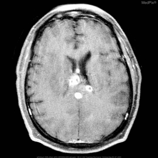
**Imaging Modality:** Axial T1-weighted contrast-enhanced Magnetic Resonance Imaging (MRI) of the brain.
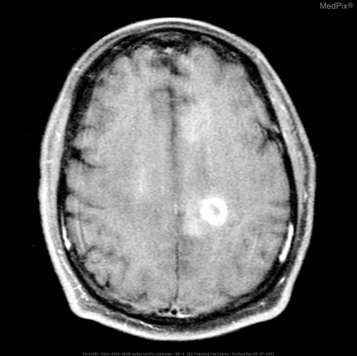
**Imaging Modality:** Axial T1-weighted post-contrast Magnetic Resonance Imaging (MRI) of the brain.
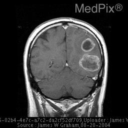
**Imaging Modality:** Coronal T1-weighted post-contrast Magnetic Resonance Imaging (MRI) of the brain.
low-grade astrocytoma diffuse glioma MRI brain
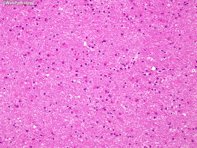
Imaging modality: light microscopy of a hematoxylin and eosin stained brain tissue section from a cerebral hemisphere tumor. Primary subject: diffuse astrocytoma, WHO Grade II (infiltrating astrocytoma). Specimen: brain parenchyma/tumor tissue. Imaging perspective: microscopic histology field. Anatomical context: central nervous system, brain (cerebral hemispheres), intra-axial glial neoplasm displaying diffuse infiltration. Visual features: moderately increased cellularity with mildly enlarged, variably hyperchromatic nuclei; mild nuclear pleomorphism; delicate, interconnected astrocytic processes creating a fibrillary background; abundant eosinophilic cytoplasm in some astrocyte-like cells; absence of overt necrosis or microvascular proliferation in this field; mitotic activity not evident at this magnification. Pathology: low-grade, infiltrative glioma consistent with WHO Grade II astrocytoma; preserves overall brain architecture; growth pattern extraparenchymal infiltration challenging complete resection; lacks features defining higher grades (no brisk mitoses, microvascular proliferation, or necrosis). Diagnostic significance: confirms low-grade astrocytic neoplasm; implications for prognosis and management; potential progression to higher-grade gliomas (III–IV) with time or treatment resistance; imaging correlation with MRI typically demonstrates non-destructive, infiltrative lesion; informs surgical planning, adjuvant radiotherapy/chemotherapy decisions; differential considerations include oligodendroglioma and pilocytic astrocytoma in specific contexts. Clinical relevance: presents with seizures/ focal deficits in adults (4th–6th decades), guiding neuro-oncologic workup and longitudinal surveillance. This image exemplifies low-grade glial tumor histology.
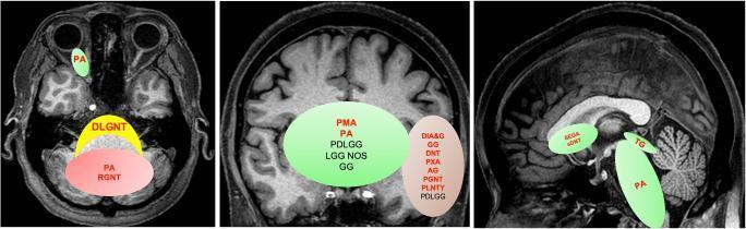
This composite educational illustration demonstrates the spatial clustering and preferred neuroanatomical locations of Pediatric Low-Grade Neuroepithelial Tumor (PLGNT) phenotypes. Three T1-weighted MRI planes (axial, coronal, and sagittal) are used as templates with overlaid color-coded regions and tumor abbreviations. Red/pink zones indicate cerebellar clustering, including Pilocytic Astrocytoma (PA) and Rosette-forming Glioneuronal Tumor (RGNT). Green zones represent midline structures: PA is shown in the optic pathway (axial) and brainstem (sagittal); Pilomyxoid Astrocytoma (PMA) and others are localized to the hypothalamic/chiasmatic region (coronal); and Tectal Glioma (TG) is at the midbrain. Brown/tan zones signify hemispheric locations, encompassing cortical and deep brain tumors such as Ganglioglioma (GG), Dysembryoplastic Neuroepithelial Tumor (DNT), Pleomorphic Xanthoastrocytoma (PXA), and Angiocentric Glioma (AG). A yellow zone in the axial view highlights the Diffuse Leptomeningeal Glioneuronal Tumor (DLGNT) along the midline leptomeningeal surfaces. The diagram serves as a diagnostic guide for neuroradiology, emphasizing that specific histological phenotypes are spatially enriched based on their underlying molecular drivers.
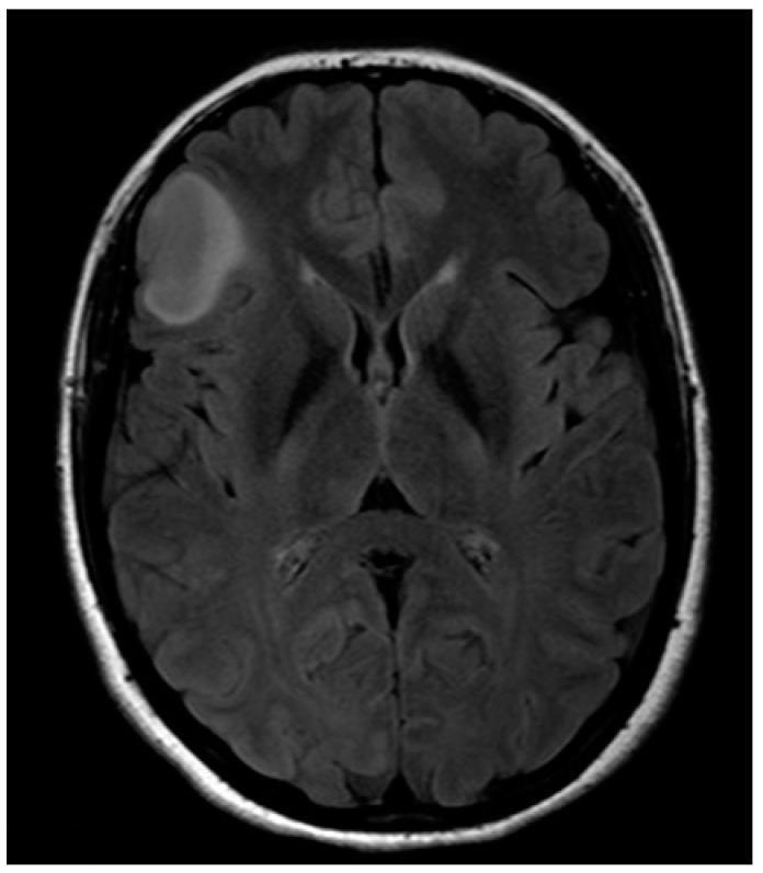
An axial MRI scan of the brain, utilizing Fluid-Attenuated Inversion Recovery (FLAIR) or T2-weighted sequencing, demonstrates a well-circumscribed hyperintense lesion in the right fronto-opercular region. The lesion is ovoid and relatively homogeneous, characteristic of a diffuse low-grade glioma, such as a low-grade astrocytoma. It is located near cortical areas involved in language and motor function, specifically the right-sided equivalent of Broca's area. While the lesion causes localized effacement of adjacent cortical sulci, there is no evidence of a significant midline shift, ventricular compression, or extensive peritumoral edema. The surrounding brain parenchyma, including the contralateral hemisphere and the ventricular system, appears structurally preserved. This diagnostic image is significant for neurosurgical planning, particularly in identifying the proximity of the tumor to eloquent white matter tracts like the inferior fronto-occipital fascicle (IFOF) and motor pathways.
meningioma MRI dural-based extra-axial mass brain
**Imaging Modality:** Coronal magnetic resonance imaging (MRI).
**Imaging Modality:** Magnetic Resonance Imaging (MRI), Coronal plane.
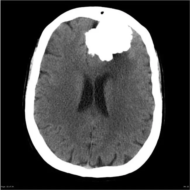
Imaging modality and technique: Non-contrast CT of the brain is the primary image, with axial sections demonstrating a well circumscribed, extra-axial mass located at the anterior cranial fossa near the frontal convexity. The lesion is markedly hyperdense due to dense calcification, with coarse calcifications producing bright foci that obscure the underlying cortex. The mass abuts the dura and appears to cause mild mass effect on the adjacent frontal lobes, but there is no midline shift or intraventricular hemorrhage. The appearance is classic for a calcified meningioma; the tumor originates from the dura and demonstrates dural-based expansion, with a possible dural tail on the MRI counterpart. The MRI image (not shown here) would typically show a lesion with variable T1/T2 signal and strong, homogeneous enhancement after gadolinium, consistent with meningioma and predominance of calcified components. Clinically, incidental discovery of a calcified meningioma aligns with a slow-growing, benign meninges-derived neoplasm. Differential considerations include psammomatous meningioma, calcified oligodendroglioma (intra-axial), calcified metastasis, and dural-based schwannoma; however, location and extra-axial behavior with dense calcifications favor meningioma. Diagnostic significance is that surgical resection often yields cure; prognosis depends on size, location, edema, and invasion; radiologic monitoring is advised.
meningioma histology whorls psammoma bodies
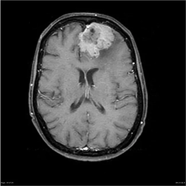
MRI with gadolinium contrast (Magnetic Resonance Imaging, post-contrast T1-weighted sequences) reveals a large extra-axial, dural-based mass projecting from the convexity of the frontal/temporal lobes. The lesion abuts the dura with an irregular but well-circumscribed silhouette and demonstrates intense, homogeneous enhancement after gadolinium administration. On axial images the mass displaces adjacent cortex and may produce mild peritumoral edema without invading the underlying brain parenchyma; a classic dural tail may be present though not required for diagnosis. The imaging pattern is typical for meningioma: a slow-growing, benign intracranial tumor arising from arachnoidal cap cells; the size can reach considerable extent in “silent” regions of cortex. Correlating histology from biopsy or resection shows meningothelial cells arranged in whorls with psammoma bodies; no evidence of brain invasion supports WHO grade I meningioma. Diagnostic significance: post-contrast, uniformly enhancing dural-based lesion with extra-axial location is highly specific for meningioma; radiologic differential includes hemangiopericytoma, dural metastasis, or dural-based schwannoma, but the presence of psammoma bodies is characteristic histology. Clinically, incidental discovery emphasizes that slow-growing meningiomas can attain large size before symptoms; management includes surgical excision with complete or subtotal resection and radiologic follow-up to monitor recurrence. These features guide biopsy decisions and surgical planning.
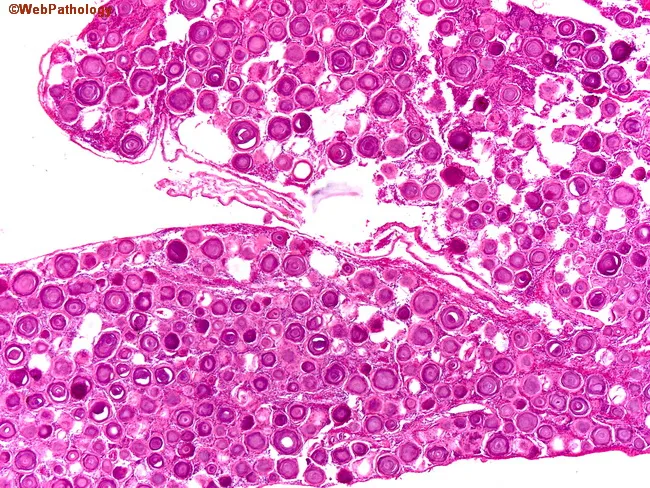
Light microscopy of a spinal meningioma specimen. Imaging modality: histology with Hematoxylin and Eosin staining. The tissue shows classic whorled sheets of uniform meningothelial cells with eosinophilic cytoplasm and oval to vesicular nuclei. Interspersed throughout the tumor are numerous psammoma bodies, concentric laminated calcifications that range from small to large; these calcifications can be basophilic or eosinophilic depending on staining and maturation. The psammomatous variant demonstrates dense sandstone-like lamination within the tumor matrix and occasional focal ossification in advanced foci. The background stroma is fibrous, with low cellular atypia and low mitotic activity, consistent with a benign WHO grade I meningioma. The overall architecture is organized into interrupted lobules and tight cellular whorls arising from meningothelial cells, with abundant extracellular collagen. The presence of multiple psammoma bodies is a diagnostic hallmark distinguishing psammomatous meningioma from other spinal neoplasms. Diagnostic significance: histopathology confirms meningothelial differentiation with psammomatous calcifications; supports a benign prognosis but may cause compressive myelopathy depending on size and location. Clinical correlation: spinal canal mass in older women presenting with progressive myelopathy or radiculopathy; treatment is surgical resection; prognosis is favorable with complete excision; radiologic correlation (MRI) typically shows dural-based enhancement.
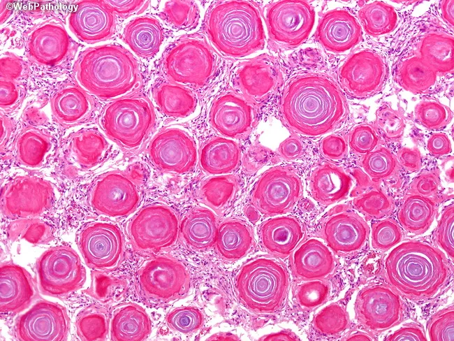
Bright-field histology of an intracranial meningioma shows densely packed meningothelial cells arranged in characteristic whorls and sheets, punctuated by numerous psammoma bodies. In this high-power view, concentric lamellations form calcified, spherical structures of varying size that appear as rings within a pink eosinophilic matrix. The psammoma bodies, composed of laminated calcium deposits, can constitute more than half of the tumor mass in psammomatous meningioma and often obscure the underlying meningothelial component. The surrounding cells display spindle-to-polygonal morphology with indistinct cytoplasmic borders, oval nuclei, and low mitotic activity, consistent with a benign, slow-growing lesion. The calcified nodules are densely eosinophilic and well delineated from the stroma, giving a gritty, laminated appearance. The background stroma is collagenous with occasional microcystic changes; vascularity is variable. This histologic pattern supports a diagnosis of psammomatous meningioma, a histologic variant of meningioma typically classified as WHO grade I. Clinical significance includes a favorable prognosis after surgical resection, with calcification often contributing to a firm, dural-based mass. Differential considerations include other calcified dural-based lesions such as ossified/psammomatous variants and less likely calcified metastases. Immunohistochemical staining would typically reveal EMA and vimentin positivity with keratin negativity, reinforcing meningothelial origin and distinguishing it from glial neoplasms. In practice.
vestibular schwannoma acoustic neuroma MRI cerebellopontine angle
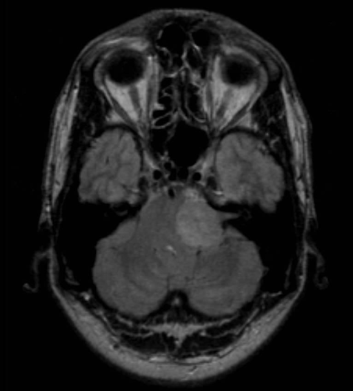
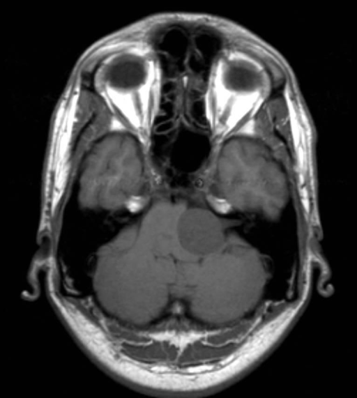
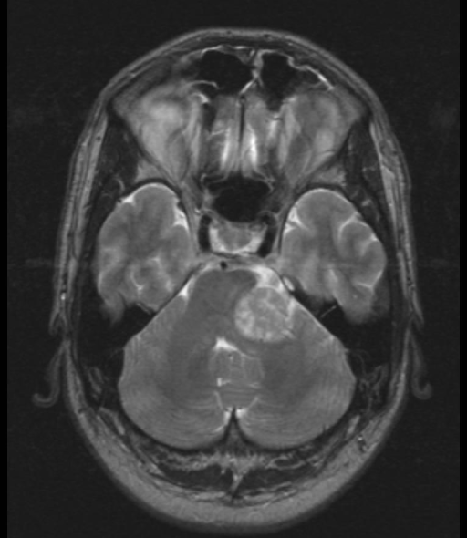
pituitary adenoma MRI sella turcica
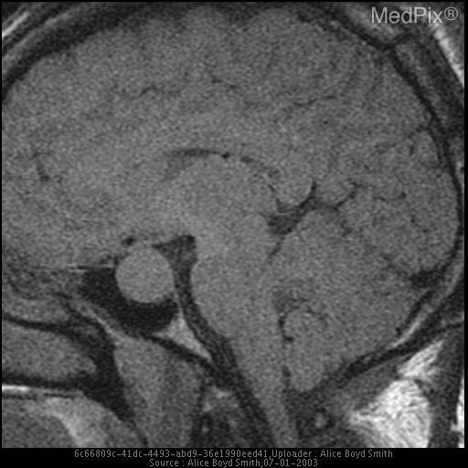
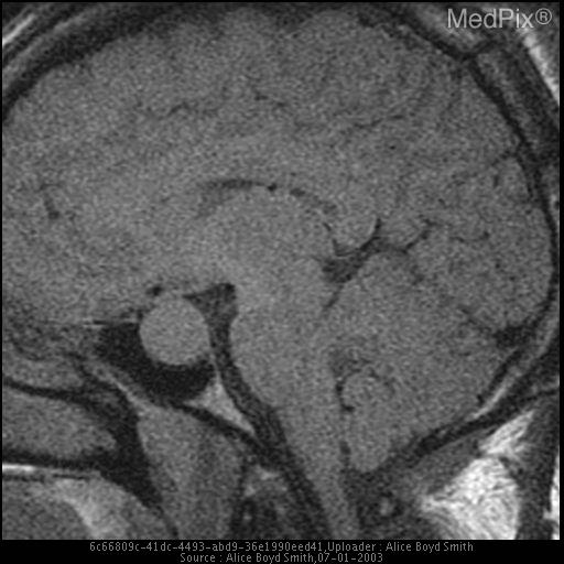
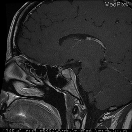
craniopharyngioma MRI brain
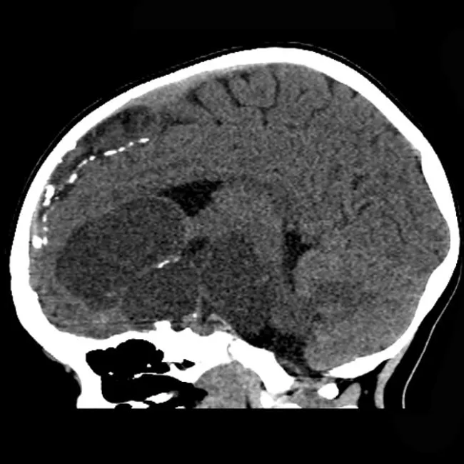
Computed Tomography (CT) of the pediatric brain, non-contrast, sagittal orientation, reveals a large suprasellar multicystic lesion with punctate and coarse calcifications. The mass occupies the sellar/suprasellar space, abutting the hypothalamic-pituitary axis, and extends into the middle cranial fossa with midline shift and displacement of the optic apparatus. The lesion displays mixed density with cystic components and calcified nodules, a hallmark of adamantinomatous craniopharyngioma in children. There may be secondary hydrocephalus from obstruction of the third ventricle. The neuroimaging phenotype includes multicystic architecture, calcifications, and suprasellar location; these findings strongly support a craniopharyngioma over other posterior fossa or suprasellar tumors. The resection specimen confirmed adamantinomatous histology, with characteristic squamous epithelium and calcified enamel-like nodules. Clinically relevant diagnosis includes pediatric brain tumor with potential endocrine dysfunction and visual compromise. Differential diagnoses to consider include germinoma and pilocytic astrocytoma in the pediatric population. This case demonstrates the quintessential radiologic signature of adamantinomatous craniopharyngioma and highlights the necessity of correlating CT findings with surgical pathology for definitive diagnosis and guiding therapeutic planning. Correlation with endocrinology imaging is essential to assess pituitary function; follow-up MRI is recommended to evaluate residual/recurrent disease; treatment typically involves subtotal resection with possible adjuvant radiotherapy and endocrinology management.
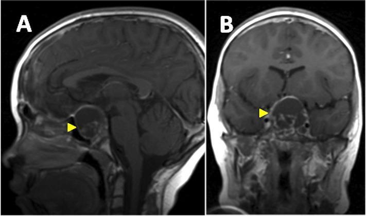
This diagnostic image consists of two panels showing T1-weighted Magnetic Resonance Imaging (MRI) of the brain with contrast. Panel A is a sagittal view, and Panel B is a coronal view. Both panels highlight a recurrent cystic lesion (indicated by a yellow arrowhead) located in the sellar and suprasellar regions, characteristic of a craniopharyngioma. The cyst is well-circumscribed, ovoid, and exhibits a hyperintense signal relative to the brain parenchyma, suggesting high protein content or contrast enhancement of the cyst wall. In the sagittal view, the mass is seen anterior to the brainstem, causing significant superior displacement and distortion of the third ventricle floor. In the coronal view, the cyst extends superiorly from the skull base towards the midline, exerting a mass effect on adjacent neuroanatomical structures including the optic chiasm region. These findings are clinically significant for monitoring post-radiotherapy recurrence of craniopharyngioma and assessing pressure effects on the ventricular system and visual pathways.
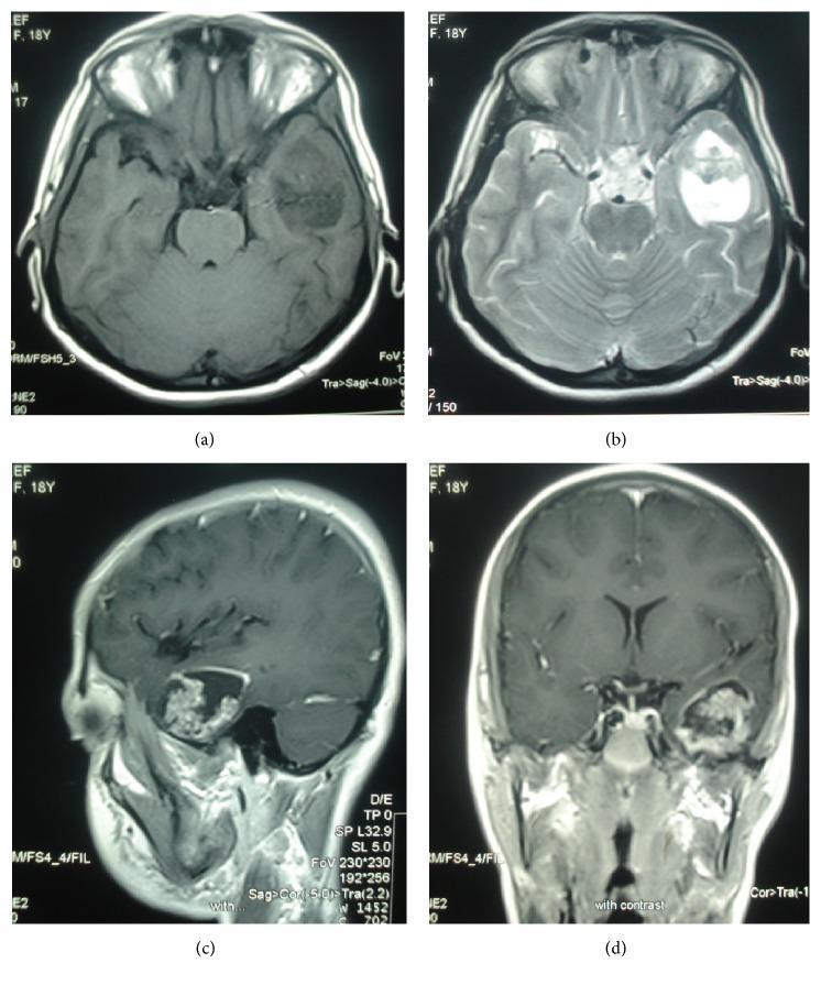
Diagnostic imaging series of a brain MRI displaying a papillary craniopharyngioma located in the left temporal lobe. The four-panel figure includes: (a) axial T1-weighted image showing a relatively hypointense, heterogeneous mass; (b) axial T2-weighted image revealing a well-defined solid-cystic lesion with high-signal cystic components and no significant surrounding vasogenic edema; (c) sagittal and (d) coronal gadolinium contrast-enhanced T1-weighted images. These post-contrast views highlight a distinct peripheral rim of enhancement around the cystic portion and nodular enhancement within the solid component. Notably, the tumor appears isolated in the temporal lobe without typical connection to the suprasellar or intrasellar regions. This case serves as a neuroimaging example of an ectopic craniopharyngioma, demonstrating its characteristic mixed composition and enhancement pattern for use in neuroradiology education and differential diagnosis of temporal lobe masses.
medulloblastoma MRI posterior fossa cerebellum pediatric brain tumor
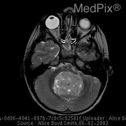
**Imaging Modality:** Magnetic Resonance Imaging (MRI), axial T2-weighted sequence.
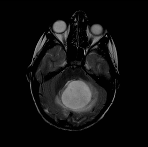
**Imaging Modality:** Magnetic Resonance Imaging (MRI), Axial view, T2-weighted/FLAIR sequence.
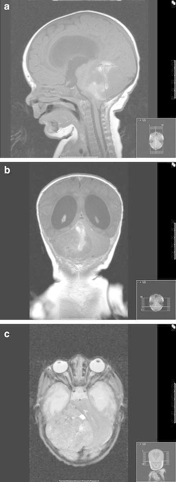
This diagnostic image consists of a three-panel MRI series (sagittal, axial, and coronal planes) of a pediatric patient's head, demonstrating a large posterior fossa tumor. The sagittal view (a) shows a heterogeneous, partially contrast-enhancing mass occupying the cerebellum and compressing the brainstem, with evidence of downward cerebellar tonsillar herniation through the foramen magnum. The coronal view (b) illustrates significant supratentorial hydrocephalus with marked enlargement of the lateral ventricles secondary to obstructive mass effect. The axial view (c) displays the tumor’s lateral extent within the right posterior fossa, causing a distinct leftward midline shift and displacement of the brainstem. These findings are characteristic of aggressive pediatric brain tumors, such as ependymoblastoma or medulloblastoma, which present with increased intracranial pressure and mass effect on critical infratentorial structures.
ependymoma MRI brain spinal cord tumor
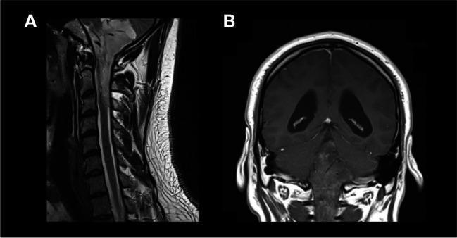
This composite diagnostic image features two MRI views (labeled A and B) demonstrating a neurosurgical case involving a posterior fossa tumor with spinal extension. Image A is a T2-weighted sagittal MRI of the cervical and upper thoracic spine. It shows the vertebral bodies, intervertebral discs, and the spinal cord. A hyperintense lesion or signal abnormality is visible extending inferiorly from the posterior fossa into the spinal canal, reaching the C1-C2 level, consistent with tumor extension. Image B is a T1-weighted coronal MRI of the brain. It illustrates significant ventriculomegaly, with prominent enlargement of the lateral ventricles. The brain parenchyma shows multiple small punctate hyperintensities, and there is evident enhancement around the cerebral hemispheres. Collectively, these images illustrate the preoperative planning for an extended craniotomy with C1-C2 laminotomy, aimed at resecting a tumor (such as an ependymoma) that originates in the fourth ventricle and extends into the upper cervical spinal canal.
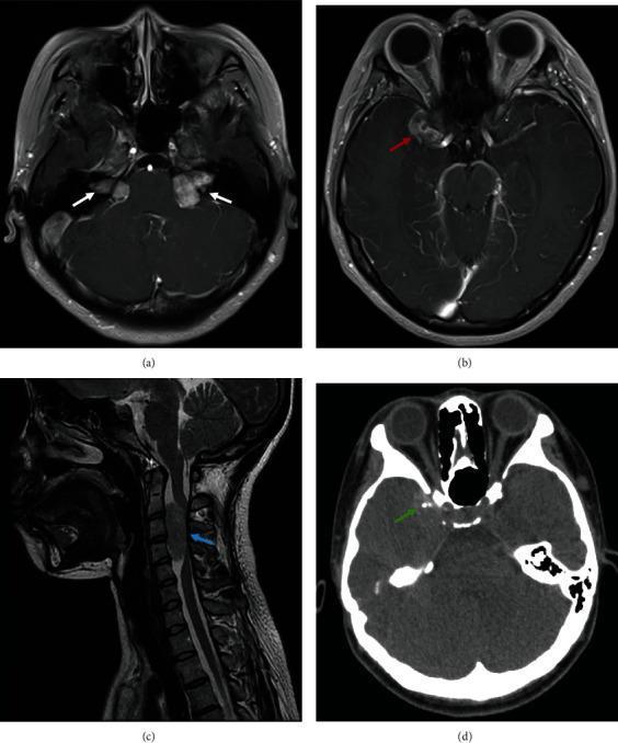
This composite figure presents multi-modal neuroimaging findings diagnostic of Neurofibromatosis Type 2 (NF2). (a) Axial contrast-enhanced MRI demonstrates pathognomonic bilateral, well-defined, intensely enhancing solid masses in the cerebellopontine angles (white arrows), consistent with vestibular schwannomas. (b) Axial enhanced MRI shows an enhancing parasellar lesion (red arrow) adjacent to the right cavernous sinus, suggestive of a meningioma. (c) Sagittal T2-weighted MRI of the cervical spine reveals an intramedullary or extramedullary hypointense focal abnormality at the C2-C4 level (blue arrow), representing a spinal cord tumor such as an ependymoma or schwannoma. (d) Non-contrast axial CT scan of the brain identifies small, hyperdense calcified lesions in the right temporal lobe (green arrow) and confirms normal contours of the bilateral optic nerves. These combined findings—bilateral acoustic neuromas, intracranial meningiomas, and spinal lesions—provide a comprehensive visual summary of the central nervous system manifestations of NF2 in a young patient.
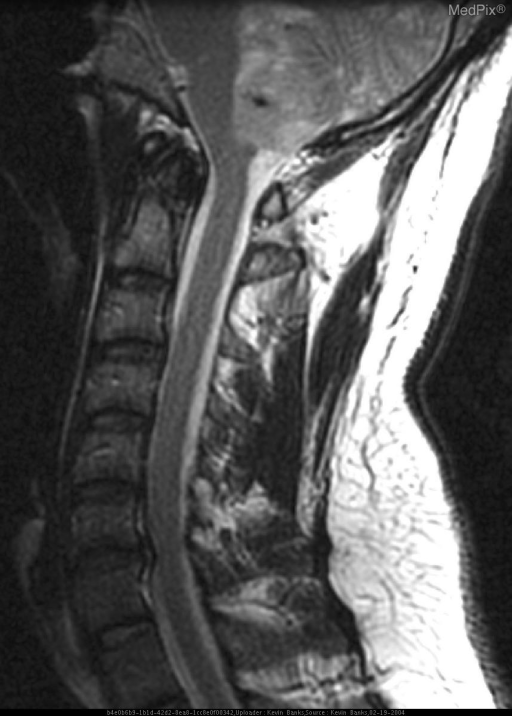
**Imaging Modality:** Sagittal Magnetic Resonance Imaging (MRI), T1-weighted sequence.
brain metastasis MRI multiple lesions
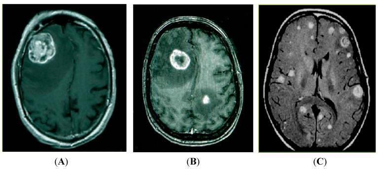
This diagnostic image consists of three axial Magnetic Resonance Imaging (MRI) scans of the human brain demonstrating varying patterns of intracranial metastasis, likely derived from lung cancer. Panel (A) shows a solitary metastasis: a large, heterogeneous, lobulated mass in the right hemisphere with significant mass effect and surrounding vasogenic edema. Panel (B) illustrates oligometastatic disease, featuring two distinct lesions; the larger anterior lesion exhibits classic peripheral ring enhancement and significant internal necrosis or cystic change, typical of rapidly growing metastatic tumors. Panel (C) displays multiple brain metastases, characterized by numerous hyperintense, smaller lesions of varying sizes disseminated throughout the parenchyma in both hemispheres. These images serve to categorize metastatic progression into solitary, oligometastatic, and multiple lesion stages, which is critical for determining neurosurgical versus radiotherapeutic treatment strategies. The modality highlights the role of contrast-enhanced MRI in identifying the breakdown of the blood-brain barrier associated with secondary CNS malignancies.
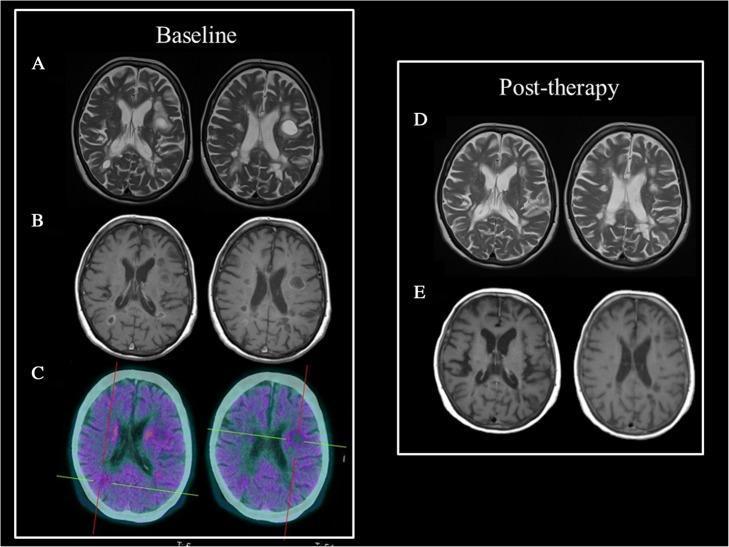
This composite of clinical diagnostic images presents a longitudinal comparison between baseline and post-therapy states of multifocal brain lesions in a patient with suspected tumefactive multiple sclerosis (TMS) or breast cancer metastasis.
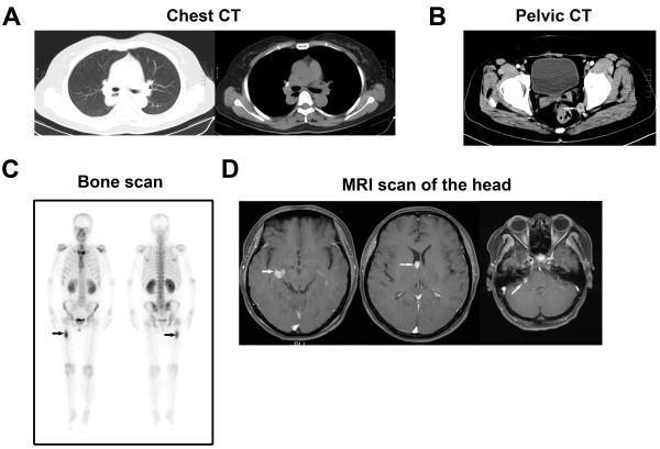
A multi-panel clinical imaging figure demonstrating widespread metastatic disease. (A) Chest CT in axial lung and mediastinal windows showing multiple pulmonary lesions in the inferior lobe of the right lung. (B) Pelvic CT displaying pelvic nodules and involvement of the subcutaneous fat layer. (C) Whole-body bone scan (scintigraphy) in anterior and posterior views showing focal increased radiopharmaceutical uptake (indicated by arrows) in the right femur, consistent with osseous metastasis. (D) Axial head MRI (T1-weighted contrast-enhanced images) revealing multiple intracranial lesions, including enhancing nodules within the brain parenchyma and the right cerebellopontine angle, suggestive of brain and leptomeningeal metastases. Collectively, these images illustrate the systemic spread of metastatic hepatocellular carcinoma (HCC) to the lungs, skeletal system, and central nervous system.
"glioblastoma"[MeSH Terms] AND temozolomide
"meningioma"[MeSH Terms] AND "WHO grade"
"medulloblastoma"[MeSH Terms] AND "molecular subgroup"
WHO 2021 classification CNS tumors meningioma grading medulloblastoma molecular subgroups WNT SHH group3 group4
Stupp protocol glioblastoma temozolomide radiotherapy median survival MGMT methylation
mkdir -p /tmp/workspace/cns-tumor-ppt/assets && ls -la /tmp/workspace/
cd /tmp/workspace/cns-tumor-ppt && python /tmp/skills/shared/scripts/fetch_images.py \ "https://cdn.orris.care/cdss_images/medpix_vqa_f303cf0f-ffb3-4634-9750-3626fba1b9b1_1766169727087.png" \ "https://cdn.orris.care/cdss_images/pmc_clinical_VQA_1ff23e200d7923f5050ab7883f299102d000ebcadb79809d0804a708fac2c3c2.jpg" \ "https://cdn.orris.care/cdss_images/Pathology_1760048798617_95609fc9-1cbe-4371-b6c8-dcf444a1d2c1.jpg" \ "https://cdn.orris.care/cdss_images/Pathology_1760048820792_5f52ec12-b4cb-4f79-8b6d-4cb46d010397.jpg" \ "https://cdn.orris.care/cdss_images/medpix_vqa_db4cab22-e499-42b9-b238-dccf0063d37b_1766169361756.png" \ "https://cdn.orris.care/cdss_images/medpix_vqa_9c1886d8-8bc6-4066-98d4-272b765efb84_1766169647271.png" \ "https://cdn.orris.care/cdss_images/pmc_clinical_VQA_6bb776b9459015f1a0569051192690ae983da1cbc2fb9db204f9cd5e2065715c.jpg" \ "https://cdn.orris.care/cdss_images/pmc_clinical_VQA_6b7e3937ff137e8d64949ba0215bcd415182388c74e47b709d0b5bffc6ca59c2.jpg" \ "https://cdn.orris.care/cdss_images/pmc_clinical_VQA_51d796d83016af04f4c068bb095e4c869d0f4ee92fc0c41c76671ba27ddeca91.jpg" \ "https://cdn.orris.care/cdss_images/pmc_clinical_VQA_37ddfacce42afab846300c45cd861a47d0d010e46618297e68f421f32769be92.jpg" \ "https://cdn.orris.care/cdss_images/pmc_clinical_VQA_63366be03229e9fac33a7697d4cedf99f0d9a67477cf614c660607dfadde1ee8.jpg" \ "https://cdn.orris.care/cdss_images/Pathology_1760048698840_bbecb3a4-3a09-4126-823c-4f614c761c2e.jpg" \ "https://cdn.orris.care/cdss_images/pmc_clinical_VQA_061cfa76b3ec08d8478fef7479518e243141d92da18dafe41a59c803885d0f21.jpg" \ "https://cdn.orris.care/cdss_images/pmc_clinical_VQA_1a4ef848ba99a85f1f7bd92735ab6b101139714fc1d28fe768b264fde46a5ed0.jpg" \ "https://cdn.orris.care/cdss_images/GLGCA_1952115_1763326322913_96048b54-9bb1-46ee-8a90-abc08264eebf.png" \ > images_result.json 2>fetch_err.log python3 -c " import json data = json.load(open('images_result.json')) print(len(data)) for d in data: print(d['url'][-40:], 'OK' if d.get('base64') else 'FAIL', d.get('error')) "
cd /tmp/workspace/cns-tumor-ppt && python3 -c " import json, base64 data = json.load(open('images_result.json')) names = ['gbm_mri','lowgrade_glioma_mri','meningioma_mri','meningioma_histo','vs_mri','pituitary_mri','cranio_mri','medullo_mri','ependymoma_mri','mets_mri','pcnsl_mri','gbm_histo','brain_anatomy','craniotomy_photo','who_diagram'] for d, name in zip(data, names): b64 = d['base64'] header, b64data = b64.split(',',1) ext = 'png' if 'png' in header else 'jpg' with open(f'assets/{name}.{ext}','wb') as f: f.write(base64.b64decode(b64data)) print(name, ext, len(b64data)) " ls -la assets/