attachment
attachment
attachment
attachment
attachment
attachment

What is this breast tru cut biopsy

analyse_media
analyse_media
analyse_media
Reading File
Finding Sources
Finding Sources
Searching Images

myofibroblastoma breast histology spindle cells bland collagen
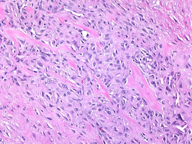
Histopathology, brightfield light microscopy, is performed on a breast tissue biopsy stained with Hematoxylin and Eosin (H&E). The image shows a classic myofibroblastoma featuring a uniform spindle cell population embedded in dense, eosinophilic collagenous stroma. Cells are fusiform with elongated, blunt-ended nuclei, scant cytoplasm, and limited mitotic activity, producing a bland cytomorphology typical of benign myofibroblastic tumors. The stromal matrix demonstrates prominent thick collagen bands arranged in interlacing fascicles, with variable cellularity. Variant patterns are recognized: collagenized forms with even denser stromal fibrosis, epithelioid variants containing polygonal to epithelioid cells that may mimic invasive carcinoma on routine sections, and myxoid variants showing a more gelatinous extracellular matrix. Immunohistochemistry (not shown) typically reveals myofibroblastic differentiation with CD34 positivity and desmin or SMA expression in a subset, while S100 is negative, aiding distinction from spindle cell sarcomas and metaplastic carcinomas. The diagnostic significance lies in distinguishing this benign stromal tumor from malignant mimics, guiding conservative surgical management and prognosis. This image demonstrates the key features of a myofibroblastoma: bland spindle cells, abundant collagen, absence of overt cytologic atypia, and low mitotic rate, consistent with benign myofibroblastic neoplasm. Recognition supports accurate diagnosis, avoids overtreatment, and informs patient prognosis and follow-up strategies in practice.
Histopathology, brightfield light microscopy, is performed on a breast tissue biopsy stained with Hematoxylin and Eosin (H&E). The image shows a classic myofibroblastoma featuring a uniform spindle cell population embedded in dense, eosinophilic collagenous stroma. Cells are fusiform with elongated, blunt-ended nuclei, scant cytoplasm, and limited mitotic activity, producing a bland cytomorphology typical of benign myofibroblastic tumors. The stromal matrix demonstrates prominent thick collagen bands arranged in interlacing fascicles, with variable cellularity. Variant patterns are recognized: collagenized forms with even denser stromal fibrosis, epithelioid variants containing polygonal to epithelioid cells that may mimic invasive carcinoma on routine sections, and myxoid variants showing a more gelatinous extracellular matrix. Immunohistochemistry (not shown) typically reveals myofibroblastic differentiation with CD34 positivity and desmin or SMA expression in a subset, while S100 is negative, aiding distinction from spindle cell sarcomas and metaplastic carcinomas. The diagnostic significance lies in distinguishing this benign stromal tumor from malignant mimics, guiding conservative surgical management and prognosis. This image demonstrates the key features of a myofibroblastoma: bland spindle cells, abundant collagen, absence of overt cytologic atypia, and low mitotic rate, consistent with benign myofibroblastic neoplasm. Recognition supports accurate diagnosis, avoids overtreatment, and informs patient prognosis and follow-up strategies in practice.

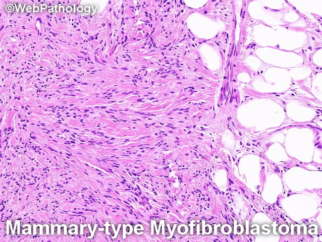
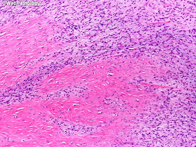
This histopathology image depicts a mammary-type myofibroblastoma of soft breast tissue, evaluated by light microscopy following formalin fixation and hematoxylin and eosin (H&E) staining. The lesion demonstrates a spindle cell-dominated stroma composed of slender, elongated myofibroblastic cells arranged in short fascicles and sweeping, interlacing patterns. Cells display bland nuclei with minimal pleomorphism and scant mitotic activity. Intervening, thick, hyalinized collagen bands create a fibrous, fibrotic background that partitions the tumor into bands and sheets. Adjacent adipose tissue is present, producing a minor adipocytic component that may be seen at the periphery, consistent with the biphasic appearance observed in some mammary-type myofibroblastomas. Immunophenotype, while not evident in H&E, classically shows CD34 positivity and absence of desmin or h-caldesmon in many cases, supporting a myofibroblastic lineage. The differential diagnosis includes dedifferentiated liposarcoma, which typically harbors MDM2/CDK4 amplification (12q13-15) and demonstrates lipogenic atypia, higher mitotic activity, and potential heterologous differentiation; mammary-type myofibroblastoma is associated with 13q/Rb family alterations and lack of aggressive features. Clinically, these tumors are benign or of low malignant potential, with excellent prognosis following local excision. Diagnostic significance centers on distinguishing from liposarcoma and other spindle cell neoplasms to guide management.
This histopathology image depicts a mammary-type myofibroblastoma of soft breast tissue, evaluated by light microscopy following formalin fixation and hematoxylin and eosin (H&E) staining. The lesion demonstrates a spindle cell-dominated stroma composed of slender, elongated myofibroblastic cells arranged in short fascicles and sweeping, interlacing patterns. Cells display bland nuclei with minimal pleomorphism and scant mitotic activity. Intervening, thick, hyalinized collagen bands create a fibrous, fibrotic background that partitions the tumor into bands and sheets. Adjacent adipose tissue is present, producing a minor adipocytic component that may be seen at the periphery, consistent with the biphasic appearance observed in some mammary-type myofibroblastomas. Immunophenotype, while not evident in H&E, classically shows CD34 positivity and absence of desmin or h-caldesmon in many cases, supporting a myofibroblastic lineage. The differential diagnosis includes dedifferentiated liposarcoma, which typically harbors MDM2/CDK4 amplification (12q13-15) and demonstrates lipogenic atypia, higher mitotic activity, and potential heterologous differentiation; mammary-type myofibroblastoma is associated with 13q/Rb family alterations and lack of aggressive features. Clinically, these tumors are benign or of low malignant potential, with excellent prognosis following local excision. Diagnostic significance centers on distinguishing from liposarcoma and other spindle cell neoplasms to guide management.

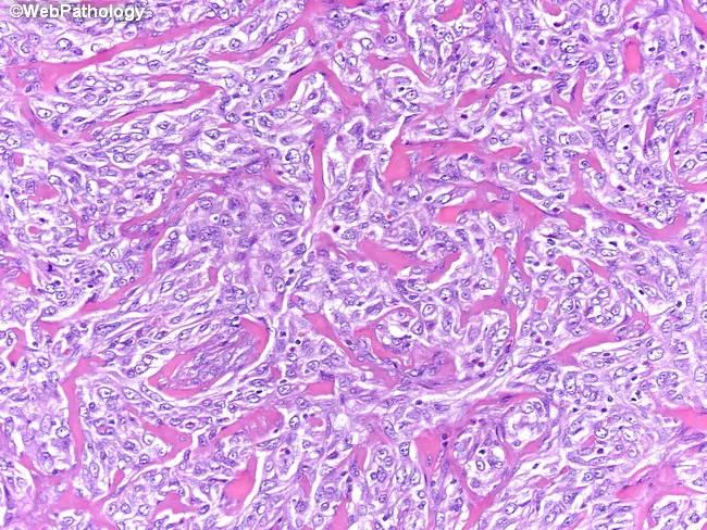
Histopathology Metaplastic spindle cell carcinoma with fibromatosis-like features of the breast. This hematoxylin and eosin (H&E) stained slide shows a combination of hypocellular and hypercellular spindle cell fascicles arranged in storiform pattern embedded within dense desmoplastic stroma containing keloid-like collagen. The spindle cells have elongated, bland to mildly pleomorphic nuclei with scant mitotic activity, low-grade cytology, and rare mitoses, consistent with a low-proliferation component. Intervening dense fibrous tissue and collagen produce a biphasic appearance, sometimes mimicking fibromatosis. In focal areas, a more cellular spindle cell proliferation is evident, but without prominent epithelial differentiation on H&E alone. Immunohistochemistry would typically demonstrate epithelial markers (cytokeratins) in spindle cells, supporting metaplastic carcinoma, and p63 or basal-type cytokeratins may be positive; vimentin is commonly positive as well. Differential diagnoses include true desmoid-type fibromatosis, spindle cell sarcomas, and other myoepithelial or myofibroblastic lesions. The diagnostic significance lies in recognizing a metaplastic process with sarcomatoid features, which informs surgical management and adjuvant therapy for breast cancer. This histology pattern should prompt correlation with clinical data, receptor status, and imaging to guide prognosis and treatment planning. Notable features include dense desmoplasia, minimal pleomorphism, and occasional inflammatory infiltrates that may complicate initial interpretation in core breast biopsies.
Histopathology Metaplastic spindle cell carcinoma with fibromatosis-like features of the breast. This hematoxylin and eosin (H&E) stained slide shows a combination of hypocellular and hypercellular spindle cell fascicles arranged in storiform pattern embedded within dense desmoplastic stroma containing keloid-like collagen. The spindle cells have elongated, bland to mildly pleomorphic nuclei with scant mitotic activity, low-grade cytology, and rare mitoses, consistent with a low-proliferation component. Intervening dense fibrous tissue and collagen produce a biphasic appearance, sometimes mimicking fibromatosis. In focal areas, a more cellular spindle cell proliferation is evident, but without prominent epithelial differentiation on H&E alone. Immunohistochemistry would typically demonstrate epithelial markers (cytokeratins) in spindle cells, supporting metaplastic carcinoma, and p63 or basal-type cytokeratins may be positive; vimentin is commonly positive as well. Differential diagnoses include true desmoid-type fibromatosis, spindle cell sarcomas, and other myoepithelial or myofibroblastic lesions. The diagnostic significance lies in recognizing a metaplastic process with sarcomatoid features, which informs surgical management and adjuvant therapy for breast cancer. This histology pattern should prompt correlation with clinical data, receptor status, and imaging to guide prognosis and treatment planning. Notable features include dense desmoplasia, minimal pleomorphism, and occasional inflammatory infiltrates that may complicate initial interpretation in core breast biopsies.

This histopathology image depicts a breast tissue specimen showing low-grade metaplastic spindle cell carcinoma, fibromatosis-like variant. Acquired via formalin fixation and paraffin embedding, the slide is stained with Hematoxylin and Eosin and examined under brightfield illumination at high magnification. The tumor target consists of plump spindle cells arranged in a storiform (cartwheel) pattern with interlacing fascicles separated by conspicuous hyalinized collagen bundles. The cells display round to oval vesicular nuclei, open chromatin, and elongated, spindle-shaped cytoplasm arranged in a slightly infiltrative, but relatively bland cytomorphology. Mitotic figures are infrequent, and nuclear atypia is mild, consistent with a low-grade appearance, yet the overall architecture indicates a malignant neoplasm with potential for local recurrence and distant metastasis. The surrounding stroma is fibrous and densely collagenous, contributing to a desmoplastic-like background. Immunophenotype may show focal cytokeratin positivity in spindle cells and p63 or high-molecular-weight cytokeratins in metaplastic carcinomas, aiding distinction from benign fibromatosis. Differential diagnoses include desmoid-type fibromatosis, myofibroblastic sarcoma, and other spindle cell neoplasms of the breast. Clinically, this lesion requires complete excision with clear margins; surveillance for recurrence and metastasis is indicated, given malignant potential despite bland histology. Correlation with imaging and clinical findings improves diagnostic accuracy and guides treatment.

This histopathology image depicts a breast tissue specimen showing low-grade metaplastic spindle cell carcinoma, fibromatosis-like variant. Acquired via formalin fixation and paraffin embedding, the slide is stained with Hematoxylin and Eosin and examined under brightfield illumination at high magnification. The tumor target consists of plump spindle cells arranged in a storiform (cartwheel) pattern with interlacing fascicles separated by conspicuous hyalinized collagen bundles. The cells display round to oval vesicular nuclei, open chromatin, and elongated, spindle-shaped cytoplasm arranged in a slightly infiltrative, but relatively bland cytomorphology. Mitotic figures are infrequent, and nuclear atypia is mild, consistent with a low-grade appearance, yet the overall architecture indicates a malignant neoplasm with potential for local recurrence and distant metastasis. The surrounding stroma is fibrous and densely collagenous, contributing to a desmoplastic-like background. Immunophenotype may show focal cytokeratin positivity in spindle cells and p63 or high-molecular-weight cytokeratins in metaplastic carcinomas, aiding distinction from benign fibromatosis. Differential diagnoses include desmoid-type fibromatosis, myofibroblastic sarcoma, and other spindle cell neoplasms of the breast. Clinically, this lesion requires complete excision with clear margins; surveillance for recurrence and metastasis is indicated, given malignant potential despite bland histology. Correlation with imaging and clinical findings improves diagnostic accuracy and guides treatment.

I now have everything needed for a thorough, well-illustrated answer.

Breast Tru-Cut Biopsy - Histopathological Analysis

What You Are Looking At

All three images show an H&E-stained core needle (tru-cut) biopsy of the breast composed almost entirely of bland spindle cells set in a fibromyxoid/collagenous stroma. There are no epithelial structures (ducts or lobules) visible in the high-power fields. This is a classic pure stromal spindle cell lesion of the breast.

Detailed Microscopic Findings

FeatureWhat Is Seen
Cell typeUniform spindle-shaped (fusiform) cells
Nuclear morphologyElongated, oval/tapered nuclei; pale, finely dispersed chromatin; inconspicuous nucleoli
AtypiaAbsent - bland cytology throughout
Mitotic figuresNot identified
ArchitectureHaphazard / loosely fascicular (short intersecting fascicles); no storiform or herringbone pattern
StromaMixed fibromyxoid - pale wavy collagen bundles interspersed between cells; no dense keloidal collagen or prominent vascularity
Epithelial elementsAbsent in these fields
Inflammatory cellsNot prominent
Image 2 (low power) shows the biopsy core architecture - a cellular spindle cell proliferation replacing breast parenchyma, abutting adipose tissue at the periphery.

Differential Diagnosis

This histomorphology in a breast tru-cut biopsy places the lesion in the bland spindle cell category, with the following main possibilities (in order of likelihood based on morphology):

1. Myofibroblastoma (most likely)

The most common benign mesenchymal tumor of the breast. Bland, uniform spindle cells in short intersecting fascicles with collagenous-to-myxoid stroma is the hallmark appearance.
Myofibroblastoma - classic histology with bland spindle cells and thick collagen bands
  • IHC profile: CD34+, Desmin+, ER/PR+ (typically), CK negative
  • Excellent prognosis; conservative excision is curative

2. Desmoid-type Fibromatosis

Locally aggressive but non-metastasizing myofibroblastic proliferation. Long sweeping fascicles, more collagenous, infiltrative border at low power.
  • IHC: Nuclear beta-catenin+, CD34-, SMA variable, CK negative
  • Tendency for local recurrence after excision

3. Pseudoangiomatous Stromal Hyperplasia (PASH) - Spindle Cell Variant

Usually shows characteristic slit-like pseudovascular spaces lined by flat myofibroblasts, which are absent in these fields but may be present in other areas of the biopsy.

4. Low-grade Metaplastic Spindle Cell Carcinoma (must be excluded)

A deceptively bland malignant mimic. Despite bland cytology, it carries malignant potential.
Metaplastic spindle cell carcinoma - fibromatosis-like variant - bland spindle cells that can mimic benign lesions
  • IHC: CK (AE1/AE3, Cam5.2, 34betaE12)+, p63+, GATA3+/-, CD34-
  • This MUST be ruled out before calling the lesion benign

5. Spindle Cell Lipoma

Bland spindle cells with myxoid stroma and "ropy" collagen - requires mature adipocytes in other fields. CD34+, S100-.

Key Points for Definitive Diagnosis

IHC panel required - morphology alone cannot definitively separate these entities on a core biopsy:
MarkerMyofibroblastomaFibromatosisMetaplastic CaPASH
CD34++--+/-
Desmin+--+/-
Beta-catenin (nuclear)-++--
AE1/AE3 / CK--++-
p63--+-
SMA+/-+/--+
ER++--+/-

Bottom Line

This biopsy shows a bland spindle cell stromal lesion - almost certainly benign, with myofibroblastoma being the top differential based on the haphazard short fascicular pattern, myxoid-collagenous stroma, and complete absence of cytological atypia or mitoses. However, IHC is mandatory to definitively exclude low-grade metaplastic spindle cell carcinoma before rendering a final benign diagnosis. Correlation with clinical details (patient age, imaging findings, lesion size and borders on ultrasound/mammogram) is also essential.
Disclaimer: This represents an educational histopathological analysis. Final diagnostic interpretation must be made by a qualified pathologist with access to clinical information, full histological sections, and IHC results.
This is a shared conversation. Sign in to Orris to start your own chat.