
Interstitial lung disease
Reading File
Now let me search the medical library for ILD content in parallel:
Finding Sources
Searching PubMed
"interstitial lung disease"[MeSH Terms] AND diagnosis AND management
Good - the main ILD chapter is in block21. Let me read the key sections in parallel:
Reading File
Reading File
Let me now search for the main ILD overview section and ILD classification/diagnosis content:
Finding Sources
Finding Sources
Reading File
Reading File
Excellent - great content. Let me fetch that CT image and read the broader ILD classification section:
Loading Image
Reading File
Finding Sources
Reading File
Loading Image
Reading File
Finding Sources
I now have rich content. Let me also quickly get the broader ILD overview and sarcoidosis/HP sections:
Reading File
I now have comprehensive content to write a thorough answer. Let me compile the full response:
Interstitial Lung Disease (ILD)
ILD refers to a heterogeneous group of diffuse parenchymal lung disorders characterized by varying degrees of inflammation and fibrosis of the alveolar walls, distal airways, and interstitium. The term is something of a misnomer - disease often extends into the alveolar spaces rather than being purely interstitial.
Classification
ILD is broadly divided into:
1. ILD of Known Cause
- Connective tissue diseases (CTD): systemic sclerosis (SSc), rheumatoid arthritis (RA), polymyositis/dermatomyositis, Sjögren's, SLE
- Drug/toxicity-induced
- Occupational/environmental exposures (hypersensitivity pneumonitis, pneumoconioses)
2. Idiopathic Interstitial Pneumonias (IIPs)
- Chronic fibrosing: IPF (UIP pattern), NSIP
- Smoking-related: RB-ILD, DIP
- Acute/subacute: COP, AIP (Hamman-Rich syndrome)
- Rare: LIP, PPFE
3. Granulomatous ILD
- Sarcoidosis
- Hypersensitivity pneumonitis (HP)
4. Other forms
- Pulmonary Langerhans cell histiocytosis (PLCH)
- LAM (lymphangioleiomyomatosis)
Key Entities
Idiopathic Pulmonary Fibrosis (IPF)
The most common ILD of unknown cause. Prevalence increases with age, estimated at 50-200 per 100,000. IPF is more common in men, typically diagnosed in the 5th-6th decade, and is frequently associated with smoking or environmental exposures. It carries a poor prognosis: approximately 50% 3-5 year survival.
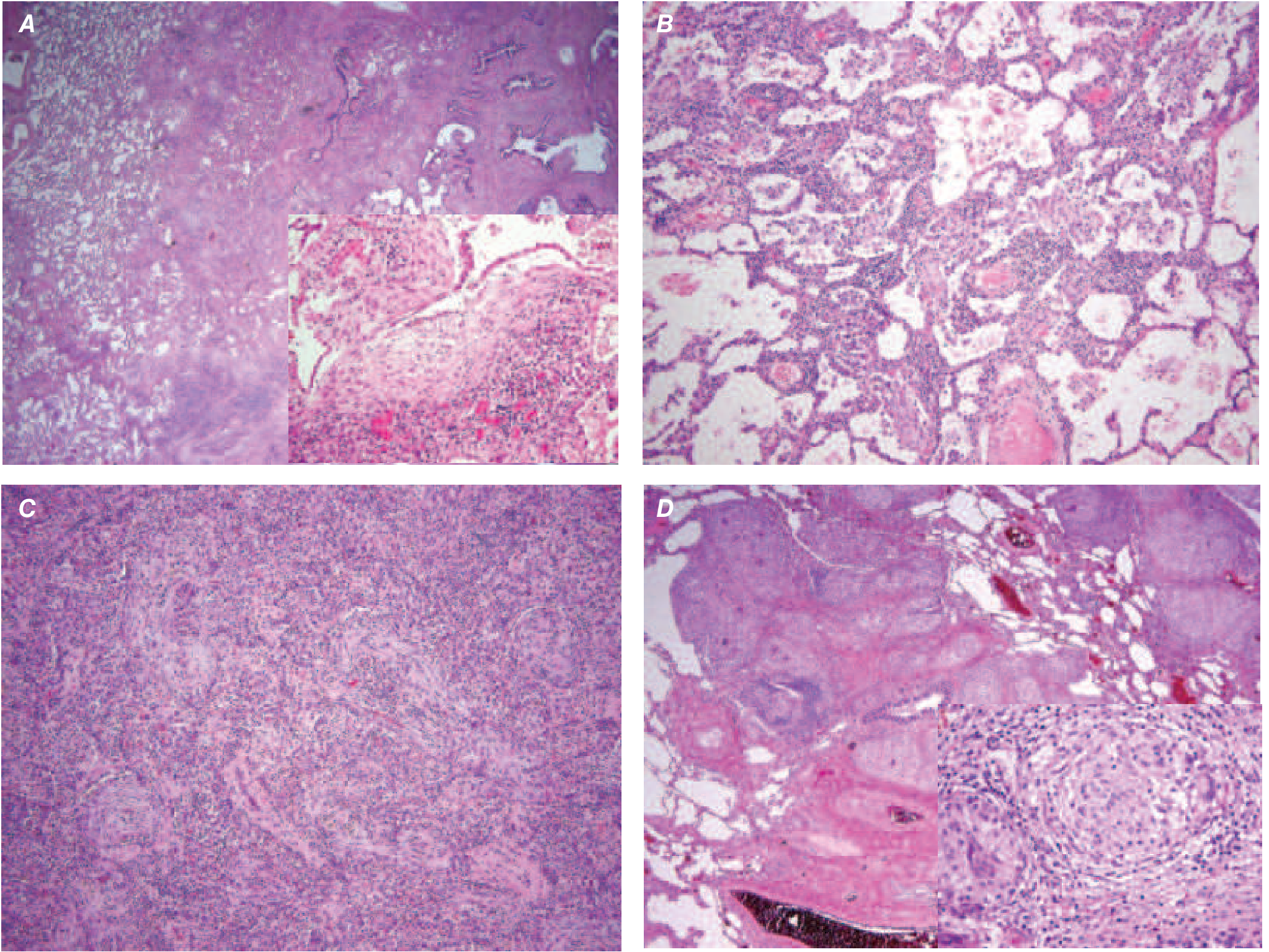
Pathology: UIP (usual interstitial pneumonia) pattern - subpleural reticulation with honeycomb changes and fibroblast foci (myofibroblast/collagen collections), alternating with areas of normal alveolar architecture (temporal and spatial heterogeneity). Fibroblast foci are the hallmark.
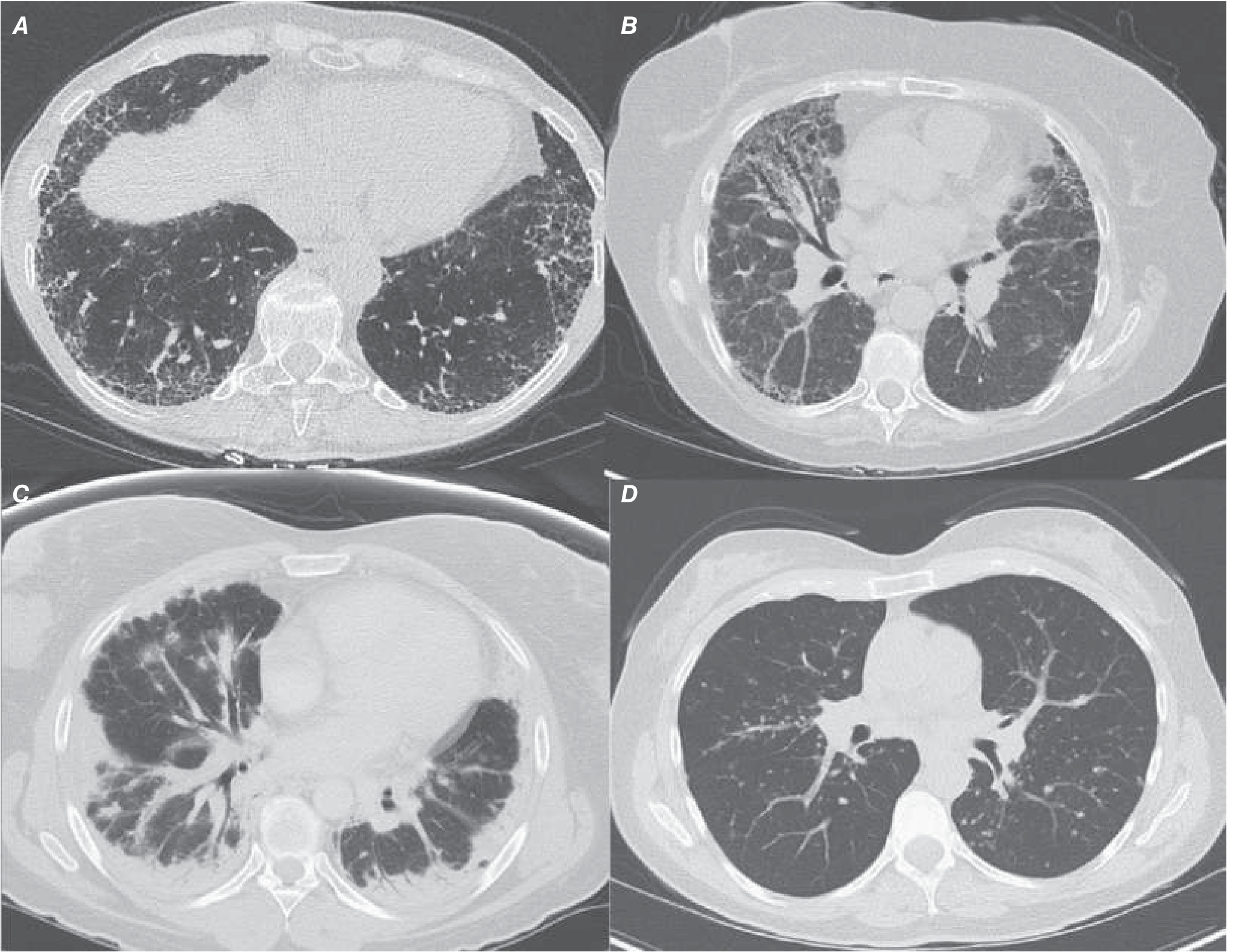
HRCT: Posterior, basilar, subpleural predominant reticular markings + honeycombing + traction bronchiectasis = "UIP pattern." Extensive ground-glass opacities, upper-lung predominance, or micronodules should raise suspicion for an alternative diagnosis.
Treatment:
- Antifibrotic therapy (pirfenidone or nintedanib) - slows FVC decline; shown in landmark 2014 trials
- Immunosuppression is contraindicated - associated with increased morbidity and mortality
- Oxygen therapy, pulmonary rehabilitation, lung transplantation for eligible patients
- Nintedanib may also reduce acute exacerbation rate
Nonspecific Interstitial Pneumonia (NSIP)
Commonly associated with CTD (especially SSc and polymyositis/dermatomyositis), also idiopathic. Diagnosed more often in nonsmoking females in their 5th decade. Better prognosis than IPF: >80% 5-year survival (cellular > fibrosing NSIP).
HRCT: Bilateral, symmetric ground-glass and reticular opacities, lower-zone predominance, traction bronchiectasis, occasional subpleural sparing. Honeycombing is uncommon (key distinguishing point from UIP).
Histology: Uniform interstitial inflammation and fibrosis; honeycomb changes usually absent; fibroblast foci rare.
Treatment: Oral steroids (prednisone), cytotoxic agents (mycophenolate, azathioprine, cyclophosphamide), biologics (rituximab, tocilizumab). Antifibrotic therapy for progressive NSIP.
Cryptogenic Organizing Pneumonia (COP)
Typically affects patients in their 50s-60s as a subacute flu-like illness - cough, dyspnea, fever, fatigue. Often misdiagnosed as pneumonia. Can be secondary to CTD (e.g., polymyositis), drugs, or malignancy.
HRCT: Patchy, sometimes migratory, subpleural consolidative opacities with ground-glass opacities. The "reversed halo" or "atoll sign" (rim of subpleural sparing) is characteristic.
Histology: Patchy organizing pneumonia with granulation tissue in small airways, alveolar ducts, and alveoli.
Treatment: Corticosteroids - often dramatic response but relapse common; minimum 6 months required. Cytotoxic/biologic agents (mycophenolate, cyclophosphamide, rituximab) for steroid-sparing.
Acute Interstitial Pneumonia (AIP / Hamman-Rich Syndrome)
A rare, often fatal disorder with acute onset of respiratory distress and hypoxemia. Prodrome of upper respiratory symptoms is common. Mortality >50% within 6 months; recurrences are common.
HRCT: Patchy bilateral ground-glass opacities with dependent consolidation.
Histology: Diffuse alveolar damage (DAD) - identical to ARDS on biopsy.
Treatment: Supportive (mechanical ventilation). No proven drug therapy; glucocorticoids are often given but of unclear benefit.
Acute Exacerbations of IIP
Not a separate disease - an accelerated phase of injury in any fibrosing ILD. Most severe in IPF. Defined as acute onset (<30 days) of respiratory distress/hypoxemia in a patient with underlying pulmonary fibrosis without alternate explanation (infection, heart failure, etc.).
Prognosis: Extremely poor - mortality >85%, survival ranges from days to months.
Treatment: Supportive. Mechanical ventilation controversial (unless bridge to transplant). Immunosuppressants commonly used without proven benefit.
Smoking-Related ILDs (RB-ILD and DIP)
Occur in active, often heavy smokers aged 40-50.
| Feature | RB-ILD | DIP |
|---|---|---|
| Histology | Pigmented macrophages in respiratory bronchioles, peribronchiolar fibrosis | More diffuse; macrophage accumulation throughout alveoli |
| HRCT | Centrilobular nodules, GGO, bronchial wall thickening | Diffuse/patchy bilateral symmetric GGO |
| Treatment | Smoking cessation (mainstay) | Smoking cessation ± steroids |
ILD Associated with Connective Tissue Disease (CTD-ILD)
ILD occurs in:
- SSc (systemic sclerosis): ~50% with diffuse disease, ~30% with limited disease. NSIP is the most common pattern. Dilated esophagus on CT is a clue. Treatment: mycophenolate (preferred over cyclophosphamide for better tolerability), tocilizumab, nintedanib. Proton pump inhibitors / antireflux surgery important (microaspiration drives progression).
- RA: Clinically evident ILD in ~10%, subclinical CT changes in 40-50%. More common in males and smokers. Most common pattern: UIP (worse prognosis than RA-NSIP). MUC5B promoter variant confers susceptibility to RA-UIP.
- Polymyositis/Dermatomyositis, Sjögren's, SLE: Less common but important.
Key fact: UIP in CTD carries better outcomes than idiopathic UIP (IPF), except in RA where UIP has a worse outcome than NSIP, and worse than UIP in other CTDs.
Diagnostic Approach
History: Occupational/environmental exposures, drug history, smoking, family history, symptoms of CTD (joint swelling, sicca symptoms, Raynaud's, skin changes, muscle weakness).
PFTs: Restrictive pattern (reduced TLC, FVC, DLCO). DLCO reduction is often disproportionate and the most sensitive early indicator.
HRCT: The cornerstone of non-invasive diagnosis.
Serologies: ANA, RF, anti-CCP, anti-Scl-70, anti-Jo-1, anti-MDA5, ANCA.
BAL: Can help exclude infection; neutrophilia (IPF/UIP), lymphocytosis (HP, NSIP, sarcoidosis), eosinophilia.
Surgical lung biopsy (VATS): Gold standard when HRCT is non-diagnostic. Now increasingly supplemented or replaced by transbronchial cryobiopsy.
CT Imaging Reference
The image below shows characteristic HRCT patterns of the major ILDs:
Histopathology Reference
Summary Table
| Disease | HRCT Pattern | Key Histology | Prognosis | Treatment |
|---|---|---|---|---|
| IPF | UIP (subpleural, basal, honeycombing) | Fibroblastic foci, temporal heterogeneity | Poor (~50% 3-5yr survival) | Pirfenidone / Nintedanib |
| NSIP | Bilateral GGO, subpleural sparing | Uniform inflammation/fibrosis, no fibroblastic foci | Good (>80% 5-yr) | Steroids, MMF, rituximab |
| COP | Migratory consolidation, atoll sign | Organizing granulation tissue in airways | Good (steroid-responsive) | Corticosteroids |
| AIP | Bilateral GGO + consolidation | DAD | Very poor (>50% mortality) | Supportive |
| RB-ILD/DIP | Centrilobular nodules, GGO | Pigmented macrophages | Good with smoking cessation | Smoking cessation |
| SSc-ILD | NSIP/UIP overlap, dilated esophagus | NSIP > UIP | Variable | MMF, nintedanib, tocilizumab |
| RA-ILD | UIP > NSIP | UIP > NSIP | Worse than other CTD-ILD | Immunosuppression |
Sources: Harrison's Principles of Internal Medicine 22E (2025), Murray & Nadel's Textbook of Respiratory Medicine
This is a shared conversation. Sign in to Orris to start your own chat.