
Wilson's disease
Kayser-Fleischer ring Wilson's disease
"Wilson disease"[MeSH Terms] AND treatment
Wilson's disease diagnosis treatment guidelines 2024
Kayser Fleischer ring cornea copper
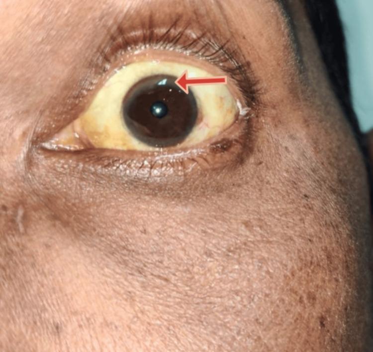
A close-up clinical photograph of a left eye and periorbital region demonstrating characteristic signs of hepatolenticular degeneration (Wilson disease). The sclera exhibits marked icterus (yellowish discoloration), consistent with hyperbilirubinemia or underlying chronic liver disease. At the peripheral margin of the cornea, a brownish-yellow Kayser-Fleischer (KF) ring is visible, particularly highlighted by a red arrow in the superior-temporal quadrant. This ring represents copper deposition in Descemet's membrane. The iris is dark brown, and the surrounding skin shows a slightly uneven pigmentation. The image serves as an educational example of ophthalmologic manifestations associated with systemic metabolic disorders and copper metabolism dysfunction.
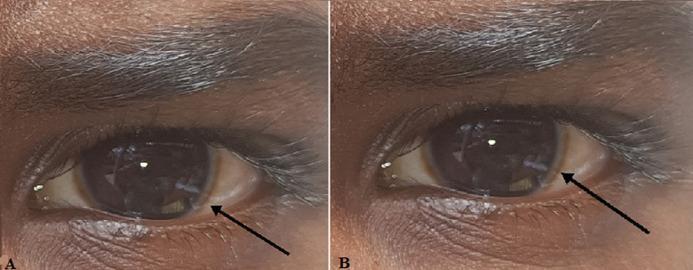
This clinical photograph consists of two panels (A and B) showing the right and left eyes of a patient with dark-pigmented skin. The primary pathological finding is a Kayser-Fleischer ring, visible as a distinct brownish-golden or greenish-brown discoloration at the periphery of the cornea near the limbus. The ring is most prominent in the superior and inferior poles, appearing as a circumferential band that encircles the dark brown iris. The sclera shows mild conjunctival vascularity but is otherwise white. The pupils are round and reactive to light. This ocular sign is a hallmark clinical manifestation of Wilson disease, resulting from copper deposition in Descemet's membrane. The image serves as a classic educational example for hepatology, neurology, and ophthalmology, illustrating the diagnostic physical exam findings associated with impaired copper metabolism.
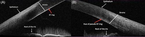
This diagnostic image consists of two panels (A and B) featuring Anterior Segment Optical Coherence Tomography (AS-OCT) cross-sections of the peripheral cornea and anterior chamber angle. The image compares the appearance of corneal deposits in different clinical conditions. Panel A illustrates a classic Kayser-Fleischer (KF) ring, characteristic of Wilson disease, visualized as a discrete hyperintense line located specifically on the Descemet membrane at the posterior corneal surface. In contrast, Panel B demonstrates a 'pseudo-KF ring,' often associated with non-Wilsonian hyperbilirubinemia, which appears as a diffuse, scattered hyper-reflective hue localized within the posterior stroma rather than a distinct line on the Descemet membrane. Both panels are labeled with key anatomical structures including the epithelium, stroma, and the root of the iris. This comparison serves as an educational tool for ophthalmologists and hepatologists to differentiate between true copper deposits and pigmentary changes due to bile pigments using high-resolution imaging.
Wilson's Disease (Hepatolenticular Degeneration)
Overview
- Incidence: ~1 in 30,000-40,000 live births; heterozygote carrier rate ~1 in 90-180 (higher with consanguinity) - Goldman-Cecil Medicine.
- Normal copper handling: 40-60% of dietary copper (2-5 mg/day) is absorbed in the duodenum/proximal small bowel; ATP7B moves it into apoceruloplasmin (forming ceruloplasmin) and into bile canaliculi for fecal excretion, the major elimination route.
- In Wilson's disease, both pathways fail: ceruloplasmin incorporation and biliary excretion are impaired, so serum ceruloplasmin and total serum copper are typically low (despite copper overload) and urinary copper excretion rises.
Clinical Presentation
- Hepatic: ranges from asymptomatic transaminase elevation to hepatomegaly, jaundice, ascites, cirrhosis, hemolytic (Coombs-negative) anemia, and acute liver failure (~5% of patients, an emergency requiring urgent transplant evaluation).
- Neurologic (typically presents after age 20): dysarthria, tremor, dystonia, chorea, rigidity, gait disturbance, a "fixed" sardonic smile, and can mimic Parkinsonism.
- Psychiatric: personality/mood change, depression (~30%), bipolar-spectrum symptoms (~20%), psychosis, and increased sensitivity to neuroleptics; often mistaken for primary psychiatric illness in teens/young adults.
- Other: renal Fanconi-like tubular dysfunction, osteoporosis/osteoarthritis, and the eponymous Kayser-Fleischer ring - golden-brown copper deposition in Descemet's membrane at the corneal limbus, best seen on slit-lamp exam. Present in ~95% of patients with neuropsychiatric disease but only 50-65% of purely hepatic presentations.
Diagnosis
- Low serum ceruloplasmin and low total serum copper
- Elevated 24-hour urinary copper excretion (>100 µg/24h)
- Kayser-Fleischer rings on slit-lamp exam
- Elevated transaminases, low albumin, elevated INR (in advanced liver disease)
- Coombs-negative hemolytic anemia, aminoaciduria, low uric acid
- Liver biopsy with quantitative hepatic copper (sometimes needed)
- ATP7B genetic testing/sequencing (identification of two disease-causing mutations confirms diagnosis)
- MRI in neurologic disease may show T2 hyperintensity in the putamen, midbrain, pons, thalamus, and cerebellum, with atrophy
Treatment
- Chelating agents - D-penicillamine or trientine (first-line historically). Penicillamine causes acute neurological worsening in 20-50% of patients with neurologic disease, and some don't recover to baseline; trientine has a lower risk of this paradoxical worsening.
- Zinc - blocks intestinal copper absorption; used for maintenance therapy or in asymptomatic/presymptomatic patients, and favored first-line in some guidelines for neurological presentations.
- Tetrathiomolybdate - an alternative with potentially lower risk of neurological deterioration, used in combination with zinc in some regimens.
- Liver transplantation - definitive cure for the metabolic defect; indicated in acute liver failure (especially with encephalopathy) or decompensated cirrhosis unresponsive to medical therapy.
- Early treatment can partially reverse MRI changes and improve neurological/psychiatric symptoms.
Recent Evidence (surfaced per protocol)
- PMID 40904321 (2025) - systematic review of cardiac ventricular dysfunction in Wilson's disease, an underrecognized manifestation.
- PMID 39206599 (2024) - systematic review/meta-analysis on adverse pregnancy outcomes and treatment effects (including penicillamine) in Wilson's disease during pregnancy.
- PMID 37311952 (2023) - meta-analysis on early neurological deterioration risk with penicillamine vs. trientine/other agents.