
Explain Atrial Septal Defect and Ventricular Septal Defect
Reading File
Finding Sources
Finding Sources
Searching PubMed
"atrial septal defect" AND "ventricular septal defect"
Searching the Web
AHA atrial septal defect ventricular septal defect congenital heart disease patient information
Finding Sources
Finding Sources
Finding Sources
Searching Images
labeled diagram comparing atrial septal defect and ventricular septal defect left-to-right shunts
Atrial Septal Defect (ASD) and Ventricular Septal Defect (VSD)
Both are congenital heart defects, meaning structural abnormalities present from birth. They are often called “holes in the heart,” but they occur in different parts of the wall separating the chambers.
Normally, pressure is higher on the left side of the heart. Therefore, blood usually passes from left to right through either defect, increasing blood flow to the lungs.
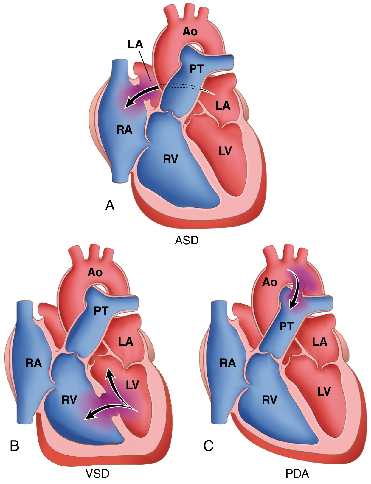
Image: ASD is an opening between the atria; VSD is an opening between the ventricles. Arrows show typical left-to-right shunting. Source: Robbins, Cotran & Kumar Pathologic Basis of Disease.
| Feature | ASD | VSD |
|---|---|---|
| Location | Septum between the two atria | Septum between the two ventricles |
| Usual shunt | Left atrium → right atrium | Left ventricle → right ventricle |
| Main effect | Volume overload of right atrium/right ventricle and pulmonary circulation | Increased pulmonary flow; larger defects can cause heart failure and pulmonary hypertension early |
| Typical presentation | Often no symptoms until later childhood or adulthood | Small defects may be silent; large defects usually present in infancy |
| Classic sound | Fixed, wide splitting of S2 with a systolic flow murmur at pulmonary area | Harsh pansystolic/holosystolic murmur at left lower sternal border |
| Natural history | Large defects generally do not close spontaneously | Many small muscular VSDs close spontaneously |
| Common treatment | Device closure or surgery if hemodynamically significant | Observation for small defects; medication, catheter closure, or surgery for significant defects |
1. Atrial Septal Defect
An ASD is a persistent opening in the wall between the left and right atria. It should not be confused with a patent foramen ovale (PFO), which is failure of the normal fetal foramen ovale to seal after birth rather than a true missing portion of the atrial septum.
How it affects circulation
Because left atrial pressure is usually higher:
Left atrium → ASD → right atrium → right ventricle → lungs
This produces extra blood flow through the right heart and pulmonary circulation. The defect itself usually does not cause cyanosis initially, because the shunted blood is already oxygenated.
Types
- Ostium secundum ASD: most common, located near the fossa ovalis. Often suitable for catheter device closure.
- Ostium primum ASD: lower atrial septum, often associated with atrioventricular septal defects.
- Sinus venosus ASD: high in the atrial septum near the vena cava and may be associated with anomalous pulmonary venous drainage.
Symptoms and signs
Small ASDs may cause no symptoms. Larger untreated ASDs can lead to:
- Exercise intolerance or shortness of breath
- Palpitations, especially atrial fibrillation or flutter in adults
- Recurrent chest infections in children
- Right-sided heart enlargement
- Pulmonary hypertension in longstanding cases
A classic examination finding is a fixed wide split S2. “Fixed” means that the separation between A2 and P2 changes little with breathing. This is particularly associated with a secundum ASD. Harrison's Principles of Internal Medicine, 22e; Fuster and Hurst's The Heart, 15e.
Diagnosis and treatment
- Echocardiography with Doppler confirms the defect, measures shunt size, and assesses right-heart enlargement.
- A small ASD without right-heart dilation may only need monitoring.
- Closure is generally considered when there is a significant left-to-right shunt causing right-heart volume overload, provided pulmonary vascular disease is not irreversible.
- Secundum ASDs are often closed by a transcatheter occluder device. Other types usually require surgery.
2. Ventricular Septal Defect
A VSD is an incomplete closure of the wall between the ventricles. It is the most common congenital heart defect overall. Robbins, Cotran & Kumar Pathologic Basis of Disease.
How it affects circulation
Because left ventricular pressure is much higher than right ventricular pressure:
Left ventricle → VSD → right ventricle → lungs
The pressure difference is larger than in an ASD. Thus, a large VSD can send a substantial volume of blood to the lungs, then back to the left atrium and left ventricle, causing pulmonary congestion and left-sided volume overload.
Types
- Perimembranous VSD: most common, near the membranous part of the septum.
- Muscular VSD: within the muscular septum; some close spontaneously.
- Inlet VSD: near the atrioventricular valves, associated with atrioventricular septal defects.
- Outlet/supracristal VSD: near the ventricular outflow tract and can affect the aortic valve.
Symptoms and signs
The severity depends mainly on defect size and pulmonary vascular resistance.
Small VSD
- Often asymptomatic
- May be detected because of a murmur
- Often has a loud, harsh murmur because the pressure gradient across a small opening is high
- Some close spontaneously
Large VSD
- Symptoms typically begin after several weeks of life, when normal postnatal pulmonary vascular resistance falls.
- Poor feeding, sweating during feeds, poor weight gain
- Fast breathing, recurrent respiratory infections
- Features of heart failure
The usual murmur is harsh pansystolic (holosystolic), best heard at the left lower sternal border. A very large VSD with little pressure difference can sometimes produce a less prominent murmur, so murmur loudness alone does not reliably grade severity.
Complications
A large untreated VSD can lead to:
- Heart failure in infancy
- Pulmonary hypertension
- Aortic valve prolapse and aortic regurgitation, especially with some outlet defects
- Infective endocarditis
- Eisenmenger syndrome: irreversible pulmonary vascular disease eventually reverses the shunt to right-to-left, producing cyanosis. Once this occurs, closing the defect can be unsafe.
Diagnosis and treatment
- Echocardiography is the main diagnostic test.
- Small defects with no adverse effects are usually observed.
- Medicines such as diuretics may be used temporarily to control heart-failure symptoms in infants with large VSDs.
- Significant VSDs may need catheter closure or surgical patch repair, ideally before irreversible pulmonary vascular disease develops.
Key difference to remember
- ASD: an atrial-level hole. It is often tolerated for many years, and may first appear with right-heart enlargement, breathlessness, or atrial arrhythmias in adulthood.
- VSD: a ventricular-level hole. A large defect has a greater pressure gradient and is more likely to produce heart failure during infancy.
Both defects are initially acyanotic because the shunt is typically left-to-right. Cyanosis is a late warning sign of severe pulmonary vascular disease and possible shunt reversal.
This is a shared conversation. Sign in to Orris to start your own chat.