
Every thing about rheumatology
rheumatology autoimmune diseases treatment guidelines
rheumatoid arthritis hand joints deformity
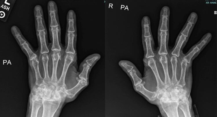
Posteroanterior (PA) view X-ray of both hands demonstrating chronic inflammatory arthritis, consistent with advanced rheumatoid arthritis. The bilateral images reveal significant involvement of the metacarpophalangeal (MCP) joints, characterized by marked joint space narrowing and subluxation where the proximal phalanges are partially displaced from the metacarpal heads. Classical ulnar deviation of the digits is present. The radiograph also shows cortical erosions and architectural distortion at the MCP and carpal joints, indicative of progressive erosive disease. The interphalangeal joints show milder changes compared to the MCP and wrist regions. These findings represent a late-stage manifestation of an inflammatory arthropathy, resulting in substantial joint deformity and malalignment. The image serves as a clinical teaching tool for recognizing radiographic hallmarks of chronic rheumatoid arthritis, including ulnar drift, subluxation, and symmetric joint space loss in the small joints of the hand.
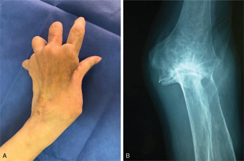
This composite educational image illustrates the clinical and radiographic features of ulnar nerve palsy in the setting of rheumatoid arthritis. Image A is a clinical photograph showing the dorsal aspect of a left hand. It demonstrates a classic 'claw hand' deformity characterized by hyperextension at the metacarpophalangeal (MCP) joints and flexion at the proximal and distal interphalangeal (IP) joints, particularly affecting the ring and small fingers. Significant atrophy of the dorsal interossei muscles is visible as hollowed depressions between the metacarpal bones. Image B is a plain anteroposterior (AP) x-ray of the corresponding elbow joint. It reveals advanced arthritic changes consistent with rheumatoid arthritis, including severe joint space narrowing, subchondral bone destruction, and marked osteophyte formation. A notable valgus deformity is present, with prominent bony projections in the medial joint space. Together, these images illustrate the correlation between joint destruction (cubital tunnel pathology) and distal neurological deficits (ulnar neuropathy).
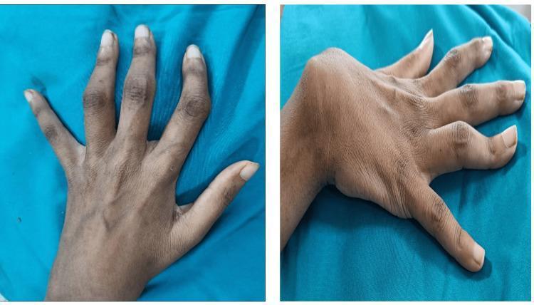
Clinical photograph showing bilateral hand and wrist deformities in a patient with chronic inflammatory arthritis. The left hand (dorsal view) exhibits classic boutonniere deformities of the index and middle fingers, characterized by flexion at the proximal interphalangeal (PIP) joints and hyperextension at the distal interphalangeal (DIP) joints. The left index finger also demonstrates significant radial deviation at the PIP joint level. The right hand (lateral-dorsal view) displays a boutonniere deformity of the ring finger and a prominent ulnar subluxation of the wrist joint, appearing as a volar and ulnar displacement of the carpus relative to the distal radius and ulna. These findings are representative of advanced joint destruction and ligamentous laxity often seen in systemic autoimmune conditions like seronegative arthritis or rheumatoid arthritis. The image serves as an educational reference for identifying characteristic musculoskeletal manifestations of systemic inflammatory diseases in the small joints of the hand.
SLE lupus malar rash butterfly face
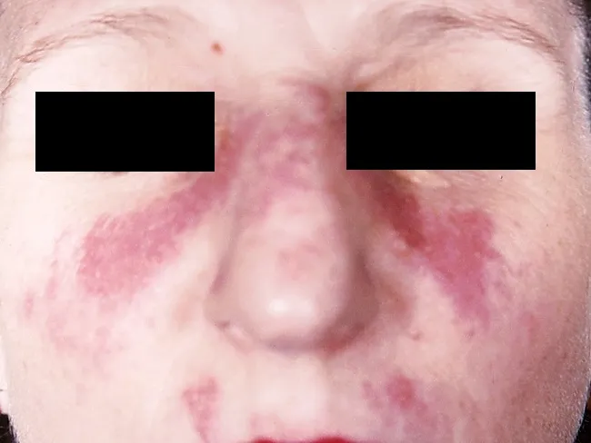
This clinical dermatology photograph displays the classic malar or butterfly rash of systemic lupus erythematosus (SLE) on the central face. Modality is clinical photography using standard white-light illumination; frontal/anterior view; color-balanced, high-resolution capture to depict superficial erythema and patchy hyperemia across the malar eminences and nasal bridge. The rash forms a bilateral, 'butterfly' distribution that typically reaches the cheeks and bridge of the nose while sparing the nasolabial folds. The observed features include confluent to patchy, erythematous macules and plaques with uniform erythema, mild perivascular edema, and subtle textural change without overt crusting or scaling in this image. The clinical morphology is characteristic for acute cutaneous lupus erythematosus; photosensitivity may exacerbate lesions. This cutaneous finding is one of the diagnostic criteria for SLE when aligned with serologic abnormalities (ANA, anti-dsDNA) and systemic features; its presence increases diagnostic probability in a compatible patient. Differential considerations include rosacea, seborrheic dermatitis, contact dermatitis, and dermatomyositis rash; however, the malar distribution and nasal bridge involvement help distinguish lupus. Clinically, this image supports SLE workup and educational reference for recognizing lupus-associated facial rash in medical students, residents, and researchers; useful for pattern-recognition training and multimodal data repository indexing. This image emphasizes clinical-context interpretation and education.
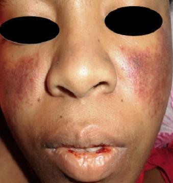
A clinical photograph of a patient's face demonstrating dermatological and mucosal manifestations of Systemic Lupus Erythematosus (SLE). A classic malar rash (butterfly rash) is present, characterized by symmetric, erythematous-to-violaceous patchy lesions over the malar eminences and the bridge of the nose, notably sparing the nasolabial folds. Additionally, the perioral region exhibits significant erythema and mucosal involvement. The lips show evidence of hemorrhagic lesions, with visible blood crusting and a small fissure on the lower lip, suggestive of vasculitis or active systemic inflammation. These visual findings are key diagnostic indicators for SLE, particularly when associated with hematological abnormalities such as thrombocytopenia. The photograph provides a clear example of cutaneous lupus manifestations for clinical diagnosis and medical education.
gout tophus uric acid crystal joint
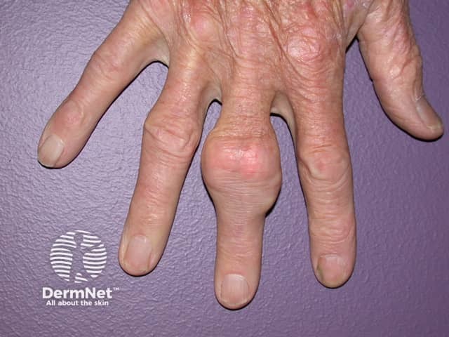
This clinical photograph demonstrates a chronic gout-related tophus involving a finger. Modality: Clinical photography of a human hand. Anatomical location: finger, likely the proximal interphalangeal joint region of the index or middle finger. Appearance: a firm, nodular subcutaneous mass projecting from the dorsal aspect of the finger, about one to two centimeters in diameter, skin overlying the lesion is thin and not broken with mild erythema; the nodule may be slightly lobulated and adherent to underlying structures. Composition: tophus composed of monosodium urate crystals deposited in soft tissue with surrounding granulomatous inflammation; chalky white material can be palpated in some cases. Clinical significance: hallmark of chronic gout; correlates with hyperuricemia and recurrent acute flares; risk of joint destruction if untreated; differential includes rheumatoid nodules, epidermal inclusion cysts, or xanthomas. Diagnostic relevance: supports diagnosis of gout in patients with elevated uric acid; can guide management including urate-lowering therapy, anti-inflammatory treatment, and consideration of imaging such as ultrasound or crystal evaluation. Usage: educational illustration for dermatology, rheumatology, and medical training; useful for patient education on chronic gout and tophi formation, assessment of soft tissue nodules, and monitoring response to therapy. Clinical correlation with gout history strengthens diagnostic confidence; early recognition prevents joint damage and guides urate-targeted therapy outcomes.
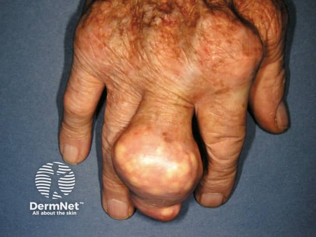
Clinical photography of a left-hand digit showing a gouty tophus. This is a soft-tissue tophus arising from chronic gout with monosodium urate crystal deposition in subcutaneous tissue around the finger joints. The image depicts a single, large, rounded, nodular mass projecting from the dorsal aspect of a finger near the metacarpophalangeal region. The lesion is firm to palpation, with a smooth, pale to whitish surface and surrounding erythema; overlying skin shows mild atrophy and telangiectasia, consistent with chronic inflammatory change. The tophus contains chalky material grossly visible within the dermal and periarticular tissues and may extend toward the joint capsule, eroding adjacent cartilage or bone in long-standing disease. The appearance supports chronic tophaceous gout, though clinical correlation with serum uric acid and prior history of gout is essential. Differential considerations include rheumatoid nodules, xanthomas, and other soft-tissue masses, but the location, size, and chalky appearance strongly favor urate depositions. Diagnostic significance lies in confirming advanced disease with tophus formation; management implications include urate-lowering therapy optimization, anti-inflammatory prophylaxis during flares, and evaluation for additional tophi in other joints. The image is valuable for rheumatology education, dermatology/podiatry teaching, and radiologic-pathologic correlation with ultrasound or dual-energy CT to quantify urate burden.
scleroderma CREST syndrome skin hands
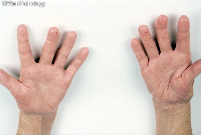
Clinical photography of both hands in dorsal view illustrating cutaneous manifestations of CREST syndrome, a limited form of systemic sclerosis. The image captures distal involvement with sclerodactyly characterized by sausage-like, firm fingers and tightly stretched skin. Telangiectatic vascular ectasia is evident on the dorsum of the hands and at digit tips, with occasional perioral telangiectasia suggested by subtle surface vessels near the lips. The skin appears pink-red and slightly glossy, reflecting chronic tightening and reduced pliability. In this presentation, Raynaud's phenomenon and calcinosis are common associated features though not directly visible in this photograph. Esophageal dysmotility, a hallmark of CREST, is clinically relevant but not demonstrable in an image; nonetheless, the clinical context remains important. The photographic field emphasizes distal-to-MCP joint distribution and the relative sparing of proximal extremities, which is typical for limited cutaneous disease. This image can be used for educational purposes to illustrate hallmark signs: scleroderma-like skin thickening, digit shortening, and superficial vascular changes without ulceration. Potential clinical applications include dermatologic assessment, rheumatology referral decision, patient education on disease phenotype, and documentation of progression or response to therapy aimed at vascular symptoms, skin tightening, and gastrointestinal involvement.
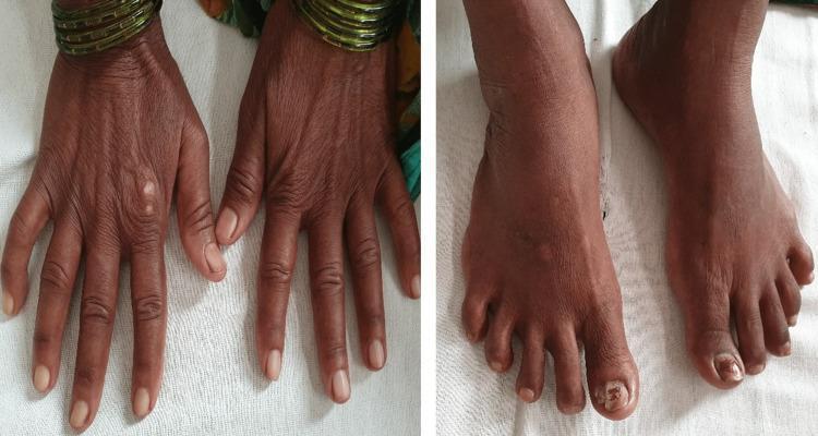
Two clinical photographs demonstrate dermatological manifestations of a systemic connective tissue disease, likely systemic sclerosis. The left panel shows the dorsal aspect of both hands, characterized by significant skin thickening and a taut, bound-down texture. The fingers exhibit tapering and fixed flexion contractures consistent with sclerodactyly. Subtle 'salt and pepper' pigmentary changes (hypopigmentation and hyperpigmentation) are visible on the dorsal surface. The right panel displays the dorsal aspect of the feet, which show similar skin tightening and thickening extending to the toes. Notable findings include xerosis (dryness) and onychodystrophy of the great toenails, suggestive of secondary fungal infection. These visual signs are classic hallmarks of the fibrotic phase of scleroderma, highlighting the loss of skin elasticity and characteristic digital changes used in the clinical diagnosis of CREST syndrome or systemic sclerosis.
osteoarthritis knee joint cartilage degeneration
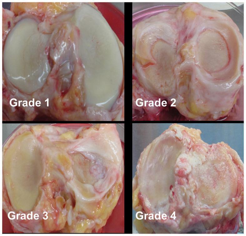
This clinical photograph displays four panels of human donor knee joint specimens illustrating the progressive stages of cartilage degeneration using the modified Collins grading system (Grades 1–4). Grade 1 shows relatively smooth articular surfaces with minor fibrillations. Grade 2 demonstrates more pronounced fibrillation and visible fissuring of the cartilage surface. Grade 3 displays significant cartilage erosion covering up to 30% of the surface, exposing subchondral bone, accompanied by the formation of osteophytes. Grade 4 represents severe osteoarthritis with over 30% of the cartilage surface eroded, extensive exposure of subchondral bone, gross geometric bony remodeling, and prominent osteophyte formation. The specimens highlight key pathological features of osteoarthritis, including loss of cartilage integrity, subchondral bone exposure, and secondary bony changes in the knee joint. This image serves as a reference for orthopedic pathology and gross morphological assessment of joint disease severity.

This diagnostic image series presents a side-by-side comparison of T2 mapping MRI pseudocolor scans of the knee joint, illustrating the progression of osteoarthritis (OA) through the Recht grading system. The sequence (a–e) demonstrates the transition from normal articular cartilage to Grade IV OA. (a) Normal: Shows smooth, continuous cartilage with a uniform yellow-green color. (b) Grade I: Displays uneven cartilage thickness and the emergence of spot-like red colorations, indicating early matrix changes. (c) Grade II: Visualizes surface frizziness and mild joint space narrowing with irregular red gradations. (d) Grade III: Shows severe contour irregularity, visible cartilage defects, and highly mixed pseudocolor distribution. (e) Grade IV: Exhibits full-thickness cartilage defects, exfoliation, significant joint space narrowing, subchondral bone exposure, and extensive osteophyte formation. Clinically, these color shifts—from uniform green to heterogeneous red—reflect alterations in water content and collagen integrity, serving as a quantitative tool for assessing cartilage degeneration and osteoarthritis severity.
vasculitis skin rash purpura leukocytoclastic
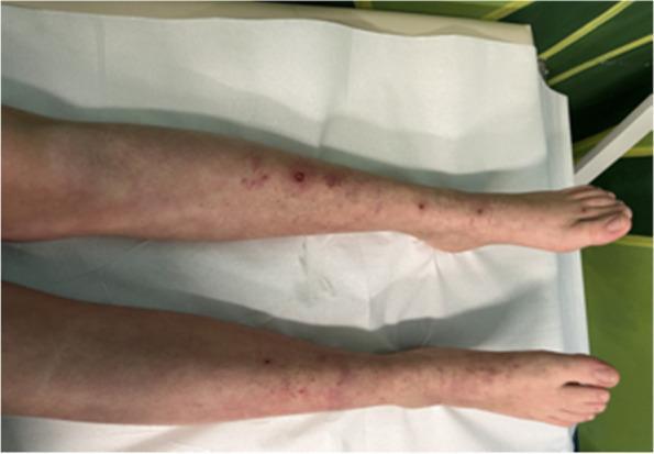
This clinical photograph displays the bilateral lower extremities of a patient, specifically from the mid-calf to the feet, showcasing the resolution phase of a dermatological condition. The skin presents with a predominantly purpuric and petechial rash. Key visible lesions include small, pinpoint reddish-purple petechiae and larger, palpable purpuric papules scattered along the anterior and lateral aspects of the legs. The morphology of the rash suggests small-vessel vasculitis, such as IgA vasculitis (formerly Henoch-Schönlein purpura), showing signs of fading following medical intervention. Some areas exhibit faint, dusky discoloration consistent with post-inflammatory changes or healing bruises. The distribution is notably symmetric and gravitates toward the distal limbs, a classic presentation for systemic leukocytoclastic vasculitis patterns. This image serves as a clinical example of cutaneous vasculitis progression and its visual response to corticosteroid therapy.
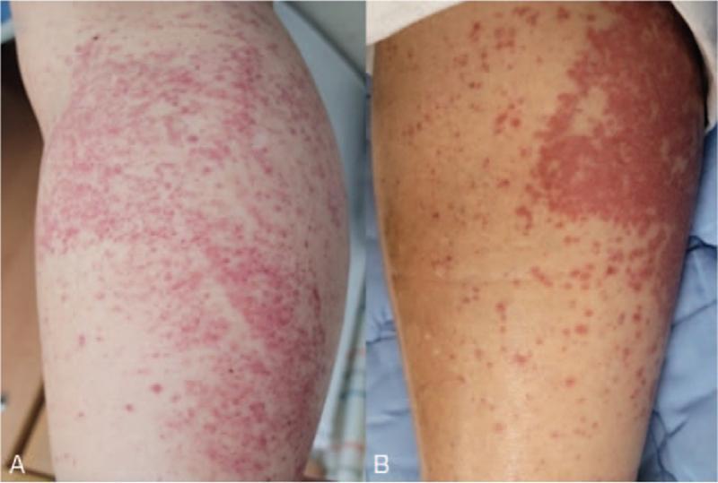
This clinical comparison photograph displays two panels (A and B) showing the progression of skin lesions on the lower leg of a patient diagnosed with IgA vasculitis (Henoch-Schönlein purpura). Panel A illustrates the acute presentation with diffuse, vibrant erythematous macules and papules that are extensively scattered and partially coalescent, forming larger inflammatory patches. Panel B shows the same anatomical region at a later stage, demonstrating a reduction in the density of the rash, though persistent palpable purpura and a localized area of intense confluent erythema remain visible. The image demonstrates the classic cutaneous manifestation of leukocytoclastic vasculitis, characterized by non-blanching red-to-purple lesions typically found in dependent areas. This visual aid is intended for medical education regarding the clinical recognition of systemic vasculitis and its dermatologic evolution during treatment.
Rheumatology - A Comprehensive Overview
1. What is Rheumatology?
2. Classification of Rheumatic Diseases
| Category | Examples |
|---|---|
| Inflammatory arthritis | Rheumatoid arthritis, psoriatic arthritis, reactive arthritis |
| Seronegative spondyloarthropathies | Ankylosing spondylitis, psoriatic arthritis, IBD-associated arthritis |
| Crystal arthropathies | Gout, pseudogout (CPPD) |
| Connective tissue diseases | SLE, scleroderma, Sjögren's syndrome, polymyositis, dermatomyositis |
| Vasculitides | GCA, Takayasu, ANCA-associated vasculitis, PAN |
| Degenerative diseases | Osteoarthritis |
| Bone diseases | Osteoporosis, Paget's disease |
| Soft tissue diseases | Fibromyalgia, bursitis, tendinopathy |
| Autoinflammatory diseases | Familial Mediterranean fever, Still's disease |
3. Rheumatoid Arthritis (RA)
Epidemiology
Pathogenesis
- Synovial T-cell activation and B-cell autoantibody production
- TNF-alpha, IL-1, IL-6, and IL-17 cytokine cascade driving synovial proliferation (pannus formation)
- Pannus erodes cartilage and subchondral bone
- Metabolic disturbances (reduced glutathione, oxidative stress, HLA-DRB1 and PTPN22 gene susceptibility) - Goldman-Cecil Medicine, p. 2794
Clinical Features
- Articular: Symmetrical small joint synovitis (MCPs, PIPs, wrists, MTPs), morning stiffness >1 hour, swan-neck deformities, boutonnière deformities, ulnar deviation
- Extra-articular manifestations (Table 243-1, Goldman-Cecil):
| System | Manifestations |
|---|---|
| Skin | Rheumatoid nodules, vasculitis, pyoderma gangrenosum |
| Heart | Pericarditis, premature atherosclerosis, valve disease |
| Lung | Pleural effusions, ILD, bronchiolitis obliterans |
| Eye | Keratoconjunctivitis sicca, scleritis, episcleritis |
| Neurologic | Entrapment neuropathy, cervical myelopathy, mononeuritis multiplex |
| Hematopoietic | Anemia, Felty syndrome (RA + splenomegaly + neutropenia) |
| Kidney | Amyloidosis |
| Bone | Osteopenia |
Diagnosis - 2010 ACR/EULAR Criteria (Score ≥6 = Definite RA)
| Domain | Score |
|---|---|
| Joint involvement: 1 large joint = 0, >10 joints (at least 1 small) = 5 | 0-5 |
| Serology: Neither RF/ACPA = 0, high positive = 3 | 0-3 |
| Acute phase reactants (CRP/ESR): abnormal = 1 | 0-1 |
| Duration of synovitis: ≥6 weeks = 1 | 0-1 |
RA - X-ray appearance
Treatment of RA
- Methotrexate - first-line anchor DMARD (7.5-25 mg/week); requires folic acid supplementation and monitoring of CBC, LFTs
- Hydroxychloroquine - antimalarial, used in mild disease and in combination
- Sulfasalazine - used especially in Europe and Asia
- Leflunomide - pyrimidine synthesis inhibitor; monitoring required
- Azathioprine - less commonly used
- Avoid without DMARDs; prednisone >10 mg/day rarely needed for articular disease
- Taper to lowest effective dose; toxic risk above 8 mg/day average
- Must use osteoporosis prophylaxis (bisphosphonates, Ca2+, Vit D) - Goldman-Cecil, p. 2795
| Drug Class | Examples |
|---|---|
| TNF inhibitors | Etanercept, adalimumab, infliximab, certolizumab, golimumab |
| IL-6 receptor antagonists | Tocilizumab, sarilumab |
| T-cell costimulation blocker | Abatacept |
| B-cell depleter | Rituximab (anti-CD20) |
| IL-1 antagonist | Anakinra |
- Tofacitinib 5 mg twice daily (JAK1/3)
- Baricitinib 2-4 mg daily (JAK1/2) - superior to adalimumab in MTX-inadequate responders
- Upadacitinib 15 mg daily (selective JAK1)
- Filgotinib 100-200 mg daily
- Monitor: CBC, LFTs; risks include infections, TB reactivation, malignancy, CV events, herpes zoster reactivation - Goldman-Cecil, p. 2796
4. Systemic Lupus Erythematosus (SLE)
Epidemiology
Pathogenesis
- Defective clearance of apoptotic debris leads to nuclear antigen exposure
- B and T cell activation against dsDNA, histones, Smith antigen, phospholipids
- Immune complex deposition activates complement (↓C3, ↓C4) and drives organ inflammation
- Type I interferon pathway plays a central role
Classification Criteria (2019 EULAR/ACR)
| Clinical Domain | Key Criteria | Points |
|---|---|---|
| Constitutional | Fever | 2 |
| Neuropsychiatric | Seizure | 5, Psychosis 3, Delirium 2 |
| Mucocutaneous | Acute cutaneous lupus (malar rash) | 6 |
| Mucocutaneous | Oral ulcers, non-scarring alopecia | 2 each |
| Musculoskeletal | Joint involvement | 6 |
| Serosal | Pleural/pericardial effusion | 5 |
| Renal | Proteinuria >0.5g/24h | 4, Lupus nephritis class III/IV = 10 |
| Immunologic | Anti-dsDNA antibody | 6 |
| Immunologic | Anti-Sm antibody | 6 |
| Complement | Low C3 AND C4 | 4 |
| Hematologic | Autoimmune hemolysis | 4, Thrombocytopenia = 4 |
Classic Malar "Butterfly" Rash
SLE - Key Autoantibodies
| Antibody | Specificity | Clinical Association |
|---|---|---|
| ANA | Sensitive screening (>95%) | Not specific |
| Anti-dsDNA | ~60% sensitive, highly specific | Disease activity, nephritis |
| Anti-Smith | ~25% sensitive, very specific | SLE-specific |
| Anti-Ro/SSA, Anti-La/SSB | - | Neonatal lupus, Sjögren's overlap |
| Antiphospholipid antibodies | - | Thrombosis, recurrent miscarriage |
| Anti-histone | - | Drug-induced lupus |
Lupus Nephritis
- Most serious complication. WHO/ISN classification I-VI based on biopsy.
- Active urine sediment (RBC casts, proteinuria) signals active nephritis.
- Treatment: High-dose prednisone (60 mg/day) or IV methylprednisolone 1g/day + cyclophosphamide or mycophenolate mofetil (MMF). MMF preferred for induction and maintenance in many centers.
- Adjunctive: ACE inhibitor/ARB for hypertension control.
SLE Treatment
- Hydroxychloroquine - cornerstone therapy for all SLE patients; reduces flares, thrombosis risk; monitor eyes every 12 months
- NSAIDs - for mild arthritis and serositis
- Glucocorticoids - for flares; minimize long-term dose
- Immunosuppressants: MMF, azathioprine, cyclophosphamide
- Biologic therapies:
- Belimumab (anti-BLyS) - approved; reduces flares and lupus nephritis
- Anifrolumab (anti-type I IFN receptor) - approved for moderate-severe SLE
- Rituximab - used off-label for refractory disease
- Contraception/Pregnancy: Avoid moderate/severe disease activity at conception. Hydroxychloroquine may reduce fetal heart block risk. - Goldman-Cecil, p. 2819-2820
5. Spondyloarthropathies (SpA)
- HLA-B27 association
- Axial skeleton involvement (sacroiliitis)
- Enthesitis (inflammation at tendon/ligament insertions)
- Asymmetric oligoarthritis
- Extra-articular features (uveitis, psoriasis, IBD)
Ankylosing Spondylitis (AS)
- Genetics: ~90% of patients are HLA-B27 positive; ERAP1 gene is second strongest risk factor; >100 genes implicated; IL-23R gene on chr. 1 also associated. - Goldman-Cecil, p. 2797
- HLA-B27 prevalence varies widely: absent in Australian Aboriginals, 1% in Japan, 7% in northern Europe, up to 50% in some western Canadian native tribes
- Clinical: Inflammatory back pain (worse at rest, better with exercise), morning stiffness >1 hour, sacroiliitis, progressive spinal fusion
- Bamboo spine on X-ray (syndesmophytes), bilateral sacroiliitis
- Modified NY Criteria: Radiographic sacroiliitis + low back pain >3 months + limited lumbar motion or chest expansion
- ASAS Criteria (2009): Axial SpA can be diagnosed by imaging arm (sacroiliitis on MRI/X-ray) or clinical arm (HLA-B27 + ≥2 SpA features)
- NSAIDs - first-line for inflammatory back pain; continuous use may slow radiographic progression
- TNF inhibitors (adalimumab, etanercept, infliximab, golimumab, certolizumab) - effective for both axial and peripheral disease
- IL-17A inhibitors (secukinumab, ixekizumab) - highly effective; particularly useful in patients who fail TNF inhibitors
- IL-23 inhibitors (risankizumab, guselkumab) - newer, effective options
- Physiotherapy is mandatory alongside drug therapy
6. Gout and Crystal Arthropathies
Gout
- Serum urate >6.8 mg/dL = hyperuricemia. MSU crystals deposit in joints, soft tissues, and kidneys.
- Crystal deposition triggers NLRP3 inflammasome activation → IL-1β release → acute inflammatory attack
- Even asymptomatic hyperuricemia may cause occult crystal deposition - Firestein & Kelley's, p. 2684
- Asymptomatic hyperuricemia
- Acute gouty arthritis (monoarticular, podagra = first MTP involvement)
- Intercritical gout (interval between attacks)
- Chronic tophaceous gout
Tophus in Chronic Gout
Treatment of Gout (Katzung's Pharmacology, p. 3104)
- Colchicine (1.2 mg then 0.6 mg 1 hour later) - effective within 24 hours; inhibits microtubule polymerization and neutrophil chemotaxis
- NSAIDs (indomethacin, naproxen) - high-dose for 5-7 days
- Corticosteroids - intra-articular or systemic; useful when NSAIDs/colchicine contraindicated (renal failure)
- Allopurinol (xanthine oxidase inhibitor) - target serum urate <6.0 mg/dL; start low (100 mg/day), titrate
- Febuxostat (selective xanthine oxidase inhibitor) - for allopurinol-intolerant patients
- Probenecid (uricosuric) - increases renal urate excretion; for underexcretors; avoid if creatinine clearance <50 mL/min
- Pegloticase (recombinant uricase) - IV every 2 weeks; for refractory tophaceous gout
- Start ULT with colchicine prophylaxis (0.6 mg daily) to prevent flares during initiation
Pseudogout (CPPD Disease)
- Calcium pyrophosphate dihydrate crystals deposit in cartilage (chondrocalcinosis)
- Crystals are positively birefringent, rhomboid-shaped
- Associated with hyperparathyroidism, hemochromatosis, hypomagnesemia, hypothyroidism
- Treatment: NSAIDs, colchicine, intra-articular steroids; no specific crystal-dissolving therapy
7. Osteoarthritis (OA)
Risk Factors
Pathophysiology
- Cartilage matrix degradation by MMPs and ADAMTS enzymes
- Chondrocyte senescence and failure of repair
- Subchondral bone remodeling → osteophytes (bony spurs)
- Synovial inflammation amplifies pain
Clinical Features
- Use-related pain (worse with activity, relieved by rest) - contrast with inflammatory arthritis (worse at rest/morning)
- No significant morning stiffness (<30 minutes)
- Heberden's nodes (DIP), Bouchard's nodes (PIP), CMC joint OA (base of thumb)
- Knee, hip, spine involvement common
OA Cartilage Degeneration - Gross Pathology
Treatment
- Weight loss and exercise (strengthening quadriceps)
- Topical NSAIDs, oral NSAIDs/COX-2 inhibitors
- Intra-articular corticosteroids (short-term pain relief)
- Intra-articular hyaluronic acid (controversial evidence)
- Duloxetine (for central sensitization component)
- Joint replacement surgery for severe, refractory disease
8. Systemic Sclerosis (Scleroderma)
Definition
Subtypes
- Limited cutaneous SSc (lcSSc): Skin involvement limited to hands, forearms, face; CREST syndrome (Calcinosis, Raynaud's, Esophageal dysmotility, Sclerodactyly, Telangiectasia); anti-centromere antibody; slower progression; pulmonary hypertension late complication
- Diffuse cutaneous SSc (dcSSc): Widespread skin thickening beyond elbows/knees; anti-topoisomerase I (anti-Scl-70) antibody; faster progression; ILD, renal crisis
CREST Syndrome Hands
Epidemiology
- Incidence 9-19 per million per year; female predominance; peak age 40-60 years
- Black patients: higher incidence, earlier onset, more diffuse disease with ILD
- Occupational risk: crystalline silica; drugs: bleomycin, pentazocine
Pathobiology
- Three pillars: immune activation, vascular damage, fibroblast activation → excess collagen
- Key cytokines: TGF-β (master fibrotic driver), PDGF, CTGF
- Genetic: family history is the strongest risk factor; 1.6% of patients have an affected first-degree relative
Treatment
- Raynaud's phenomenon: Calcium channel blockers (nifedipine), phosphodiesterase-5 inhibitors (sildenafil), IV prostacyclin (iloprost) for severe ischemia
- ILD: Mycophenolate mofetil (first-line), cyclophosphamide, nintedanib (anti-fibrotic)
- Pulmonary arterial hypertension: ERA (bosentan), PDE5-inhibitors (sildenafil), prostacyclin analogues, selexipag
- Scleroderma renal crisis: ACE inhibitors (captopril) - mandatory; can be life-saving
- GI involvement: Proton pump inhibitors, prokinetics
9. Vasculitides
| Vessel size | Disease |
|---|---|
| Large vessel | Giant cell arteritis (GCA), Takayasu's arteritis |
| Medium vessel | Polyarteritis nodosa (PAN), Kawasaki disease |
| Small vessel | ANCA-associated (GPA, MPA, EGPA), IgA vasculitis (HSP), cryoglobulinemic vasculitis |
Giant Cell Arteritis (GCA)
- Most common vasculitis in adults >50 years
- Affects superficial temporal artery, aorta and branches
- Symptoms: temporal headache, scalp tenderness, jaw claudication, visual loss (ischemic optic neuropathy - EMERGENCY)
- Lab: ESR typically >50 mm/hr, CRP elevated
- Diagnosis: temporal artery biopsy (skip lesions - biopsy ≥2 cm length)
- Treatment: High-dose prednisone (40-60 mg/day) IMMEDIATELY if visual involvement suspected; do NOT wait for biopsy. Tocilizumab (IL-6 inhibitor) as steroid-sparing agent.
ANCA-Associated Vasculitis
- GPA (Granulomatosis with Polyangiitis): Upper/lower respiratory tract + kidneys; c-ANCA (anti-PR3); saddle-nose deformity, alveolar hemorrhage
- MPA (Microscopic Polyangiitis): Kidney + lung; p-ANCA (anti-MPO)
- EGPA (Eosinophilic GPA/Churg-Strauss): Asthma + eosinophilia + vasculitis; p-ANCA
- Treatment: Cyclophosphamide + high-dose steroids for induction; rituximab now preferred for GPA/MPA induction. Maintenance: azathioprine or rituximab.
IgA Vasculitis (Henoch-Schönlein Purpura)
- Triad: palpable purpura + arthritis + abdominal pain; ± nephritis (IgA nephropathy pattern)
- Most common vasculitis in children; self-limiting in most cases
10. Other Important Conditions
Sjögren's Syndrome
- Autoimmune exocrinopathy: lymphocytic infiltration of salivary and lacrimal glands
- Primary Sjögren's vs. Secondary (with RA, SLE, SSc)
- Features: dry eyes (keratoconjunctivitis sicca), dry mouth (xerostomia), fatigue, arthralgia
- Antibodies: Anti-Ro/SSA (most sensitive, ~75%) and Anti-La/SSB
- Complications: lymphoma (MALT type, 5-8x increased risk), parotid enlargement, renal tubular acidosis, peripheral neuropathy
- Treatment: Artificial tears, pilocarpine/cevimeline for sicca, hydroxychloroquine
Polymyositis (PM) and Dermatomyositis (DM)
- Inflammatory myopathies with proximal muscle weakness
- DM: characteristic skin findings - heliotrope rash (periorbital), Gottron's papules (over MCP/PIP joints), V-sign/shawl sign
- Antibodies: anti-Jo-1 (anti-synthetase syndrome: myositis + ILD + mechanic's hands + fever + arthritis)
- Elevated CK, aldolase, LDH, AST
- MRI of muscles shows edema; biopsy confirms diagnosis
- DM carries increased risk of underlying malignancy (especially older patients)
- Treatment: High-dose glucocorticoids + methotrexate or azathioprine; IVIG for refractory cases
Polymyalgia Rheumatica (PMR)
- Age >50 (typically >70); bilateral shoulder girdle and hip girdle aching and stiffness
- Markedly elevated ESR (>40 mm/hr) and CRP
- Closely associated with GCA (occurs in 15-20% of PMR patients)
- Dramatically responsive to low-dose prednisone (15-20 mg/day) - diagnostic and therapeutic
- Diagnosis of exclusion (rule out infection, malignancy, inflammatory arthritis)
Reactive Arthritis (formerly Reiter's Syndrome)
- Triggered by infection (urogenital: Chlamydia; GI: Salmonella, Shigella, Campylobacter, Yersinia)
- Classic triad: arthritis + urethritis + conjunctivitis ("can't see, can't pee, can't bend the knee")
- HLA-B27 association; mostly resolves in months; some develop chronic SpA
- Treatment: NSAIDs; antibiotics if active infection; sulfasalazine for chronic cases
11. Key Laboratory Investigations in Rheumatology
| Test | Significance |
|---|---|
| ANA | Screening for SLE, SSc, Sjögren's, mixed CTD |
| Anti-dsDNA | SLE (high specificity), correlates with disease activity |
| Anti-Sm | SLE-specific |
| RF (IgM) | RA (70-80% positive), Sjögren's, other CTDs |
| Anti-CCP (ACPA) | RA - more specific than RF; predicts erosive disease |
| Anti-Ro/SSA, Anti-La/SSB | Sjögren's, neonatal lupus, subacute cutaneous lupus |
| Anti-centromere | Limited SSc (CREST) |
| Anti-Scl-70 | Diffuse SSc; associated with ILD |
| Anti-Jo-1 | Anti-synthetase syndrome (PM/DM + ILD) |
| c-ANCA (anti-PR3) | GPA |
| p-ANCA (anti-MPO) | MPA, EGPA |
| Complement (C3, C4) | Low in active SLE (consumed by immune complexes) |
| Antiphospholipid antibodies | APS: thrombosis + recurrent miscarriage |
| Serum uric acid | Gout (>6.8 mg/dL) |
| Synovial fluid analysis | Distinguishes inflammatory vs. non-inflammatory vs. septic; crystal identification |
| CRP, ESR | Nonspecific markers of inflammation |
| CK, aldolase | Inflammatory myopathies |
12. Key Imaging in Rheumatology
| Modality | Use |
|---|---|
| Plain radiographs | Joint space narrowing, erosions (RA), syndesmophytes (AS), chondrocalcinosis (CPPD), osteophytes (OA) |
| MRI | Early sacroiliitis (bone marrow edema), synovitis, tendon/cartilage assessment |
| Musculoskeletal ultrasound | Synovitis (power Doppler), enthesitis, effusions, crystal deposits, guided injections |
| CT | Bone erosions, ILD in SSc/RA, pulmonary vasculitis |
| DEXA scan | Bone mineral density (osteoporosis monitoring) |
| Dual-energy CT | Gout - urate crystal burden quantification |
| PET-CT | GCA (large vessel involvement), malignancy screening in DM |
13. Key Medications in Rheumatology
DMARDs Summary
| Drug | Mechanism | Main Uses | Monitoring |
|---|---|---|---|
| Methotrexate | Folate antagonist, anti-inflammatory | RA, PsA, vasculitis | CBC, LFTs, creatinine |
| Hydroxychloroquine | Lysosomotropic agent | SLE, RA, Sjögren's | Ophthalmology (retinal toxicity) |
| Sulfasalazine | Anti-inflammatory, anti-folate | RA, SpA | CBC, LFTs |
| Leflunomide | Pyrimidine synthesis inhibitor | RA, PsA | CBC, LFTs, BP |
| Mycophenolate mofetil | Purine synthesis inhibitor | Lupus nephritis, SSc-ILD | CBC |
| Cyclophosphamide | Alkylating agent | Severe SLE, vasculitis, SSc | CBC, urinalysis (hemorrhagic cystitis), bladder cancer risk |
| Azathioprine | Purine analog | Maintenance RA, SLE, vasculitis | CBC, LFTs; check TPMT |
| Colchicine | Tubulin inhibitor | Gout, pericarditis, FMF | GI side effects; renal/hepatic dose adjust |
| Allopurinol | Xanthine oxidase inhibitor | Gout prevention | Allopurinol hypersensitivity syndrome (check HLA-B*5801 in Asian patients) |
Biologic/Targeted Agents
| Class | Examples | Key Indications |
|---|---|---|
| TNF inhibitors | Etanercept, adalimumab, infliximab | RA, SpA, PsA, IBD-related arthritis |
| IL-6 inhibitors | Tocilizumab, sarilumab | RA, GCA, systemic JIA |
| IL-17A inhibitors | Secukinumab, ixekizumab | AS, PsA, nr-AxSpA |
| IL-23 inhibitors | Guselkumab, risankizumab | PsA, SpA |
| IL-12/23 inhibitors | Ustekinumab | PsA, IBD-related |
| CTLA-4-Ig (costimulation blockade) | Abatacept | RA |
| Anti-CD20 | Rituximab | RA, ANCA vasculitis, SLE (off-label) |
| Anti-BLyS | Belimumab | SLE, lupus nephritis |
| Anti-IFN receptor | Anifrolumab | Moderate-severe SLE |
| JAK inhibitors | Tofacitinib, baricitinib, upadacitinib | RA, SpA, PsA |
| IL-1 inhibitors | Anakinra, canakinumab, rilonacept | Gout, adult-onset Still's, FMF |
| Uricase | Pegloticase | Refractory chronic gout |
14. Emerging Therapies (2025-2026)
- CAR-T cell therapy targeting CD19 is showing remarkable early responses in severe refractory SLE, antisynthetase syndrome, and systemic sclerosis - achieving drug-free remission in small series
- Microbiome-immunosuppression interactions: A 2025 systematic review (PMID 40883126) found that gut microbiota composition influences response to immunosuppressive therapy in systemic autoimmune rheumatic diseases
- TYK2 inhibitors (deucravacitinib) - under study for SLE and PsA
- IL-2-based therapies (low-dose IL-2) to restore regulatory T cells - phase II trials in SLE
- Obinutuzumab (enhanced anti-CD20) - trials in lupus nephritis
15. Fibromyalgia
- Pathophysiology: central sensitization (abnormal pain processing in CNS)
- Tender points replaced by Widespread Pain Index (WPI) and Symptom Severity Scale (SSS) in 2010 ACR criteria
- No laboratory or imaging abnormalities
- Treatment: multidisciplinary - aerobic exercise (best evidence), cognitive-behavioral therapy, duloxetine, milnacipran, pregabalin/gabapentin, amitriptyline (low-dose)
16. Approach to the Patient with Joint Disease
- Is it articular or periarticular?
- How many joints? (mono-, oligo-, polyarthritis)
- Which joints? (small vs. large; axial vs. peripheral)
- Symmetric or asymmetric?
- Is there inflammation? (warmth, swelling, prolonged morning stiffness)
- Acute or chronic? (gout = acute; OA = chronic)
- Are there extra-articular features?
| Category | WBC/µL | PMNs | Appearance | Causes |
|---|---|---|---|---|
| Non-inflammatory | <2,000 | <25% | Clear, viscous | OA, trauma |
| Inflammatory | 2,000-100,000 | >50% | Turbid | RA, SpA, gout, pseudogout |
| Septic | >50,000 | >75% | Purulent | Bacterial infection |
| Hemorrhagic | RBCs | - | Bloody | Trauma, hemarthrosis, anticoagulation |
Summary Table: Key Distinguishing Features
| Disease | Key Antibodies | HLA | X-ray/Imaging | Hallmark Clinical Feature |
|---|---|---|---|---|
| RA | RF, Anti-CCP | HLA-DRB1 | Symmetric erosions, joint space narrowing | Symmetrical small joint synovitis |
| SLE | ANA, anti-dsDNA, anti-Sm | HLA-DR2/DR3 | Usually no erosions | Multi-system + malar rash |
| Ankylosing Spondylitis | None specific | HLA-B27 (90%) | Bamboo spine, sacroiliitis | Inflammatory back pain, young men |
| Psoriatic Arthritis | None specific | HLA-B27 | "Pencil-in-cup" deformity | Psoriasis + arthritis + nail changes |
| Gout | - | - | Punched-out erosions with overhanging edge | Acute podagra, tophi |
| Scleroderma (diffuse) | Anti-Scl-70 | HLA-DR11 | ILD, calcinosis | Raynaud's + skin thickening |
| Scleroderma (limited) | Anti-centromere | - | Calcinosis | CREST syndrome |
| Sjögren's | Anti-Ro/SSA, Anti-La/SSB | HLA-DR3 | - | Sicca (dry eyes/mouth) |
| PM/DM | Anti-Jo-1, anti-Mi-2 | HLA-DR3 | - | Proximal weakness; Gottron's papules (DM) |
| GPA | c-ANCA (anti-PR3) | - | Pulmonary cavities | Upper/lower respiratory + nephritis |
- Goldman-Cecil Medicine International Edition, chapters 243-249
- Firestein & Kelley's Textbook of Rheumatology, 11th edition
- Rheumatology, 2-Volume Set, Elsevier 2022
- Katzung's Basic and Clinical Pharmacology, 16th edition, chapter 36
- CAR-T in autoimmune disease - Wang et al., Medicine 2025 (PMID 41305772)
- Microbiome and immunosuppression - Franco-Domingo et al., Reumatologia Clinica 2025 (PMID 40883126)