I now have comprehensive data from multiple authoritative textbooks. Let me compile the full answer.
Succinylcholine (Suxamethonium) - Complete Reference
1. Classification & Synonyms
Succinylcholine (also called suxamethonium; trade names: Anectine, Quelicin) is the only depolarizing neuromuscular blocking drug (NMBD) currently in clinical use. It belongs to the class of short-acting, depolarizing agents at the nicotinic acetylcholine receptor.
2. Chemical Structure
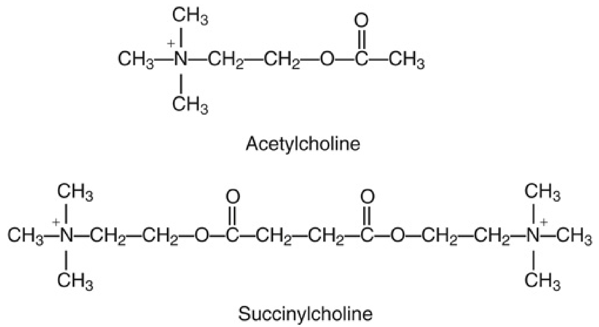
Succinylcholine consists of two acetylcholine molecules linked through their acetate methyl groups (Fig. 24.1, Miller's Anesthesia). It is a small, flexible, bisquaternary ammonium compound with very low lipid solubility, giving it a small volume of distribution. Like ACh, it activates cholinergic receptors at the neuromuscular junction and muscarinic autonomic sites.
3. Mechanism of Action
Succinylcholine binds to and activates (not blocks) the nicotinic acetylcholine receptor (nAChR) at the motor end plate, causing initial depolarization - seen clinically as muscle fasciculations. Unlike ACh, it is not rapidly hydrolyzed at the NMJ (no acetylcholinesterase is present there), so it persists and causes a sustained depolarization.
This sustained depolarization produces Phase I (depolarizing) block:
- The muscle membrane around the end plate remains depolarized
- Sodium channels enter an inactivated state
- Surrounding muscle cannot be re-excited → flaccid paralysis
- Characteristics: no fade on train-of-four (TOF), no post-tetanic facilitation, not reversed by anticholinesterases (in fact, worsened)
With high doses or repeated/prolonged exposure, a Phase II (desensitization) block can develop, which takes on characteristics resembling nondepolarizing block (fade on TOF, post-tetanic facilitation, partial reversal by neostigmine). This occurs because the receptor undergoes conformational change to a desensitized state.
- Miller's Anesthesia, 10e
- Morgan and Mikhail's Clinical Anesthesiology, 7e
4. Pharmacokinetics
| Parameter | Value |
|---|
| Onset (IV) | ~60 seconds (complete block with 1 mg/kg) |
| Duration (IV) | 9-13 min (90% recovery); clinically 4-6 min |
| ED95 | 0.51-0.63 mg/kg (Kopman estimates < 0.3 mg/kg) |
| Elimination half-life | ~47 seconds |
| Volume of distribution | Small (very low lipid solubility) |
| Metabolism | Plasma butyrylcholinesterase (pseudocholinesterase) |
Metabolism pathway: Succinylcholine → succinylmonocholine (weak NMBD) → succinic acid + choline
Only ~10% of IV succinylcholine reaches the NMJ - the rest is hydrolyzed in plasma before getting there. Because no butyrylcholinesterase exists at the NMJ, block terminates by diffusion away from the NMJ back into plasma, where it is then hydrolyzed.
5. Dosing
Adults
| Route | Dose |
|---|
| IV (intubation) | 0.3-1.1 mg/kg (typical RSI dose: 1-1.5 mg/kg) |
| IM | 3-4 mg/kg; max 150 mg |
| Maintenance (IV bolus) | 0.04-0.07 mg/kg Q5-10 min PRN |
| Continuous infusion | Not recommended (risk of Phase II block) |
Pediatric (Harriet Lane Handbook, 23rd ed.)
| Patient | IV dose | IM dose |
|---|
| Infant | 2-3 mg/kg | 4-5 mg/kg (<6 mo) |
| Child | 1-2 mg/kg | 4 mg/kg; max 150 mg |
| Adolescent | 1-1.5 mg/kg | 3-4 mg/kg; max 150 mg |
Children require higher mg/kg doses than adults because of their larger volume of distribution. Duration IV: 4-6 min; IM: 10-30 min.
6. Clinical Uses
- Rapid Sequence Intubation (RSI) - the primary indication; gold standard for providing fast, reliable intubation conditions
- Electroconvulsive therapy (ECT) - attenuates convulsion intensity
- Laryngospasm treatment (IV or IM)
- Short procedures requiring brief muscle relaxation
- When a "cannot intubate, cannot oxygenate" scenario must be avoided (ultra-short duration means spontaneous ventilation returns quickly if intubation fails)
After succinylcholine for intubation, a nondepolarizing NMBD is typically given to maintain relaxation. Prior succinylcholine enhances the depth and duration of subsequent nondepolarizing block.
7. Butyrylcholinesterase (Pseudocholinesterase) Variants
This is one of the most clinically important topics related to succinylcholine.
Dibucaine Number
Dibucaine (a local anesthetic) inhibits normal pseudocholinesterase by 80% but inhibits atypical enzyme by only 20%. The dibucaine number = percentage of enzyme inhibition.
| Genotype | Dibucaine Number | Block Duration |
|---|
| Normal homozygote | ~80 | Normal (9-13 min) |
| Heterozygote (1 normal + 1 atypical) | 40-60 | Mildly prolonged (20-30 min) |
| Atypical homozygote (1 in 3000) | ~20 | Very prolonged (4-8 hours) |
The atypical (dibucaine-resistant) allele produces enzyme with 1/100th the normal affinity for succinylcholine. Other variants: fluoride-resistant and silent (no activity) alleles.
Factors that Reduce Butyrylcholinesterase Activity
(Cause modest prolongation, usually 2-20 min)
- Liver disease, advanced age, malnutrition, pregnancy, burns
- Drugs: echothiophate, neostigmine, pyridostigmine, cyclophosphamide, metoclopramide, phenelzine (MAOIs), esmolol, oral contraceptives, bambuterol, pancuronium
Important: Even reducing butyrylcholinesterase to 20% of normal (severe liver disease) only extends apnea from ~3 min to ~9 min. Atypical genotype is the main cause of clinically significant prolonged block.
A
2026 systematic review in BJA (PMID: 42120224) has characterized genotype-phenotype relationships in butyrylcholinesterase deficiency in detail.
8. Cardiovascular Effects
Bradycardia
- Results from stimulation of muscarinic receptors in the SA node
- More pronounced after a second dose (can cause sinus arrest)
- Children are particularly susceptible to profound bradycardia even after the first dose
- Atropine pretreatment is mandatory in children (minimum 0.1 mg IV)
Nodal (Junctional) Rhythms
- Due to relatively greater muscarinic stimulation at the SA node, allowing the AV node to take over
- More common after the second dose; can be prevented by prior d-tubocurarine
Ventricular Dysrhythmias
- Succinylcholine lowers the ventricular threshold for catecholamine-induced dysrhythmias
- Circulating catecholamines increase ~4-fold and K+ by ~1/3 after succinylcholine
- Risk is increased by: hypoxia, hypercarbia, digitalis, tricyclics, MAOIs, halothane
9. Hyperkalemia
- Normal subjects: plasma K+ rises by ~0.5 mEq/L (well tolerated)
- Mechanism: activation of postsynaptic AChR → Na+ influx → K+ efflux
- Patients with renal failure are NOT inherently more susceptible (unless uremic neuropathy present) - safe if baseline K+ is normal
- Life-threatening hyperkalemia occurs in patients with:
- Major burns (after acute phase)
- Massive trauma
- Extensive denervation (spinal cord injury, stroke with upper motor neuron lesion)
- Prolonged immobilization
- Skeletal muscle myopathies (Duchenne, Becker)
- Severe intra-abdominal infection
- Peripheral neuropathies (Guillain-Barre)
The mechanism: after denervation/upregulation, extrajunctional AChRs proliferate across the entire muscle membrane. Succinylcholine activates all of them simultaneously → massive K+ efflux → cardiac arrest
Treatment of severe hyperkalemia from succinylcholine:
- Immediate hyperventilation
- IV calcium chloride 500-1000 mg or calcium gluconate over 3 min
- Insulin + glucose (adults: 10 U regular insulin in 50 mL of 50% dextrose; children: 0.15 U/kg in 1 mL/kg 50% dextrose)
10. Other Side Effects
| Effect | Details |
|---|
| Muscle fasciculations | Universal; due to initial depolarization |
| Myalgias | Postoperative muscle pain (most common in ambulatory patients); fasciculations tear muscle fibers; pre-treatment with small dose of nondepolarizing NMBD reduces fasciculations |
| Increased intraocular pressure (IOP) | Peaks 2-4 min after injection; concern for open globe injury |
| Increased intragastric pressure | Partially offset by increased lower esophageal sphincter tone; aspiration risk not clearly increased |
| Increased intracranial pressure | May transiently increase ICP |
| Malignant hyperthermia (MH) | Triggers MH in susceptible individuals (RYR1 or CACNA1S gene mutations); treat with dantrolene; absolute contraindication if MH history |
| Anaphylaxis | Most common trigger of perioperative anaphylaxis in some studies |
| Masseter spasm | Trismus; may be an early sign of MH |
| Prolonged Phase II block | With repeated doses or high-dose infusions |
11. Contraindications
Absolute
- Personal or family history of malignant hyperthermia
- Skeletal muscle myopathies (Duchenne, Becker) - risk of rhabdomyolysis and cardiac arrest
- Receptor upregulation states (denervation, burns >24-48 hrs, prolonged immobility, UMN lesion) - risk of fatal hyperkalemia
- Known hypersensitivity
Relative
- Hyperkalaemia or risk thereof
- Open globe injury (raises IOP, though this is debated)
- Penetrating eye injuries
The FDA has issued a Black Box Warning for succinylcholine use in pediatric patients (except for emergency intubation) due to risk of acute rhabdomyolysis and hyperkalemia with cardiac arrest in undiagnosed myopathies.
12. Prevention of Side Effects
| Side Effect | Prevention |
|---|
| Fasciculations/myalgia | "Precurarization" - small dose of nondepolarizing NMBD (e.g., vecuronium 0.01 mg/kg) 3 min before |
| Fasciculations (antagonism) | Small dose of nondepolarizing blocker can also antagonize depolarizing effect |
| Bradycardia | Atropine pretreatment (mandatory in children) |
| Phase II block | Avoid repeated doses/infusions |
13. Drug Interactions
| Drug | Effect on succinylcholine block |
|---|
| Anticholinesterases (neostigmine, pyridostigmine) | Prolong block (inhibit butyrylcholinesterase) |
| Organophosphates (echothiophate) | Prolong block |
| Aminoglycosides | Potentiate |
| Inhalational anesthetics | Potentiate |
| Phenothiazines, quinidine, β-blockers, amphotericin B | Prolong block |
| Cyclophosphamide, lithium, diuretics | Prolong block |
| Diazepam | May decrease neuromuscular blocking effect |
| Bambuterol | Marked inhibition of butyrylcholinesterase - significant prolongation |
| Prior succinylcholine → subsequent vecuronium | Enhances vecuronium block duration |
14. RSI Context & Alternatives
Succinylcholine is the traditional gold standard for RSI due to its unmatched onset/offset profile. The main competitor is rocuronium at high doses (1.2 mg/kg), which can be reversed rapidly with sugammadex - making high-dose rocuronium + sugammadex a viable alternative, especially when succinylcholine is contraindicated.
A
2025 systematic review (PMID: 39931560) on pharmacological interventions in Duchenne muscular dystrophy anesthesia specifically highlights why succinylcholine must be avoided in this population.
15. Quick Summary
| Feature | Value |
|---|
| Class | Depolarizing NMBD |
| Mechanism | nAChR agonist → sustained depolarization |
| Onset | ~60 sec (IV) |
| Duration | 4-13 min (IV) |
| Metabolism | Butyrylcholinesterase |
| Primary use | RSI, ECT, laryngospasm |
| Key contraindications | MH susceptibility, myopathy, receptor upregulation, hyperkalemia |
| Black box warning | Pediatric use (except emergencies) |
| Only depolarizing agent | Yes - unique in current clinical practice |
Sources: Miller's Anesthesia 10e | Morgan & Mikhail's Clinical Anesthesiology 7e | Barash's Clinical Anesthesia 9e | Harriet Lane Handbook 23e | Rosen's Emergency Medicine | Katzung's Basic & Clinical Pharmacology 16e