
Sex determination and age determination of hip bone
Reading File
This is a broad anatomy/forensic topic. Let me search the library in parallel for both sex determination and age determination from the hip bone.
Finding Sources
Finding Sources
Finding Sources
Finding Sources
Finding Sources
Finding Sources
Found relevant forensic medicine textbooks. Let me read the key sections now in parallel.
Reading File
Reading File
Good data found. Now let me read the hip bone-specific sex determination table and the age determination from hip bone sections in parallel.
Reading File
Finding Sources
Reading File
Finding Sources
Now let me get the age determination content and the hip bone sex determination table from the Essentials textbook:
Reading File
Finding Sources
Reading File
Reading File
Now let me get the ossification/age determination data specific to the hip bone from the Dikshit textbook:
Reading File
Loading Image
Now let me fetch a couple more key images and look for the ossification table for age from the innominate bone:
Loading Image
Reading File
Now let me get the ossification centres / age determination from the hip bone (Dikshit textbook) and fetch key pelvic images:
Reading File
Loading Image
I now have enough data to compile a comprehensive answer. Let me also fetch the innominate bone greater sciatic notch image and the male/female pelvis images:
Loading Image
Loading Image
I now have comprehensive material from both textbooks. Here is the full, detailed answer:
Sex Determination and Age Determination from the Hip Bone (Innominate Bone)
Sources: The Essentials of Forensic Medicine and Toxicology, 36th ed. (2026); P C Dikshit Textbook of Forensic Medicine and Toxicology
Why the Hip Bone is So Important
The hip bone (innominate bone = ilium + ischium + pubis) is the single most valuable bone for both sex and age determination in forensic skeletal analysis.
According to Krogman's accuracy table:
| Material Available | Accuracy |
|---|---|
| Entire skeleton | 100% |
| Pelvis + skull | 98% |
| Pelvis alone | 95% |
| Skull alone | 90-92% |
| Long bones alone | 80-85% |
Without the pelvis, accuracy drops to ~50%. The pelvis alone gives ~95% accuracy in adults - it is by far the most reliable single bone.
PART 1: SEX DETERMINATION FROM HIP BONE
Sex differences in the skeleton generally only appear after puberty - except in the pelvis, where some differences are evident even in children and fetuses. The greater sciatic notch is the single most important criterion for sex determination in children and adolescents.
The fundamental functional basis: the female pelvis is shaped for parturition (wider, shallower, more circular), while the male pelvis is shaped for locomotion (deeper, narrower, more funnel-like).
Detailed Comparison Table: Male vs. Female Hip Bone / Pelvis
| Feature | Male | Female |
|---|---|---|
| General framework | Deep, funnel-shaped; massive, rough | Shallow, bowl-shaped; less massive, smooth |
| True pelvis | Narrow, deep, funnel-shaped | Wide and shallow |
| Pelvic brim (inlet) | Heart-shaped | Circular/oval |
| Pelvic outlet | Smaller | Larger |
| Ilium | Curve more prominent; more sloped; less rounded margins | Curve less prominent; less sloped; more rounded margins |
| Iliac crest | More prominent | Less prominent |
| Anterior superior iliac spine (ASIS) | Not widely separated | Widely separated |
| Pre-auricular sulcus | Not prominent, narrow, shallow | Prominent, broad, deep |
| Acetabulum | Wide and deep; diameter ~52 mm; directed laterally | Small and narrow; diameter ~46 mm; directed anterolaterally |
| Subpubic angle | 70°-75° (acute); arch is V-shaped | ~90° (obtuse); arch is U-shaped |
| Greater sciatic notch | Smaller, deeper, narrower; less than a right angle (acute) | Wider, larger, shallower; about a right angle (obtuse) |
| Sciatic notch index (width/depth) | 4-5 | 5-6 |
| Obturator foramen | Large, oval-shaped with base upwards | Small, triangular, apex directed forwards |
| Ischial tuberosity | More or less inverted | Everted (turned outwards) |
| Pubic body | Narrow and triangular | Broad and square |
| Symphysis pubis (height) | Higher, deeper, narrower | Lower, wider, rounded |
| Dorsal pubic margin | Smooth margins; no parturition pits | Irregular; parturition pits (scars of parturition) present in parous females |
| Symphyseal block shape | Triangular | Rectangular |
| Pelvic index (AP/transverse × 100) | Higher | Lower |
| Ischiopubic index (ischial length/pubic length × 100) | 73-94 | 91-115 |
| Kell index (acetabulum SA/ilium SA × 100) | Higher | Lower |
Key Individual Landmarks on the Innominate Bone
1. Greater Sciatic Notch - Most reliable single feature
- Male: narrow, deep, acute angle (< 90°)
- Female: wide, shallow, obtuse (≈ 90°)
- Best criterion in children when other features are absent
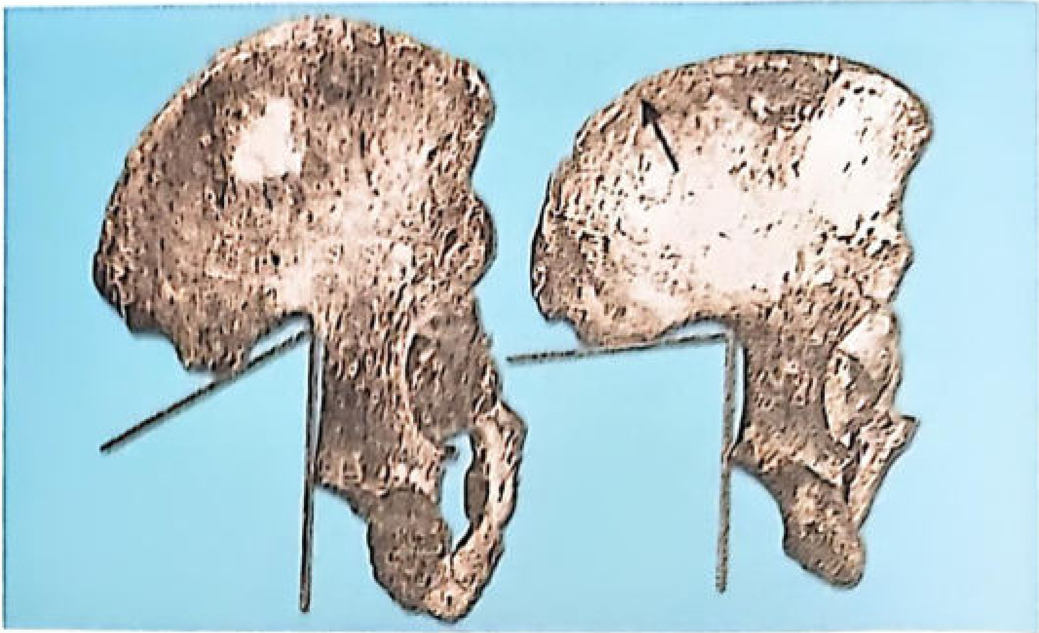
Fig: Innominate bone - the greater sciatic notch angle is acute in the male (left) and wider/obtuse in the female (right).
2. Subpubic Angle
- Male: 70-75°, V-shaped arch
- Female: ~90°, U-shaped arch
3. Pre-auricular Sulcus
- A groove on the posterior ilium, adjacent to the sacroiliac joint
- Male: absent or barely visible
- Female: broad, deep, well-marked (related to ligamentous attachment during pregnancy)
4. Parturition Pits
- On the dorsal surface of the pubic bone near the symphysis
- Exclusive to females who have given birth (parous females)
5. Obturator Foramen
- Male: large, oval
- Female: small, triangular
6. Acetabulum
- Male: ~52 mm diameter; faces laterally
- Female: ~46 mm diameter; faces anterolaterally
Helpful Indices
| Index | Formula | Male | Female |
|---|---|---|---|
| Sciatic notch index | Width / Depth | 4-5 | 5-6 |
| Ischiopubic index | Ischial length / Pubic length × 100 | 73-94 | 91-115 |
| Pelvic index | AP diameter / Transverse diameter × 100 | Higher | Lower |
Female has a higher ischiopubic index because her pubic rami are relatively longer.
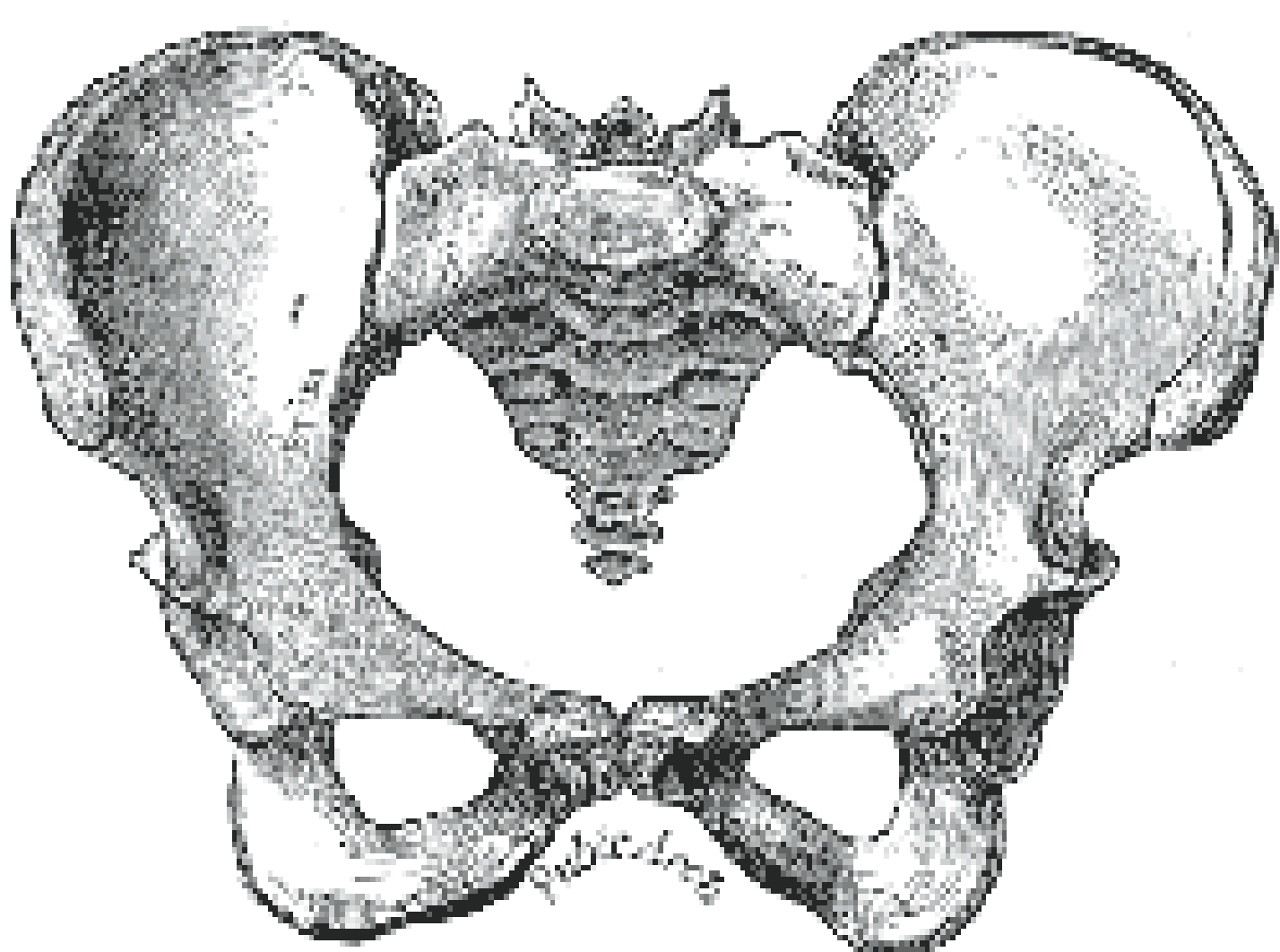
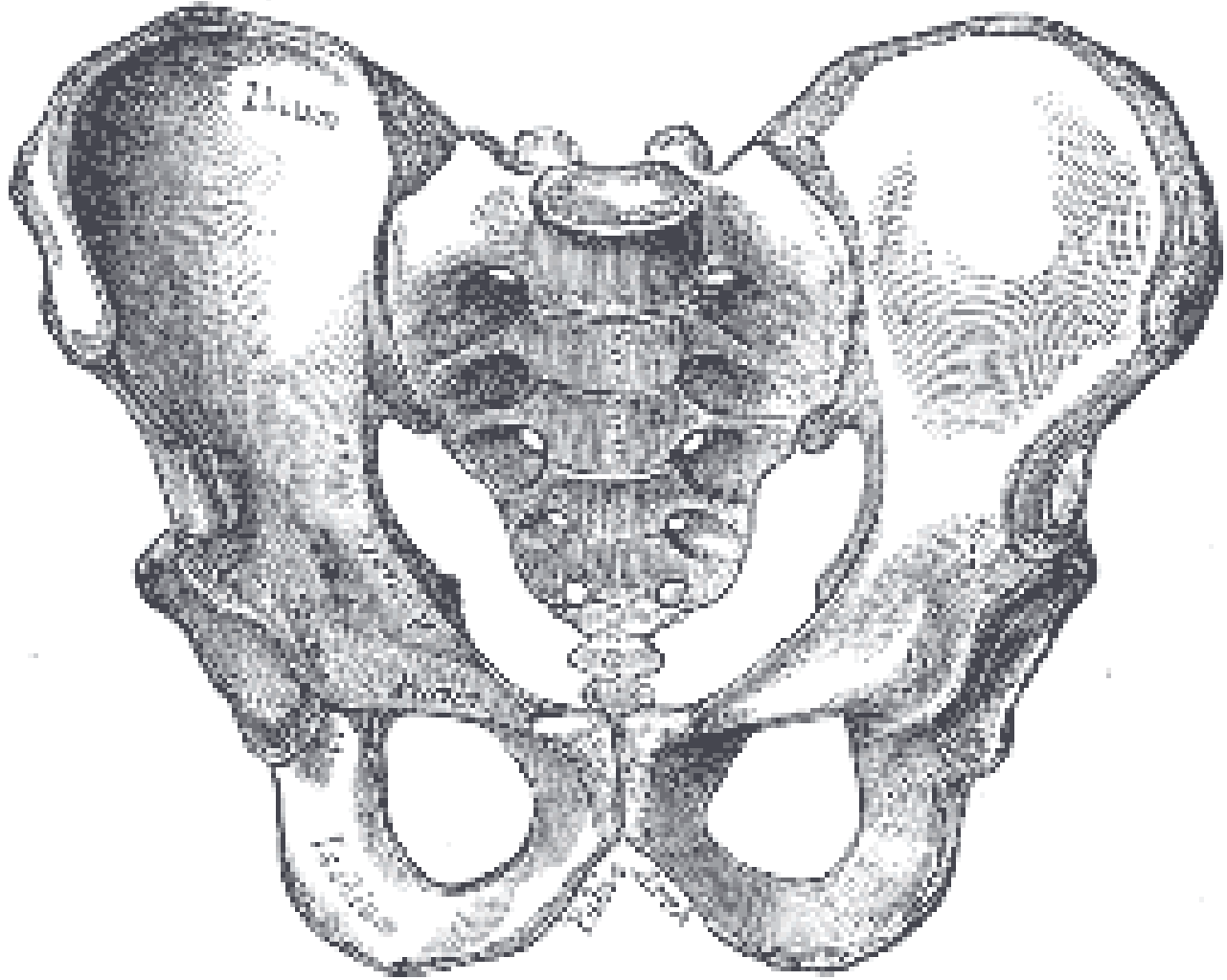
Male vs. Female Pelvis - Photographic Reference
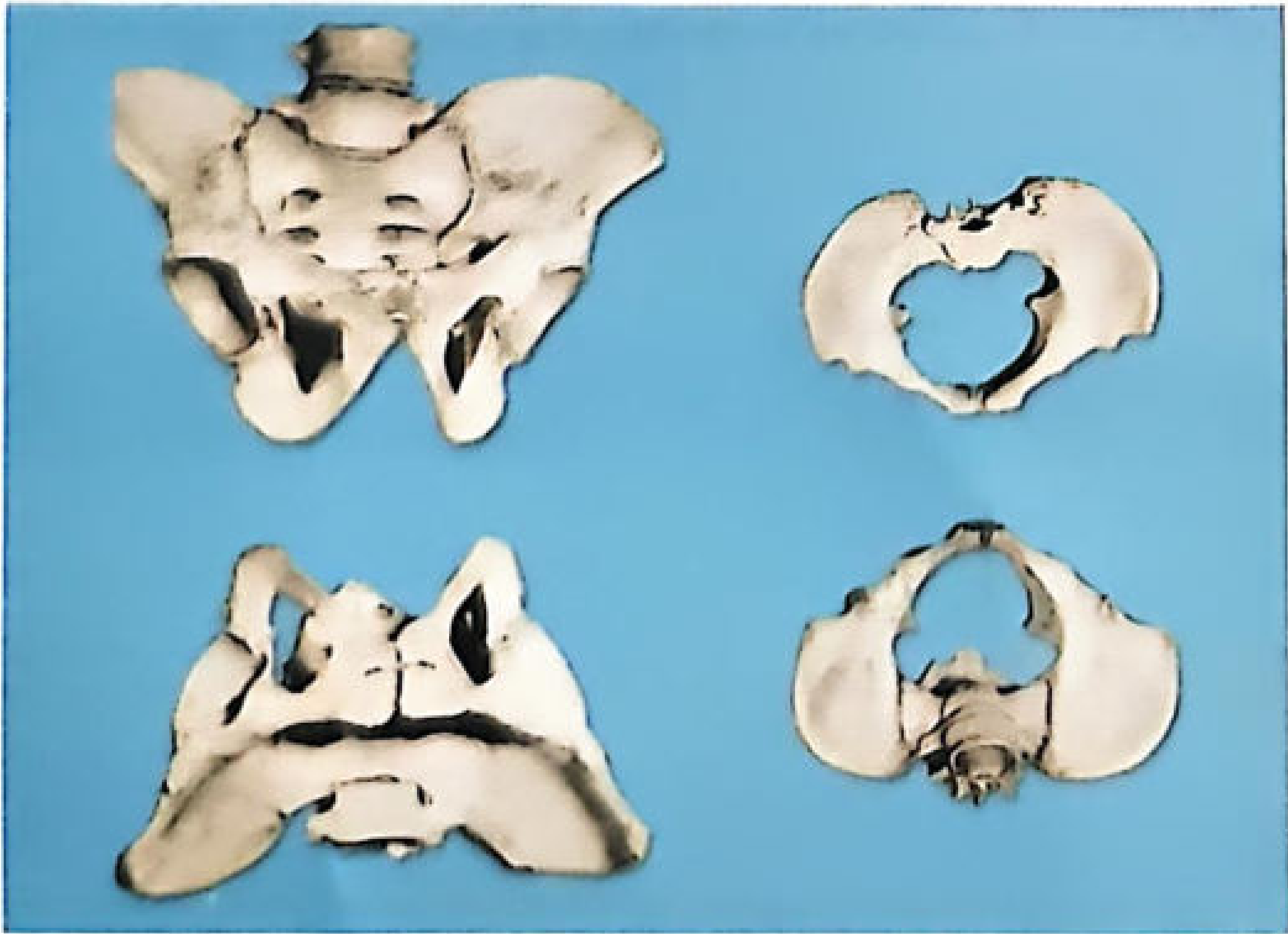
Fig: Male pelvis (upper) vs. female pelvis (lower). Note the V-shaped subpubic angle and smaller pelvic outlet in male, contrasted with U-shaped subpubic angle and larger outlet in female.
Special Features in Females Only
- Chilotic line: Anthropometric line in the pelvis; its index is used in sex identification.
- Sternal index: Higher in females (54.3 vs 46.2 in males - not hip bone, but supplementary)
- Female skeletal measurements are on average 94% of male size (Pfitner's table), varying from 91-98% across different dimensions.
PART 2: AGE DETERMINATION FROM HIP BONE
Age from the hip bone is assessed primarily through ossification centres and their fusion times. The hip bone is the composite of three primary bones (ilium, ischium, pubis) that fuse in a specific sequence.
Ossification Centres and Fusion Timetable
The three bones of the hip (ilium, ischium, pubis) meet at the acetabulum (triradiate cartilage). Additional secondary ossification centres appear at puberty.
| Ossification Event | Age |
|---|---|
| Ilium - primary centre appears | ~8th week of intrauterine life |
| Ischium - primary centre appears | ~4th month of intrauterine life |
| Pubis - primary centre appears | ~5th month of intrauterine life |
| Ischium and pubis fuse (inferior ramus) | 5-8 years |
| Three bones meet in acetabulum (triradiate cartilage closes) | 15-17 years |
| Iliac crest epiphysis - appears | ~15-16 years |
| Iliac crest epiphysis fuses | 20-25 years |
| Ischial tuberosity epiphysis appears | ~15-17 years |
| Ischial tuberosity epiphysis fuses | ~20-25 years |
| Acetabular epiphysis | fuses ~18-20 years |
| Anterior inferior iliac spine (AIIS) epiphysis | appears ~14 years; fuses ~18-20 years |
Pubic Symphysis Changes with Age (Todd's Method)
The pubic symphysis face changes progressively with age and is one of the most studied age indicators in forensic anthropology:
| Phase | Age Range | Features of Symphyseal Face |
|---|---|---|
| 1 | ~18-19 years | Billowing surface (ridges and furrows), no rim |
| 2 | ~20-21 years | Ridges beginning to fuse, active remodeling |
| 3 | ~22-24 years | Ridges almost obliterated, dorsal plateau forming |
| 4 | ~25-26 years | Complete dorsal plateau, ventral rampart begins |
| 5 | ~27-30 years | Complete dorsal and ventral plateaus, oval outline |
| 6 | ~30-35 years | Complete oval symphyseal face, smooth surface |
| 7 | ~35-39 years | Slight erosion, lipping at margins |
| 8 | ~39-44 years | Marked erosion, rim disruption |
| 9 | ~45-50 years | Progressive degeneration |
| 10 | ~50+ years | Irregular, pitted, eroded surface |
Practical Age Milestones from Hip Bone
| Age | Finding |
|---|---|
| Birth to 5 years | Three separate bones (ilium, ischium, pubis) visible as separate ossifications |
| 5-8 years | Ischium and pubis fuse inferiorly |
| 15-17 years | Triradiate cartilage fuses - three bones now form one innominate |
| 20-25 years | Iliac crest epiphysis fuses - marks completion of skeletal maturity |
| After 25 | Pubic symphysis morphology used (degenerative changes) |
| After 40 | Progressive degenerative changes on auricular surface of ilium |
Auricular Surface of Ilium (Age Estimation in Older Adults)
The auricular surface (where the sacroiliac joint is) also changes with age:
- Young adults: granular, billowing texture
- Middle age: transverse organisation, increasing density
- Old age: dense, irregular, porosity, lipping at margins
This method (Lovejoy et al.) is particularly useful for estimating age in individuals over 50 years.
Key Points to Remember
- Pelvis alone = 95% accuracy for sex determination (Krogman).
- Greater sciatic notch is the single most reliable feature, usable even in children.
- Subpubic angle: V-shape (70-75°) = male; U-shape (~90°) = female.
- Pre-auricular sulcus and parturition pits are female-specific.
- For age: triradiate cartilage fusion (15-17 years) and iliac crest fusion (20-25 years) are the most useful milestones.
- Pubic symphysis morphology is the gold standard for age in adults (Todd's 10-phase system).
- Sex differences in the pelvis begin appearing before birth; they are detectable in fetuses with ~75-80% accuracy.
The Essentials of Forensic Medicine and Toxicology, 36th ed., Table 4.5, pp. 79-80 | P C Dikshit Textbook of Forensic Medicine and Toxicology, Table 6.10, pp. 82-83
This is a shared conversation. Sign in to Orris to start your own chat.