
10 mark answer on nerve injuries
Reading File
Now let me search the medical library for nerve injury content in parallel.
Finding Sources
Finding Sources
Finding Sources
Reading File
Reading File
Those were surgical context entries. Let me search for the core nerve injury classification content (Seddon/Sunderland) and Rockwood & Green which is most relevant for a 10-mark exam answer.
Reading File
Finding Sources
Excellent! Found the key classification section. Let me read it in full, plus the neurology textbook table.
Reading File
Reading File
Now let me read more of the Sabiston section on clinical evaluation, diagnosis, and management of nerve injuries.
Reading File
Finding Sources
Reading File
Reading File
I now have comprehensive material. Let me fetch the Wallerian degeneration diagram from Bailey & Love for visual inclusion.
Loading Image
I now have all the material needed for a comprehensive 10-mark answer. Let me compose it.
Nerve Injuries
1. Introduction
A nerve injury is any disruption to the structural or functional integrity of a peripheral nerve, resulting in impaired motor, sensory, or autonomic function. Injuries range from a transient conduction block to complete nerve transection and are classified by the degree of structural damage to the nerve's components (axon, myelin, endoneurium, perineurium, epineurium).
2. Anatomy of a Peripheral Nerve
Understanding classification requires knowledge of nerve architecture:
- Endoneurium - connective tissue sheath surrounding each individual axon
- Perineurium - surrounds a fascicle (bundle of axons)
- Epineurium - outer sheath enclosing the entire nerve trunk
3. Classification of Nerve Injuries
A. Seddon Classification (1943)
Seddon described three types based on injury severity:
| Type | Structures Damaged | Key Features | Prognosis |
|---|---|---|---|
| Neuropraxia | Myelin only | Focal conduction block; axon intact | Full recovery within days to 6 weeks |
| Axonotmesis | Myelin + axon | Basal lamina intact; Wallerian degeneration occurs distally | Recovery possible but slower; depends on level |
| Neurotmesis | All structures including epineurium | Complete transection or severe disorganization | Poor without surgical repair |
Classic example of neuropraxia: Wrist drop from prolonged compression of the radial nerve at the spiral groove of the humerus ("Saturday night palsy").
B. Sunderland Classification (Further Subdivision)
Sunderland extended Seddon's system into 5 grades to better capture the spectrum of axonotmesis and neurotmesis:
| Grade | Seddon Equivalent | Structures Damaged |
|---|---|---|
| I | Neuropraxia | Myelin only; focal demyelination |
| II | Axonotmesis | Myelin + axon; endoneurium, perineurium, and epineurium intact |
| III | Neurotmesis | Myelin, axon, endoneurium disrupted; perineurium and epineurium intact |
| IV | Neurotmesis | Myelin, axon, endoneurium, perineurium disrupted; epineurium intact |
| V | Neurotmesis | Complete transection of all structures |
| VI (MacKinnon) | Mixed | Combined injury with elements of different grades in the same nerve |
(Sabiston Textbook of Surgery, Table 41.5)
4. Pathophysiology: Wallerian Degeneration and Regeneration
When axonal continuity is lost (Sunderland grades II-V), Wallerian degeneration occurs distal to the injury site:
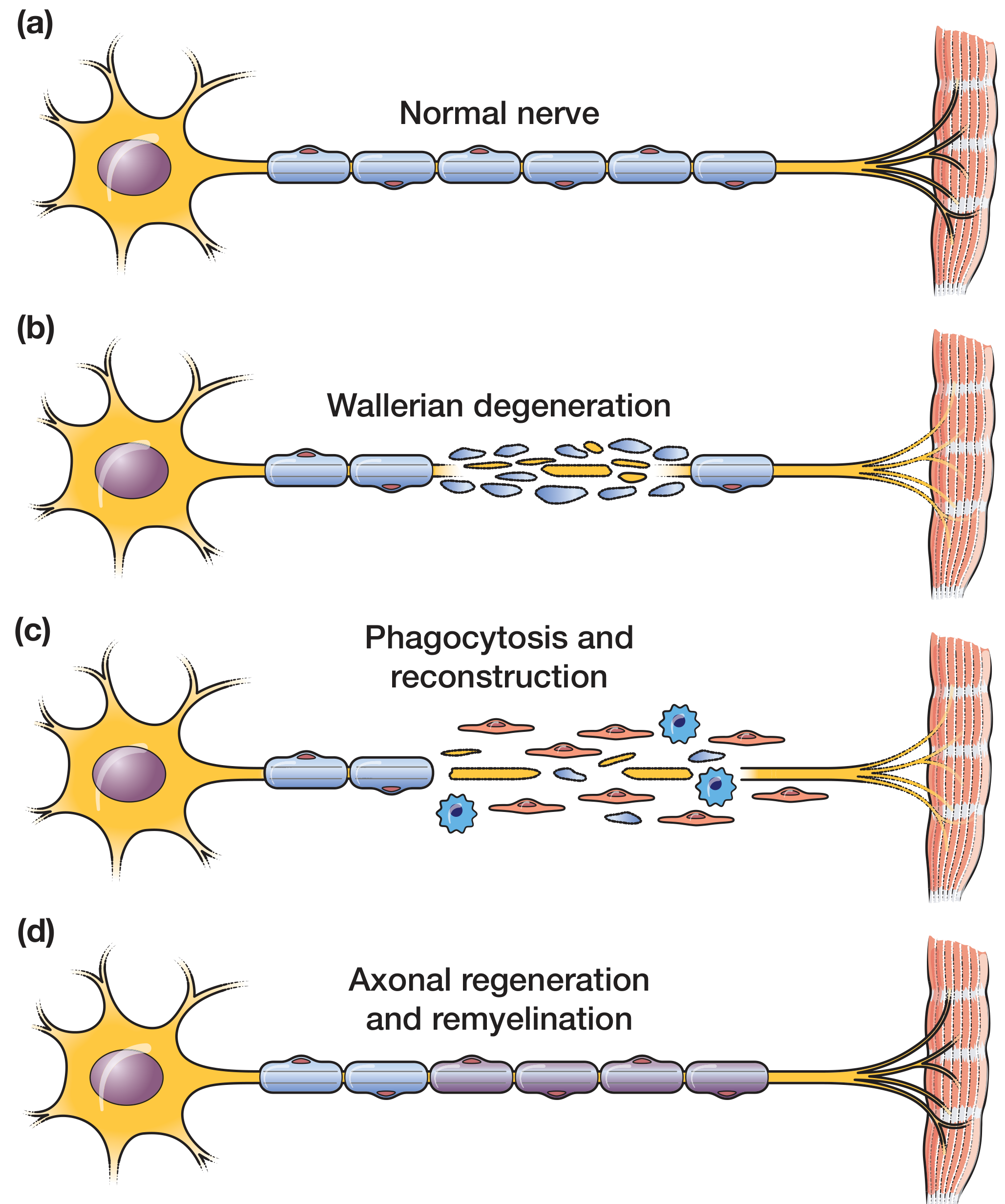
Figure: (a) Normal nerve; (b) Wallerian degeneration - breakdown of axon and myelin distal to injury; (c) Phagocytosis by macrophages recruited by Schwann cells; (d) Axonal regeneration and remyelination guided by bands of Büngner.
Key steps:
- The axon and myelin distal to the injury break down within 24-72 hours
- Schwann cells dedifferentiate and form bands of Büngner - tubes guiding axon regrowth
- Macrophages recruited by Schwann cells scavenge myelin debris
- Regenerating axons grow at approximately 1 mm/day (or 1 inch/month) from the proximal stump
- Proximal nerve also undergoes retrograde degeneration back to the nearest node of Ranvier
- Neurotropism (guided by growth factors and the extracellular matrix) directs the regenerating axon toward its target (Bailey & Love's Short Practice of Surgery, p.48)
5. Causes of Nerve Injury
| Mechanism | Example |
|---|---|
| Compression | Tourniquet, cast, external pressure |
| Traction/stretch | Brachial plexus during shoulder dislocation |
| Laceration | Knife wound, iatrogenic (scalpel) |
| Crush | Fractures, industrial injuries |
| Ischemia | Prolonged tourniquet, compartment syndrome |
| Injection injury | Intramuscular injection near sciatic nerve |
| Radiation | Post-radiation plexopathy |
6. Clinical Features
- Motor: Weakness or paralysis in the distribution of the nerve
- Sensory: Numbness, paraesthesia, hyperaesthesia in the cutaneous territory
- Autonomic: Loss of sweating, vasomotor changes
- Tinel's sign: Percussion over the site of nerve injury (or regenerating front) produces electric shock/pins-and-needles sensation distally - useful to track recovery
7. Investigations
- Nerve conduction studies (NCS): Identify conduction block (neuropraxia) vs. axonal loss; after transection, conduction persists for up to 72 hours until Wallerian degeneration is complete
- Electromyography (EMG): Fibrillations and fasciculations confirm denervation; begin at 14 days post-injury; voluntary motor units on EMG indicate reinnervation. Optimally ordered 3 weeks post-injury to differentiate neuropraxic from more severe injuries
- High-resolution ultrasound: Assesses nerve continuity and fascicular architecture; useful in the acute setting
- MRI neurography: Best for deep or proximal nerve injuries (brachial/lumbosacral plexus); visualizes edema, neuroma, and nerve-in-continuity lesions (Sabiston Textbook of Surgery)
8. Management
Conservative (Non-operative)
- Indicated for neuropraxia and many axonotmesis injuries
- Expectant management with physiotherapy and splinting
- Recovery of neuropraxia expected within 6 weeks; axonotmesis may take months depending on distance to target
- Serial EMG/NCS at 3 weeks and 3-6 months to monitor recovery
- Radial nerve palsy with humeral shaft fracture: initial non-operative management preferred; spontaneous recovery in majority (Rockwood & Green, p.1440)
Operative
Indicated for:
- Complete transection (neurotmesis), sharp laceration injuries
- No signs of recovery at 3-6 months
- Nerve entrapment, concomitant vascular injury, open fractures, gunshot wounds
Surgical options:
| Technique | Indication |
|---|---|
| Primary neurorrhaphy (direct repair) | Tension-free approximation; sharp wounds; gaps <5 mm |
| Nerve graft (autograft) | Gaps >5 mm; sural nerve most common donor |
| Nerve conduit | Gaps <10 mm; avoids donor site morbidity |
| Nerve transfer | Proximal injuries with long reinnervation distances; reroutes expendable donor nerve |
| Tendon transfer | Late presentation; irreversible muscle fibrosis (>12-18 months) |
Principles of repair: tension-free anastomosis; operating microscope; 8-0 to 10-0 monofilament suture; epineural or grouped fascicular repair. Reinnervation of muscle before 12-18 months is necessary to avoid irreversible atrophy. (Sabiston, p.808)
9. Motor Recovery Grading (MRC Scale)
| Grade | Description |
|---|---|
| M0 | No contraction |
| M1 | Perceptible contraction of proximal muscles |
| M2 | Perceptible contraction of proximal and distal muscles |
| M3 | Contraction against gravity |
| M4 | Contraction against resistance |
| M5 | Full recovery |
The minimum functional goal is M3 (antigravity strength). (Sabiston Textbook of Surgery)
10. Prognosis
- Neuropraxia: Excellent - full and spontaneous recovery within hours to 6 weeks
- Axonotmesis: Good if the level of injury is distal; recovery follows Wallerian degeneration at ~1 mm/day; less predictable with higher-grade injury
- Neurotmesis: Poor without surgery; even with repair, complete functional recovery is rarely achieved. Factors affecting prognosis include patient age, level of injury, mechanism, time to repair, and gap length
Sources: Bradley and Daroff's Neurology in Clinical Practice, Table 64.1 | Sabiston Textbook of Surgery, Table 41.5 and pp.806-809 | Bailey & Love's Short Practice of Surgery 28th Ed., p.48 | Rockwood & Green's Fractures in Adults 10th Ed., p.1440
This is a shared conversation. Sign in to Orris to start your own chat.