
Explain Rotator cuff , shoulder complex biomechanics
rotator cuff muscles anatomy shoulder
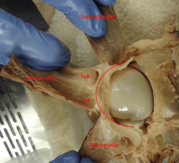
Anatomical Dissection: This clinical photograph provides a posterior view of a dissected left human shoulder, focusing on the rotator cuff complex. The supraspinatus, infraspinatus, and teres minor muscles are reflected laterally to reveal the underlying humeral head and its capsule. A curved red line overlay highlights the rotator cable, a thick fibrous band that spans the avascular zone of the rotator cuff. The infraspinatus muscle is further subdivided with labels 'Sup.' and 'Inf.', representing the superior and inferior portions of the muscle, respectively. The image demonstrates the varying degrees of adherence between the infraspinatus and the rotator cable, illustrating the functional anatomy of the shoulder's capsuloligamentous structures. This material is useful for studying musculoskeletal anatomy, specifically the insertions of the rotator cuff and the biomechanical role of the rotator cable in distributing forces across the shoulder joint.
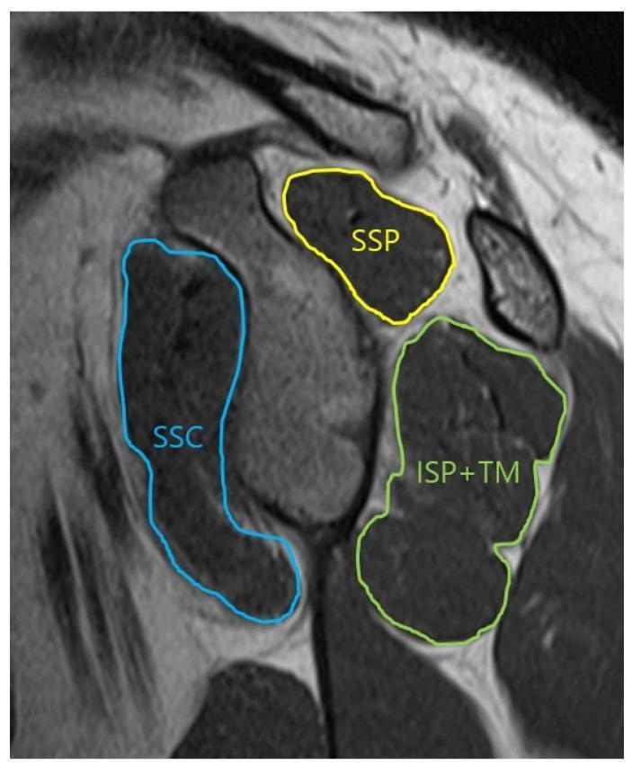
This oblique-sagittal T2 Turbo Spin Echo (TSE) MRI scan demonstrates the musculoskeletal anatomy of the shoulder in a glenoid face view, specifically for assessing the cross-sectional area of the rotator cuff muscles. The image features color-coded annotations outlining the muscular boundaries: the supraspinatus (SSP) is outlined in yellow in the superior position; the subscapularis (SSC) is traced in blue anteriorly; and the infraspinatus combined with the teres minor (ISP+TM) is outlined in green in the posterior-inferior quadrant. The musculature appears as intermediate to dark gray signal intensity, allowing for the evaluation of muscle volume and potential fatty infiltration or atrophy. This imaging view is clinically significant for orthopedic evaluation and preoperative planning in cases of rotator cuff tears or shoulder instability, providing a clear visualization of the muscular support surrounding the glenohumeral joint.
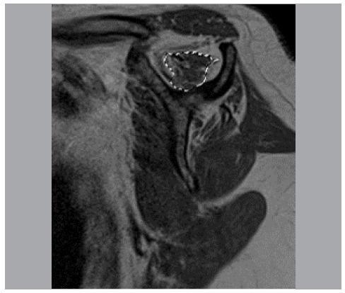
This diagnostic image is a T1-weighted oblique sagittal MRI of the shoulder, specifically at the level where the scapular spine is continuous with the scapular body, forming a characteristic 'Y' shape. The image demonstrates the cross-sectional anatomy of the rotator cuff muscles. Fat is visualized with high signal intensity (bright white), while the muscle tissue shows intermediate signal intensity (gray). A white dotted line is used as an annotation to trace the area of the supraspinatus muscle belly within the supraspinatus fossa. This specific slice and tracing method are clinically relevant for assessing muscular trophism and fatty infiltration, often utilized in the evaluation of rotator cuff tears and postoperative recovery. Key visible structures include the supraspinatus, infraspinatus, and subscapularis muscles, providing a clear view for volumetric and qualitative analysis of muscle mass.
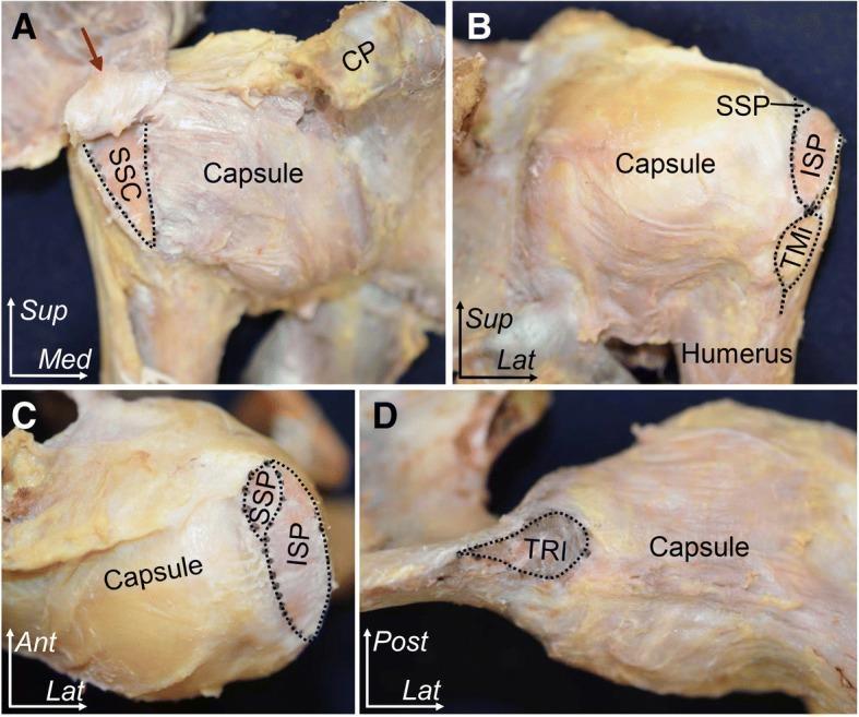
This composite of four clinical cadaveric photographs (A–D) illustrates the anatomy of the human shoulder joint, focusing on the insertions of the rotator cuff muscles and the articular capsule. The images use black dotted lines to delineate footprint locations and arrows to indicate specific ligamentous connections. (A) Anterior aspect: Displays the subscapularis (SSC) insertion on the lesser tubercle and its superior relationship to the coracohumeral ligament (red arrow) and coracoid process (CP). (B) Posterior aspect: Highlights the sequential insertions of the supraspinatus (SSP), infraspinatus (ISP), and teres minor (TMi) along the greater tubercle of the humerus. (C) Superior aspect: Provides a bird's-eye view of the SSP and ISP footprints, emphasizing their anteroposterior orientation atop the humeral head. (D) Inferior aspect: Demonstrates the origin of the long head of the triceps brachii (TRI) at the infraglenoid tubercle and its proximity to the inferior articular capsule. These views collectively demonstrate the footprint morphology and spatial relationships critical for orthopedic surgery and rotator cuff repair.
shoulder complex biomechanics scapulohumeral rhythm force couple
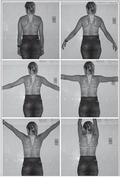
A series of six clinical photographs in black and white, capturing a posterior view of a human subject to demonstrate scapular kinematics during shoulder abduction. The sequence displays progressive bilateral arm abduction in the frontal plane at standardized intervals: 0 degrees (pendant position), 30 degrees, 60 degrees, 90 degrees, 120 degrees, and maximum abduction (approximately 170-180 degrees). Anatomical surface markers are visible on the subject's back, specifically positioned at the superior and inferior angles of the scapulae to facilitate biophotogrammetry analysis. The images illustrate the scapulohumeral rhythm, showing the upward and lateral rotation of the scapula as the humerus is elevated. This clinical imaging series is typical for orthopedic or physical therapy assessments of scapular dyskinesis, shoulder range of motion, and biomechanical studies of the shoulder complex.
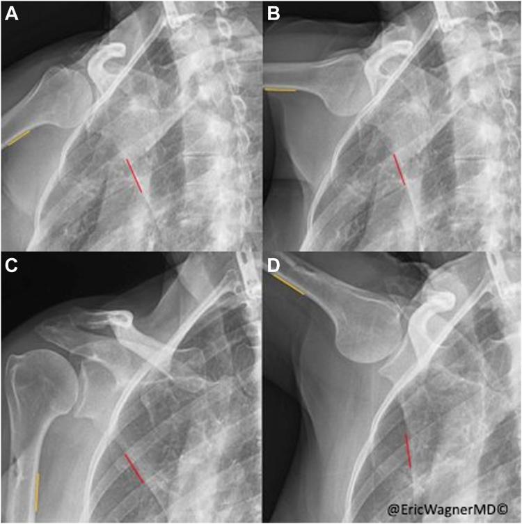
This composite diagnostic image features four clips from dynamic digital radiography (DDR) of the right shoulder, illustrating scapulohumeral rhythm (SHR). Images A and B show preoperative states at minimal and maximal humeral abduction, while C and D show the same intervals postoperatively. The images demonstrate the relationship between the humerus, scapula, and thorax. Yellow lines annotate the humerothoracic angle, while red lines represent the scapulothoracic angle measurement zones. In the preoperative sequence (A-B), there is significant humeral abduction but minimal change in the scapulothoracic angle, indicative of serratus anterior palsy and dysfunctional kinematics. In the postoperative sequence (C-D), following a pectoralis major transfer, there is a visible increase in scapular rotation accompanying humeral abduction. This visual comparison highlights the restoration of coordinated scapular movement and a normalized scapulohumeral rhythm. Key anatomical landmarks include the humeral head, glenoid cavity, coracoid process, and rib cage.
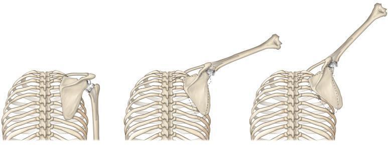
This medical illustration presents a comparative sequence of three panels demonstrating modified scapulohumeral rhythm in a patient with a reverse total shoulder arthroplasty (rTSA). The diagram shows the rib cage, scapula, and humerus during progressive arm elevation: at rest, at approximately 45 degrees, and at 90 degrees of abduction. Visible prosthetic components include a convex glenosphere mounted on the scapula and a corresponding concave humeral liner. A dashed line overlay indicates the native scapular position for comparison. The illustration highlights the biomechanical shift where, in rTSA, the scapula undergoes greater upward rotation relative to the humerus to compensate for altered joint mechanics and rotator cuff insufficiency. This increased scapular contribution is shown as a compensatory mechanism to achieve functional range of motion, providing a visual explanation of how prosthetic design influences dynamic musculoskeletal kinematics in orthopedic rehabilitation.
Rotator Cuff & Shoulder Complex Biomechanics
1. The Shoulder Complex: An Overview
| Articulation | Type | Key Role |
|---|---|---|
| Glenohumeral (GH) | Ball-and-socket (diarthrodial) | Primary motion joint |
| Scapulothoracic (ST) | Physiological (not true synovial) | Scapular positioning and rotation |
| Acromioclavicular (AC) | Plane synovial | Links clavicle to scapula |
| Sternoclavicular (SC) | Saddle joint with intra-articular disc | Only bony link of upper limb to axial skeleton |
- Miller's Review of Orthopaedics, 9th Ed.
- Rheumatology 2-Volume Set (Kelley's), Elsevier
2. Rotator Cuff: The Four Muscles (SITS)
Supraspinatus (S)
- Origin: Supraspinous fossa (dorsal scapula, above spine)
- Insertion: Superior facet of greater tuberosity
- Nerve: Suprascapular nerve (C5-C6)
- Blood supply: Suprascapular artery (from thyrocervical trunk)
- Function: Initiates abduction (0-15°), compresses and centers humeral head
- Key detail: Two distinct tendinous bundles (superficial and deep). Full-thickness tears disrupt both; partial tears involve only one. The avascular "critical zone" near the insertion makes it most prone to degeneration.
Infraspinatus (I)
- Origin: Infraspinous fossa (dorsal scapula, below spine)
- Insertion: Posterior-superior facet of greater tuberosity (interdigitates with supraspinatus)
- Nerve: Suprascapular nerve (C5-C6)
- Function: External rotation, posterior dynamic stabilization
Teres Minor (T)
- Origin: Lateral border of scapular body
- Insertion: Inferior facet of greater tuberosity (lowest/most posterior)
- Nerve: Axillary nerve (C5-C6)
- Function: External rotation (works with infraspinatus), inferior stabilization
Subscapularis (S)
- Origin: Subscapular fossa (anterior scapular surface)
- Insertion: Lesser tuberosity
- Nerve: Upper and lower subscapular nerves (C5-C6) - from posterior cord
- Blood supply: Subscapular artery (largest branch of axillary artery)
- Function: Internal rotation, anterior stabilization; largest of the four cuff muscles
- Rockwood & Green's Fractures in Adults, 10th Ed., 2025
- Imaging Anatomy, Vol. 3 (THIEME)
3. Key Biomechanical Concepts
3a. Force Couple Mechanism
- In the coronal plane: Supraspinatus + infraspinatus + teres minor (superior-posterior force) pair with subscapularis (anterior-inferior force) to compress the head into the glenoid
- In the transverse plane: Anterior cuff (subscapularis) and posterior cuff (infraspinatus + teres minor) create a compressive couple
- The result: humeral head depression and stabilization - without the cuff, deltoid activation would translate the head superiorly and impinge it against the acromion
"Function: depress and stabilize the humeral head against the glenoid; force-couple larger shoulder muscles to maintain humeral head center of rotation" - Miller's Review of Orthopaedics, 9th Ed.
3b. Concavity-Compression
3c. Glenohumeral Ligaments as Static Restraints
- Inferior GHL (anterior band + axillary pouch + posterior band) - primary restraint to anterior dislocation with the arm at 90° abduction + external rotation
- The capsule and ligaments work in concert with the rotator cuff, whose tendons blend into the capsule and reinforce it dynamically
4. Scapulohumeral Rhythm
- Total arm elevation = ~180° (120° GH + 60° ST)
- Early (0-30°): Variable, setting phase - scapula seeks a stable position
- Mid range (30-90°): Approximately 2:1 GH:ST ratio
- Above 90°: The proportion of scapulothoracic motion increases relative to glenohumeral motion - the scapula must rotate more to keep the glenoid oriented upward
"As elevation increases above 90 degrees, so does the proportion of scapulothoracic motion relative to glenohumeral motion." - Rheumatology 2-Volume Set (Kelley's)
- Upward rotation (glenoid faces superiorly)
- Posterior tilting
- External rotation
- Lateral translation along thoracic wall
- Trapezius (upper, middle, lower) + Serratus anterior = force couple for scapular upward rotation
- Rhomboids + Levator scapulae for retraction/downward rotation
5. Subacromial Space and Impingement
- Above: Acromion, coracoacromial ligament, AC joint (coracoacromial arch)
- Below: Supraspinatus tendon + subacromial/subdeltoid bursa
- Dimension: Fixed - any enlargement of contents (tendon swelling, bursal inflammation, bony spurs) produces impingement
- Gray's Anatomy for Students
6. Rotator Interval
- Coracohumeral ligament (CHL) - superficial, limits inferior translation and external rotation
- Superior glenohumeral ligament (SGHL) - deep, contributes to inferior stability
- Long head of biceps tendon - passes between the two ligaments
7. Sternoclavicular and Acromioclavicular Joints
- Only true bony link of upper limb to axial skeleton
- Diarthrodial saddle joint with intra-articular disc
- Posterior capsule/ligament = primary AP restraint; anterior = primary restraint against superior displacement
- Costoclavicular ligament provides additional stability
- Motion: elevation/depression, protraction/retraction, rotation (~50° total)
- Plane synovial joint stabilized by AC ligaments (horizontal stability) and coracoclavicular ligaments (vertical stability - trapezoid + conoid)
- During arm elevation, the clavicle rotates ~40° at the SC joint, which is transmitted as scapular rotation through the AC joint
8. Clinical Pearls: Biomechanical Implications
| Concept | Implication |
|---|---|
| Rotator cuff depresses humeral head | Full-thickness tear → superior migration → cuff arthropathy |
| 2:1 scapulohumeral rhythm | Tight posterior capsule → altered rhythm → impingement |
| Internal rotators > external rotators | Seizures/electric shock → posterior dislocation |
| Supraspinatus avascular zone | Most common site of degenerative tears |
| Rotator cable distributes forces | Tears in the cable region are more functionally significant |
| Posterior capsule tightness (GIRD) | Forces anterosuperior shift → internal impingement in throwers |
9. Rotator Cuff Tear Continuum (Disease Progression)
- Mild impingement (subacromial crowding)
- Supraspinatus tendinopathy / partial tear (articular > bursal side in young athletes; bursal in older patients)
- Full-thickness tear (crescent, U, L, or massive contracted)
- Massive tear (>5 cm, involving ≥2 tendons)
- Rotator cuff tear arthropathy - end-stage, with loss of head depression mechanism, superior migration, and glenohumeral arthritis
"Bursa-side tears are considered more ominous (mnemonic: 'Bursa is bad')" - Miller's Review of Orthopaedics 9th Ed.
- Miller's Review of Orthopaedics, 9th Ed.
- Rockwood & Green's Fractures in Adults, 10th Ed. (2025)
- Gray's Anatomy for Students
- Rheumatology, 2-Volume Set (Kelley's, Elsevier)
- Imaging Anatomy Vol. 3 - Bones, Joints, Vessels and Nerves (THIEME)
- Campbell's Operative Orthopaedics, 15th Ed. (2026)