I now have all the material needed. Let me write the comprehensive answer.
Ovarian Tumors: Benign, Malignant, FIGO Staging, and Management of Carcinoma Ovary
(30 Marks - PG Obs & Gynec Answer)
INTRODUCTION
The ovary is a unique structure in that it gives rise to a wider variety of tumors than any other organ in the body. This diversity stems from the three fundamental cell populations in the ovary:
- Müllerian (coelomic) epithelium - surface epithelial-stromal tumors (65-70%)
- Germ cells - germ cell tumors (15-20%)
- Sex cord-stromal cells - sex cord-stromal tumors (5-10%)
CLASSIFICATION OF OVARIAN TUMORS
WHO Classification (Histological - Table 1)
| Category | Cell of Origin | Major Types |
|---|
| Epithelial-Stromal | Müllerian/coelomic epithelium | Serous, Mucinous, Endometrioid, Clear cell, Brenner, Mixed |
| Germ Cell | Primordial germ cells | Teratoma, Dysgerminoma, Yolk sac tumor, Choriocarcinoma |
| Sex Cord-Stromal | Gonadal mesenchyme | Granulosa cell, Theca cell, Sertoli-Leydig cell tumors |
| Secondary (Metastatic) | Various primary sites | Krukenberg tumor (from stomach/colon) |
Each epithelial type is further subdivided into:
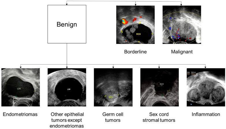
- Benign (well-differentiated, minimal proliferation, no invasion)
- Borderline (low malignant potential - increased proliferation but no stromal invasion)
- Malignant (stromal invasion present, cytological atypia)
PART I: BENIGN OVARIAN TUMORS
A. Benign Epithelial Tumors
1. Serous Cystadenoma
- Commonest benign ovarian tumor (25% of all benign)
- Gross: Unilocular/multilocular cyst, smooth outer surface, thin wall, straw-colored watery fluid
- Histology: Single layer of ciliated tubal-type columnar epithelium (salpingeal type); psammoma bodies (concentric calcifications) may be seen
- Size: Usually 5-15 cm; bilateral in ~15%
- Papillary excrescences on inner wall may be present; outer surface excrescences suggest malignant transformation
2. Mucinous Cystadenoma
- Second most common benign ovarian neoplasm (8-10% of epithelial tumors)
- Gross: Multilocular, huge - can fill entire abdominal cavity; mucoid gelatinous content
- Histology: Tall columnar mucin-secreting epithelium resembling endocervix or intestinal goblet cells; intracytoplasmic mucin
- Bilateral: Only 2-5%
- Complication: Rupture can cause pseudomyxoma peritonei - mucinous material throughout peritoneal cavity
Pseudomyxoma peritonei: The peritoneal cavity fills with gelatinous mucin surrounded by fibrous tissue. Most commonly it is secondary to a well-differentiated appendiceal mucinous neoplasm. - Berek & Novak's Gynecology
3. Brenner Tumor
- Rare; composed of transitional (urothelial-type) epithelium within dense fibrous stroma
- Usually small, solid, unilateral, and incidental finding
- Cut surface: resembles fibroma
- Almost always benign; malignant Brenner rare
4. Endometrioid Cystadenoma (Benign)
- Lined by endometrium-like epithelium; rarely purely benign (most are borderline or in context of endometriosis)
B. Functional/Non-Neoplastic Cysts (Benign)
| Type | Features |
|---|
| Follicular cyst | Unruptured Graafian follicle; <8 cm; lined by granulosa cells; resolves spontaneously |
| Corpus luteal cyst | Post-ovulation; hemorrhage possible ("chocolate"; may rupture) |
| Theca-lutein cysts | Bilateral; seen in molar pregnancy / ovarian hyperstimulation; very elevated hCG |
| Polycystic ovary | Multiple follicular cysts; associated with androgen excess, anovulation |
C. Benign Germ Cell Tumors
Mature Cystic Teratoma (Dermoid Cyst)
- Most common ovarian tumor in young women (20-30 years)
- Bilateral in 10-15%
- Gross: Unilocular cyst; thick sebaceous material, hair, teeth, bone fragments; Rokitansky protuberance (solid area)
- Histology: Stratified squamous epithelium with sebaceous glands, hair follicles, cartilage, thyroid tissue, neural elements - derivatives of all three germ layers
- Karyotype: Normal female 46,XX
- Complications: Torsion (most common), rupture, infection, hemolytic anemia, and malignant transformation (~1% - usually to squamous cell carcinoma)
- Tumor marker: May show elevated AFP if neural elements present; elevated CA-125 if skin/sebaceous elements rupture
"Occasionally associated with clinically important paraneoplastic syndromes, such as inflammatory limbic encephalitis, which may remit following removal of the tumor." - Robbins Pathology
Struma Ovarii
- Monodermal teratoma composed predominantly of thyroid tissue
- May cause hyperthyroidism; rarely malignant
D. Benign Sex Cord-Stromal Tumors
Fibroma
- Most common benign sex cord-stromal tumor
- Solid, white-gray, rubbery; produces fibrous tissue
- Meigs syndrome: Fibroma + ascites + right-sided pleural effusion
- Treatment: Simple oophorectomy (curative)
Thecoma
- Rare; contains lipid-laden theca cells; produces estrogen
- Post-menopausal; associated with endometrial hyperplasia
PART II: MALIGNANT OVARIAN TUMORS (CARCINOMA OVARY)
Epidemiology
- 3rd most common gynecological malignancy in India (after Ca cervix and Ca endometrium)
- Leading cause of gynecological cancer death (due to late presentation)
- Peak incidence: 50-70 years
- 5-year survival (all stages): ~46%; Stage I: ~90%; Stage IV: <20%
Risk Factors
| Increased Risk | Decreased Risk |
|---|
| BRCA1/BRCA2 mutations | OCP use |
| Family history (1st degree) | Multiple pregnancies |
| Nulliparity, early menarche, late menopause | Breastfeeding |
| Lynch syndrome / HNPCC | Tubal ligation |
| HRT (long-term) | Salpingectomy / oophorectomy |
| Talc exposure | |
A. Epithelial Ovarian Cancer (EOC) - 65-70% of all
Molecular Pathogenesis - Type I vs Type II (Berek & Novak)
| Feature | Type I (Low-grade) | Type II (High-grade) |
|---|
| Origin | Serous borderline tumors, endometriosis | Fallopian tube fimbriae (STIC), inclusion cysts |
| Growth | Slow | Rapid |
| Genetic mutations | KRAS, BRAF, ERBB2 | TP53 (nearly universal), BRCA1/2 |
| Genetic stability | Stable | Unstable (chromosomal) |
| Stage at diagnosis | Often early | Usually advanced |
| Histotypes | Low-grade serous, mucinous, endometrioid | High-grade serous, carcinosarcoma |
Histological Subtypes of EOC
1. Serous Adenocarcinoma (75-80% of EOC)
- Two grades: Low-grade (Type I, indolent) and High-grade (Type II, aggressive)
- High-grade serous is the most lethal; nearly all have TP53 mutations
- Microscopy: Papillae lined by cytologically malignant cells; necrosis; psammoma bodies
- Serous tubal intraepithelial carcinoma (STIC): Precursor in fallopian tube fimbria
2. Mucinous Carcinoma (5%)
- Bilateral in only 8-10%; confined to ovary in 95-98%
- Must exclude metastasis from GI tract (primary vs. Krukenberg)
- Microscopy: Irregular glandular spaces lined by tall columnar cells with abundant mucinous cytoplasm
3. Endometrioid Carcinoma (10%)
- Arises from endometriosis; associated with synchronous endometrial cancer in 15-20%
- Better prognosis than serous
4. Clear Cell Carcinoma (5%)
- Müllerian origin; arises from endometriosis
- High resistance to platinum chemotherapy; worse prognosis
- Microscopy: "Hobnail" cells, glycogen-rich clear cytoplasm
5. Undifferentiated/Unclassified
- High grade, poor prognosis; anaplastic cells
B. Malignant Germ Cell Tumors (15-20%)
Predominantly in young girls and adolescents; excellent response to chemotherapy
| Tumor | Marker | Features |
|---|
| Dysgerminoma | LDH, PLAP | Most common malignant GCT; equivalent of testicular seminoma; 10-15% bilateral; radiosensitive and chemo-sensitive |
| Yolk Sac Tumor (Endodermal sinus) | AFP (elevated) | Schiller-Duval bodies histologically; aggressive; most common in children |
| Immature Teratoma | AFP, CA-125 | Primitive neuroepithelium; graded 1-3; younger women |
| Choriocarcinoma | β-hCG (very high) | Extremely rare; aggressive; hematogenous spread; may cause precocious puberty |
| Embryonal carcinoma | AFP + β-hCG | Highly malignant; mixed patterns |
C. Sex Cord-Stromal Tumors (5-10%)
Granulosa Cell Tumor (GCT)
- Low-grade malignancy; bilateral in <5%
- Produces estrogen → endometrial hyperplasia/carcinoma (risk)
- Histology: Call-Exner bodies (rosette pattern around pink material - pathognomonic)
- Tumor marker: Inhibin, AMH
- Adult type: Perimenopausal women; late recurrence (10-30 years later)
- Juvenile type: Children and adolescents; isosexual precocious puberty
Sertoli-Leydig Cell Tumor (Androblastoma)
- Produces androgens → virilization (amenorrhea, clitoromegaly, voice deepening, hirsutism)
- Rare; usually in young women <30 years
- Mostly unilateral; low-grade malignancy
D. Metastatic Tumors (Krukenberg Tumor)
- Bilateral ovarian metastases (usually from stomach or colon)
- Histology: Signet-ring cells containing mucin within cellular stroma
- Poor prognosis
PART III: CLINICAL FEATURES OF CARCINOMA OVARY
Symptoms (Ovarian Cancer Symptom Index)
- Pelvic or abdominal pain
- Abdominal bloating/distension
- Difficulty eating / early satiety
- Urinary urgency or frequency
- Increased abdominal girth (ascites)
Signs
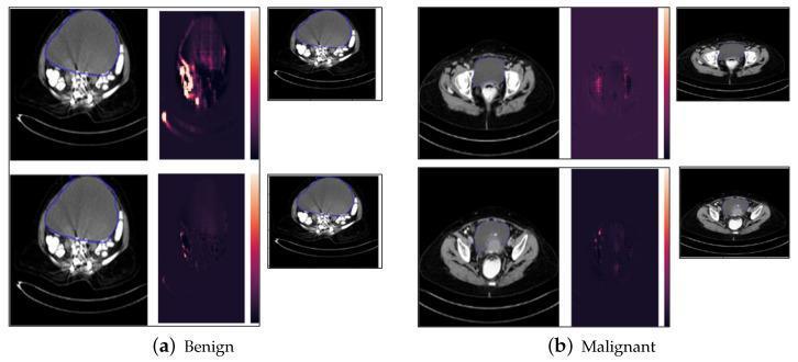
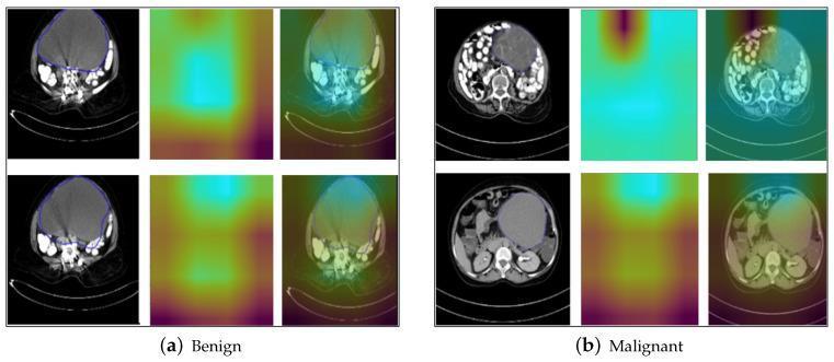
- Pelvic mass: solid, irregular, fixed, bilateral - highly suggestive of malignancy
- Ascites
- Upper abdominal mass (omental cake)
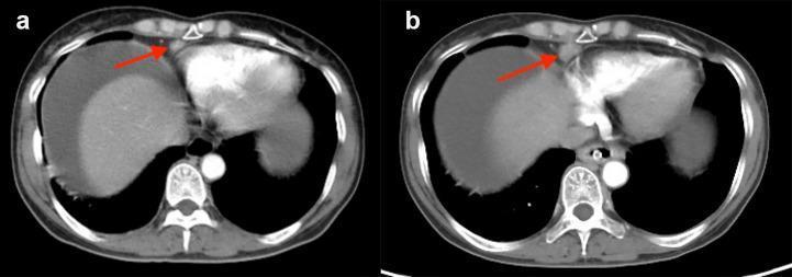
- Pleural effusion (in advanced disease)
Investigation
| Investigation | Significance |
|---|
| CA-125 | Elevated in 80-90% advanced serous; >200 U/mL in postmenopausal = 96% PPV; less specific premenopausal |
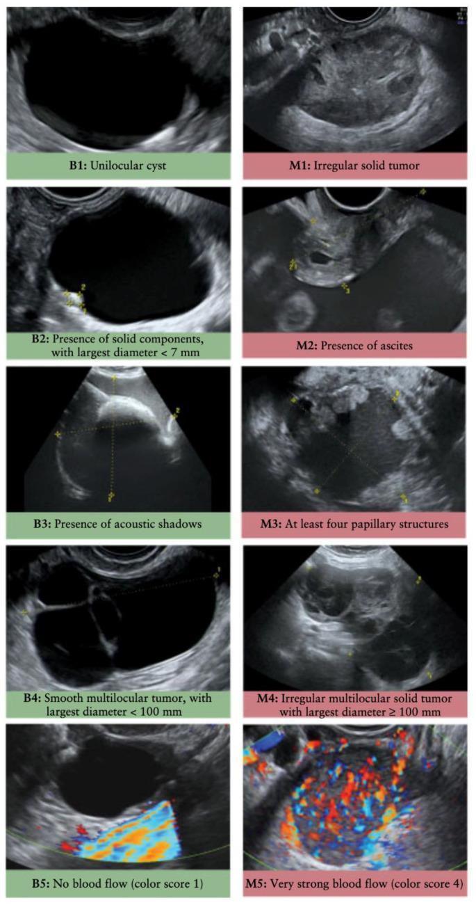
| Transvaginal USG | Morphology assessment; complex mass with solid areas, septae, papillary projections, ascites |
| CT abdomen-pelvis | Staging, nodal assessment, peritoneal deposits |
| MRI pelvis | Superior soft tissue characterization |
| AFP, β-hCG, LDH | For germ cell tumors in young women |
| Inhibin | Granulosa cell tumors |
| Chest X-ray/CT | Pleural effusion (Stage IVA) |
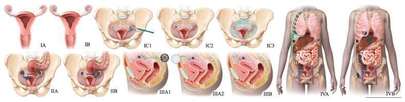
PART IV: FIGO STAGING OF OVARIAN CANCER (2014 Revised)
The 2014 FIGO staging applies to ovarian, fallopian tube, and primary peritoneal carcinomas:
STAGE I - Tumor confined to ovaries / fallopian tubes
| Stage | Definition |
|---|
| IA | One ovary/tube; capsule intact; no surface tumor; negative washings |
| IB | Both ovaries/tubes; capsule intact; no surface tumor; negative washings |
| IC1 | Surgical spill intraoperatively |
| IC2 | Preoperative capsule rupture OR tumor on ovarian/tubal surface |
| IC3 | Malignant cells in ascites or peritoneal washings |
STAGE II - Pelvic extension or primary peritoneal cancer
| Stage | Definition |
|---|
| IIA | Extension and/or implants on uterus and/or tubes |
| IIB | Extension to other pelvic intraperitoneal tissues (bladder, sigmoid, rectum) |
STAGE III - Peritoneal metastases beyond pelvis AND/OR retroperitoneal lymph node metastases
| Stage | Definition |
|---|
| IIIA1 | Positive retroperitoneal lymph nodes only (cytologically/histologically confirmed) |
| IIIA1(i) | Nodal metastasis ≤10 mm in greatest dimension |
| IIIA1(ii) | Nodal metastasis >10 mm in greatest dimension |
| IIIA2 | Microscopic extrapelvic (above the pelvic brim) peritoneal involvement ± positive retroperitoneal nodes |
| IIIB | Macroscopic peritoneal metastasis beyond pelvis ≤2 cm ± positive retroperitoneal nodes |
| IIIC | Macroscopic peritoneal metastasis beyond pelvis >2 cm ± positive retroperitoneal nodes (including liver/spleen capsule) |
STAGE IV - Distant metastases (excluding peritoneal metastases)
| Stage | Definition |
|---|
| IVA | Pleural effusion with positive cytology |
| IVB | Parenchymal metastases (liver/spleen parenchyma), extra-abdominal organ involvement (including inguinal lymph nodes and lymph nodes outside abdominal cavity) |
Key 2014 change: Retroperitoneal lymph node metastasis alone is now Stage IIIA1 (not Stage IIIC) if there is no peritoneal spread beyond the pelvis. Pleural effusion with positive cytology is Stage IVA (not IVB).
5-Year Survival by Stage:
| Stage | 5-Year Survival |
|---|
| I | ~90% |
| II | ~70% |
| III | ~39% |
| IV | <20% |
PART V: MANAGEMENT OF CARCINOMA OVARY
General Principles
- Surgery is the cornerstone - for both diagnosis (histological confirmation) AND treatment (cytoreduction)
- Most patients (~75%) present at Stage III/IV
- Treatment depends on: Stage, histology, grade, age, performance status, desire for fertility
Surgical Staging Procedure (For Apparently Early Disease)
Performed via vertical midline incision:
- Evacuation of ascites / peritoneal washings (diaphragm, right and left paracolic gutters, pelvis)
- Total abdominal hysterectomy + bilateral salpingo-oophorectomy (TAH + BSO)
- Infragastric omentectomy
- Retroperitoneal pelvic and para-aortic lymph node dissection
- Appendicectomy (especially for mucinous tumors)
- Random biopsies of peritoneal surfaces (diaphragm undersurface, cul-de-sac, paracolic gutters, pelvic side walls, bladder peritoneum)
- Excision of all suspicious lesions/adhesions
- Bailey & Love's Short Practice of Surgery, p.1615
A. Management by Stage
Stage I (Early Disease)
Stage IA/IB Grade 1-2 (Low Risk):
- TAH + BSO + surgical staging
- No adjuvant chemotherapy required
- Fertility-sparing surgery acceptable in young women with Stage IA, Grade 1-2: unilateral salpingo-oophorectomy + staging; uterus and contralateral ovary preserved
Stage IA/IB Grade 3 OR Stage IC (High Risk):
- TAH + BSO + staging
- Adjuvant chemotherapy: 3-6 cycles of carboplatin + paclitaxel
Prognostic factors for high risk Stage I: high grade, ruptured capsule, surface excrescences, ascites, positive peritoneal cytology, dense adhesions, aneuploid tumor - Berek & Novak's Gynecology
Stage II
- TAH + BSO + staging + cytoreduction
- Adjuvant chemotherapy: 6 cycles of carboplatin (AUC 5-6) + paclitaxel (175 mg/m² IV every 3 weeks)
Stage III-IV (Advanced Disease) - PRIMARY TREATMENT
Cytoreductive Surgery (Debulking Surgery)
The goal is to achieve optimal cytoreduction - residual disease ≤1 cm (ideally no gross residual disease = R0)
Components:
- TAH + BSO
- Infragastric omentectomy
- Peritonectomy
- Bowel resection if necessary (colorectal resection with reanastomosis)
- Diaphragmatic resection/stripping
- Splenectomy and/or distal pancreatectomy if required
"In a meta-analysis of 81 studies, Bristow et al. documented that each 10% increase in cytoreduction equaled a 5.5% increase in median survival." - Berek & Novak's Gynecology
Post-operative Chemotherapy (First Line):
- Standard IV regimen: Carboplatin AUC 5-6 + Paclitaxel 175 mg/m² IV every 3 weeks x 6 cycles
- Intraperitoneal (IP) therapy: For optimally debulked Stage III - IP cisplatin/carboplatin + paclitaxel; NCI clinical alert (2006) endorsed IP therapy as superior in survival for optimally cytoreduced patients
- Dose-dense weekly paclitaxel (80 mg/m² weekly) + carboplatin AUC 5-6 every 3 weeks: superior PFS vs. standard in Japanese trial (JGOG 3016)
Neoadjuvant Chemotherapy (NACT) + Interval Debulking Surgery (IDS):
- When patient is not a surgical candidate upfront (unresectable disease, poor performance status)
- Fagotti Score ≥8 at laparoscopy predicts suboptimal cytoreduction → proceed to NACT
- Protocol: 3 cycles NACT → IDS → 3 cycles adjuvant chemotherapy
- Supported by EORTC and CHORUS trials (non-inferior to primary debulking for selected patients)
Targeted Therapy:
- Bevacizumab (anti-VEGF monoclonal antibody):
- GOG218 and ICON7 trials: Added to 6 cycles CP, then maintenance for 15 months
- 3.8-month improvement in PFS; no OS benefit except in high-risk subgroup (ascites, Stage IVB)
- Dose: 7.5-15 mg/kg IV every 3 weeks
- Toxicities: Hypertension, proteinuria, bowel perforation (2%), fistula
- PARP inhibitors (Olaparib, Niraparib, Rucaparib):
- Maintenance therapy after platinum response
- Greatest benefit in BRCA1/2 mutation carriers (SOLO-1 trial: olaparib vs. placebo after first-line treatment in BRCA-mutated advanced ovarian cancer - 3-year PFS 60% vs 27%)
B. Management of Germ Cell Tumors
- BEP chemotherapy (Bleomycin + Etoposide + Cisplatin) x 3-4 cycles - standard for malignant GCT
- Fertility preservation is possible and usually achievable - unilateral salpingo-oophorectomy + staging + chemotherapy
- Dysgerminoma: Highly radiosensitive and highly chemosensitive; excellent prognosis even for advanced disease
- Yolk sac tumor: AFP monitoring for recurrence
C. Management of Recurrent Disease
| Platinum Sensitivity | Definition | Treatment |
|---|
| Platinum-refractory | Progression while on platinum | Non-platinum agents (topotecan, gemcitabine); clinical trial |
| Platinum-resistant | Progression within 6 months | Non-platinum ± bevacizumab; consider clinical trial |
| Platinum-sensitive | Recurrence >6 months | Platinum + paclitaxel ± bevacizumab ± PLD; consider secondary debulking if >12 months interval and complete clinical response; maintenance PARP inhibitor |
DESKTOP3 trial: Secondary cytoreduction + chemotherapy vs. chemotherapy alone for platinum-sensitive recurrence (>6 months interval) showed survival benefit when strict entry criteria met. - Schwartz's Principles of Surgery
D. Role of Radiation Therapy
- Limited role in ovarian cancer
- Whole-abdominal radiation: High morbidity (30% bowel obstruction); largely abandoned
- May be used for palliative purposes in localized recurrence
E. Surveillance After Treatment
- Clinical examination + CA-125 every 3 months x 2 years, then every 6 months x 3 years
- CT scan when clinically indicated or CA-125 rising
- BRCA testing of all patients with high-grade serous carcinoma
SUMMARY TABLE: Key Points for Exam
| Feature | Serous Ca | Mucinous Ca | Dysgerminoma | Granulosa Cell |
|---|
| % of malignant | 75-80% | 5% | Most common GCT | Most common SCST |
| Marker | CA-125 | CA-125, CEA | LDH, PLAP | Inhibin, AMH |
| Molecular | TP53 (HG), KRAS/BRAF (LG) | KRAS | Isochromosome 12p | FOXL2 mutation |
| Histology | Papillae, psammoma bodies | Glands, mucin | Large cells, fibrous septa | Call-Exner bodies |
| Special features | Fallopian tube origin | GI metastasis excluded | Bilateral 10-15%; chemo sensitive | Late recurrence; estrogen |
CONCLUSION
Ovarian tumors represent the most diverse neoplasms in human pathology. Benign tumors (80% of all) are predominantly epithelial cystadenomas and germ cell teratomas. Malignant ovarian cancer - dominated by high-grade serous carcinoma - carries high mortality owing to silent early disease and advanced stage at diagnosis. The 2014 revised FIGO staging provides prognostic stratification with critical substages for IC, IIIA1, and the IVA/IVB distinction. Management centers on maximal surgical cytoreduction followed by platinum-taxane chemotherapy, with targeted agents (bevacizumab, PARP inhibitors) in selected patients. Early detection remains the unmet need in ovarian oncology.
Sources: