
Detailed anatomy of sternum
Reading File
Finding Sources
Searching the Web
detailed anatomy of sternum parts joints clinical significance
Reading File
Reading File
Reading File
Loading Image
Loading Image
Reading File
Finding Sources
Finding Sources
Finding Sources
Reading File
Reading File
Reading File
Loading Image
Finding Sources
Finding Sources
Loading Image
I now have all the information needed for a comprehensive answer. Here is the full detailed anatomy of the sternum:
Detailed Anatomy of the Sternum
The sternum is a flat, slightly anteriorly convex bone that forms the anterior midline of the thoracic cage. It has multiple indentations (costal notches) along its lateral borders. In the adult it consists of three distinct parts.
Overview Diagram (Anterior View)
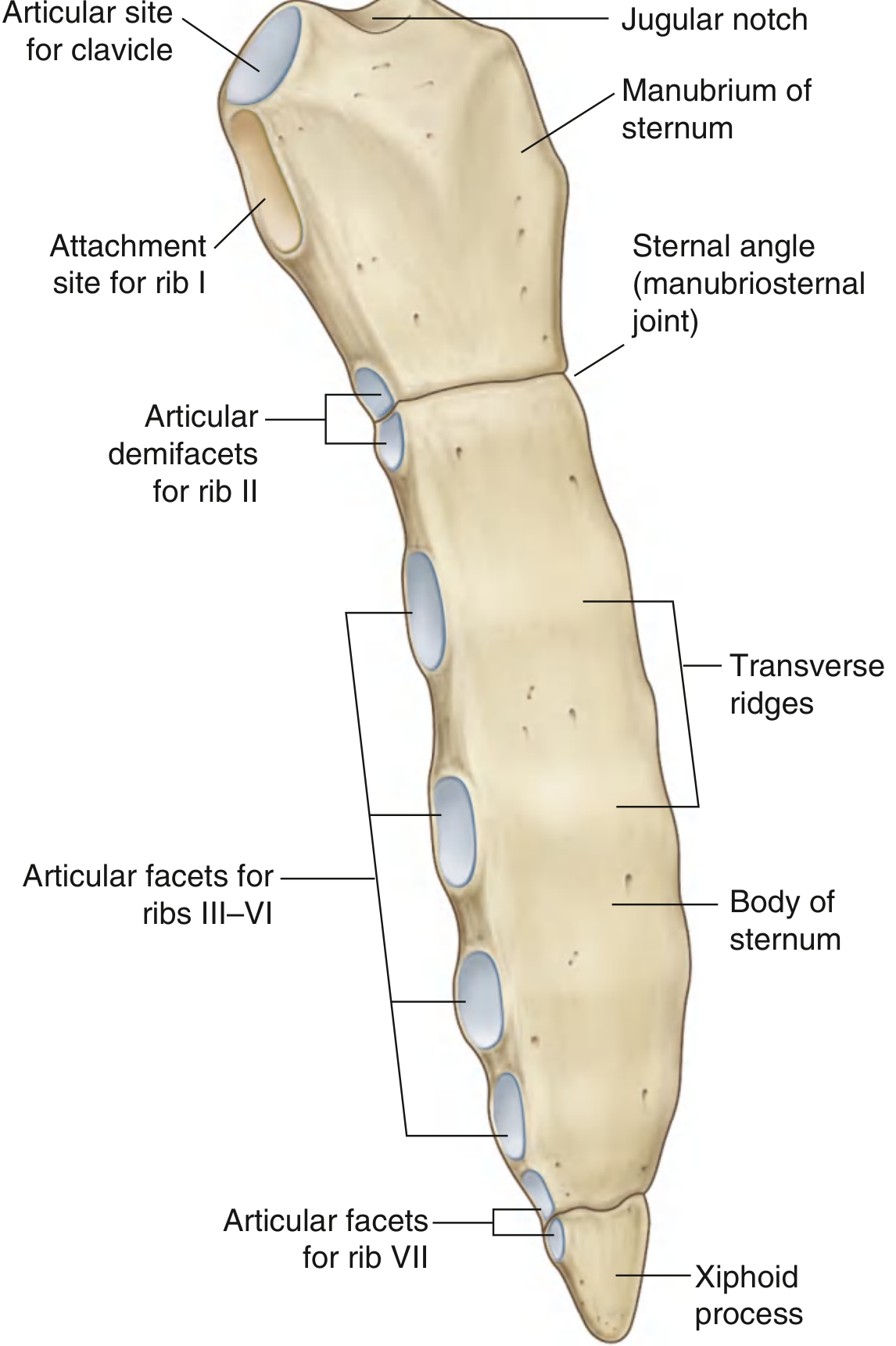
1. Parts of the Sternum
A. Manubrium
The manubrium is the broadest and most superior part of the sternum. It forms part of the bony framework of both the neck and the thorax.
Superior border:
- Bears the jugular (suprasternal) notch in the midline - a clearly palpable depression that marks the inferior margin of the jugular fossa.
- On either side of the jugular notch is a large oval fossa - the clavicular notch - for articulation with the medial end of the clavicle (sternoclavicular joint).
Lateral borders:
- Immediately inferior to the clavicular notch is the first costal notch, a shallow concavity for synchondrosis (fibrocartilaginous union) with the first costal cartilage. This joint is not synovial.
- At the lower end of the lateral border is a demifacet for the upper half of the second costal cartilage.
Inferior border:
- Joins with the body of the sternum at the manubriosternal joint (see Joints section below), forming the sternal angle.
B. Body of the Sternum
The body is flat, narrow, and longitudinally oriented. It is longer than the manubrium.
Anterior surface:
- Often marked by transverse ridges - lines of fusion between the segmental embryological elements called sternebrae, from which the body develops.
Lateral margins (costal notches):
- Superiorly, each lateral margin has a demifacet for the inferior half of the second costal cartilage (completing the articulation with rib II, shared with the manubrium).
- Below this are four complete facets for the costal cartilages of ribs III to VI.
- At the inferior end of the body is a demifacet for the upper portion of the seventh costal cartilage.
Inferior border:
- Joined to the xiphoid process at the xiphisternal joint.
C. Xiphoid Process
The xiphoid is the smallest and most inferior part of the sternum. It is highly variable in shape - it may be wide, thin, pointed, bifid, curved, or perforated. It begins as a cartilaginous structure; ossification commences during childhood but it may never completely ossify even in adults.
- Each upper lateral margin bears a demifacet for the inferior end of the seventh costal cartilage.
- It has no costal cartilage attached to it directly - only the demifacets for rib VII.
- It serves as an attachment for the linea alba, diaphragm, and some abdominal muscles.
2. Lateral View
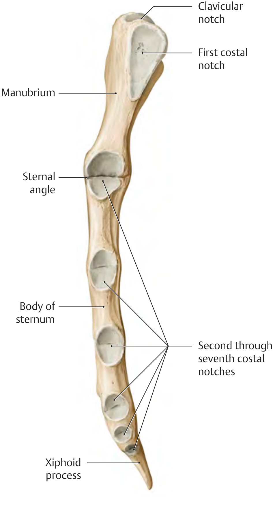
The lateral view clearly shows the sternal angle where the manubrium angles slightly posteriorly relative to the body. It also shows the costal notches for ribs 2 through 7 arranged along the lateral border.
3. Joints of the Sternum
Sternocostal Joints
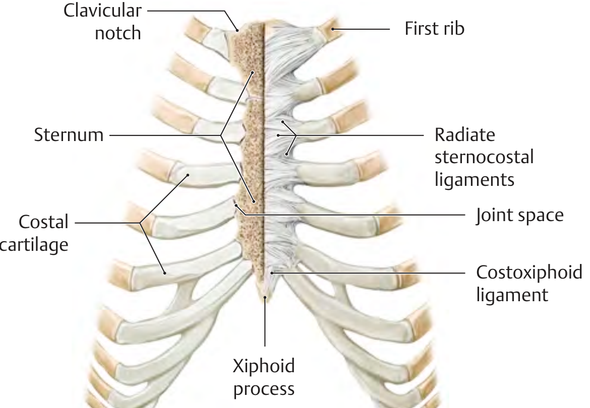
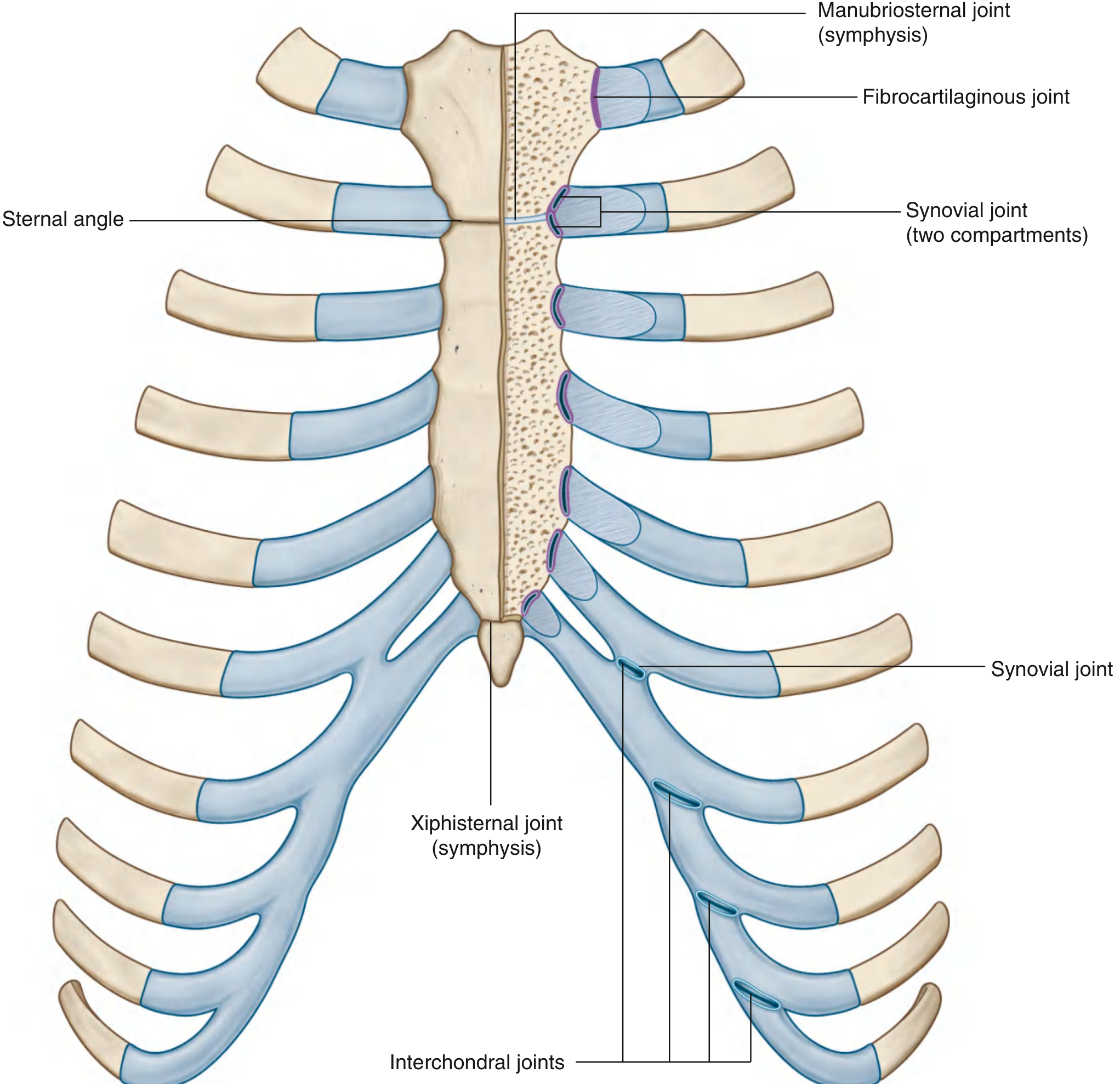
The sternocostal joints connect the costal cartilages of ribs I-VII to the sternum. They are of two types:
| Rib | Type of Joint | Notes |
|---|---|---|
| I | Synchondrosis (fibrocartilaginous) | No joint space; not synovial |
| II | Synovial with two compartments | Divided by an intra-articular ligament at the manubriosternal junction |
| III-V | Synovial | Thin capsule, reinforced by sternocostal ligaments |
| VI-VII | Synchondroses | No joint space |
Radiate sternocostal ligaments radiate from the perichondrium of each costal cartilage to the anterior surface of the sternum, blending with the periosteum to form the dense sternal membrane.
Manubriosternal Joint (Angle of Louis)
- Type: Secondary cartilaginous joint (symphysis) filled with fibrocartilage.
- The manubrium angles posteriorly on the body, creating the palpable sternal angle (angle of Louis / angle of Ludwig).
- Only slight angular movements occur here during respiration.
Clinical significance of the sternal angle:
- Marks the articulation of rib II with the sternum - the reference rib for counting intercostal spaces (rib I is hidden under the clavicle).
- Lies on a horizontal plane passing through the intervertebral disc between T4 and T5.
- This plane marks the:
- Junction of superior and inferior mediastinum
- Superior border of the pericardium
- End of ascending aorta / beginning of aortic arch
- End of aortic arch / beginning of thoracic aorta
- Bifurcation of the trachea (carina)
- Level just superior to the pulmonary trunk
The sternal angle is also used in estimating venous pressure - it lies approximately 5 cm above the mid-right atrium.
Xiphisternal Joint
- Type: Symphysis (secondary cartilaginous joint).
- Joins the body of the sternum to the xiphoid process.
- Commonly ossifies with advancing age.
4. Muscle Attachments
| Muscle / Structure | Attachment Site |
|---|---|
| Pectoralis major (sternocostal part) | Anterior surface of body and manubrium |
| Sternocleidomastoid | Superior surface of manubrium |
| Sternohyoid | Posterior surface of manubrium |
| Sternothyroid | Posterior surface of manubrium |
| Transversus thoracis | Posterior surface of body |
| Diaphragm | Xiphoid process (posterior) |
| Rectus abdominis | Xiphoid process (anterior) |
| External oblique | Xiphoid process |
| Linea alba | Xiphoid process |
5. Blood Supply
The sternum is supplied by the internal thoracic (internal mammary) arteries, which run 1 cm lateral to the sternal edges on each side. Periosteal and nutrient branches penetrate the bone. This anatomy is important surgically - the internal thoracic artery is harvested for coronary artery bypass grafting, and median sternotomy must respect these vessels.
6. Nerve Supply
Sensory innervation to the periosteum and overlying skin is provided by the anterior cutaneous branches of the intercostal nerves (T1-T6), which penetrate the internal intercostal muscle, external intercostal membrane, and pectoralis major to reach the anterior chest wall.
7. Development (Embryology)
From - The Developing Human, Clinically Oriented Embryology:
- A pair of vertical mesenchymal bands, the sternal bars, develops ventrolaterally in the body wall.
- Chondrification occurs as these bars migrate medially.
- By 10 weeks, they fuse craniocaudally in the median plane to form cartilaginous models of the manubrium, sternebrae, and xiphoid process.
- The manubrium develops from mesenchyme between the clavicles, with contributions from neural crest cells (endochondral ossification).
- Centers of ossification appear craniocaudally before birth, except for the xiphoid process, which ossifies during childhood.
- The xiphoid process may never completely ossify.
Congenital anomalies result from incomplete or asymmetric fusion of the sternal bars:
- Bifid sternum (cleft sternum) - partial or complete failure of midline fusion
- Sternal foramen - a benign midline perforation, important to recognize before acupuncture or sternal biopsy to avoid inadvertent cardiac injury
- Pectus excavatum (funnel chest) - posterior depression of the lower sternum; may be associated with Marfan syndrome or congenital heart disease (particularly ASD)
- Pectus carinatum (pigeon chest) - anterior protrusion; may be congenital or acquired
8. Clinical Relevance
| Application | Detail |
|---|---|
| Sternal biopsy | Rich red bone marrow throughout adult sternum; body is preferred site for aspiration |
| Median sternotomy | Standard access for cardiac surgery; the sternum is split and later wired together |
| CPR | Compressions applied to the lower half of the sternal body |
| Intraosseous access | Manubrium used for IO cannulation in adults |
| Sternal fracture | Usually from high-energy anterior chest trauma; often associated with underlying cardiac contusion |
| Sternal angle (Louis) | Universal landmark for counting ribs and locating mediastinal structures |
| CT of sternum | Best imaging modality - eliminates overlapping structures, detects destruction, allows soft tissue (parasternal/internal mammary zone) assessment |
Sources: Gray's Anatomy for Students (9780323934237, p. 182-185); THIEME Atlas of Anatomy - General Anatomy and Musculoskeletal System (9781626237186, p. 154-155); The Developing Human - Clinically Oriented Embryology (9780323611541, p. 920-921); Grainger & Allison's Diagnostic Radiology (9780323760751)
This is a shared conversation. Sign in to Orris to start your own chat.