
Jg apparatus 5 marker and also diagram
python3 -c " from pptx import Presentation prs = Presentation('/tmp/workspace/attachments/3f2e2ddd-1e8c-49b8-acea-15e6c99a4045/renal system 2020.pptx') for i, slide in enumerate(prs.slides, 1): texts = [] for shape in slide.shapes: if shape.has_text_frame and shape.text_frame.text.strip(): texts.append(shape.text_frame.text.strip()) if texts: combined = ' | '.join(texts) if 'juxtaglomerular' in combined.lower() or 'jg apparatus' in combined.lower() or 'JG' in combined or 'macula densa' in combined.lower() or 'renin' in combined.lower(): print(f'=== SLIDE {i} ===') for t in texts: print(t) print() "
python3 -c " from pptx import Presentation from pptx.util import Inches import os prs = Presentation('/tmp/workspace/attachments/3f2e2ddd-1e8c-49b8-acea-15e6c99a4045/renal system 2020.pptx') # Check slides 37 and 63 for images for slide_num in [37, 63]: slide = prs.slides[slide_num - 1] print(f'=== SLIDE {slide_num} ===') for shape in slide.shapes: print(f' Shape: {shape.shape_type}, name={shape.name}') if hasattr(shape, 'image'): img = shape.image ext = img.ext fname = f'/tmp/workspace/slide{slide_num}_img.{ext}' with open(fname, 'wb') as f: f.write(img.blob) print(f' -> Saved image: {fname} ({len(img.blob)} bytes)') if shape.has_text_frame: print(f' Text: {shape.text_frame.text[:100]}') "
Juxtaglomerular (JG) Apparatus
Definition
Components (3 Cell Types)
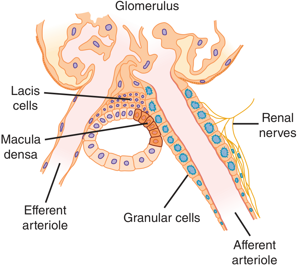
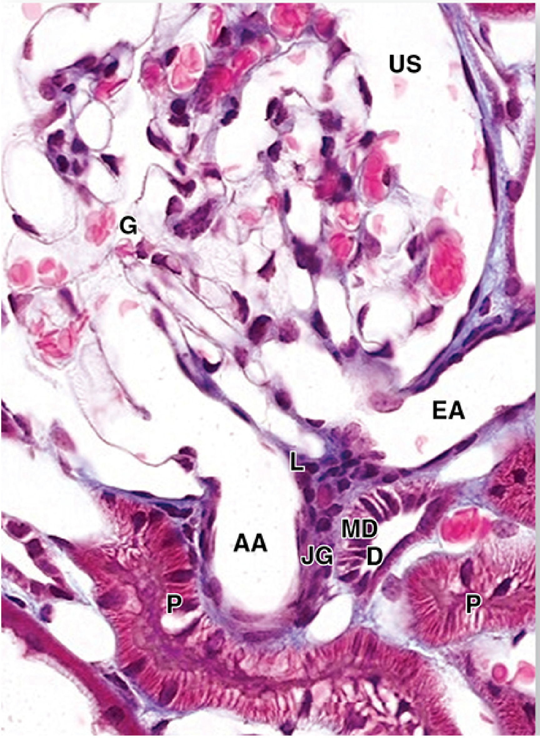
1. Juxtaglomerular (Granular) Cells
- Modified smooth muscle cells in the media of the afferent arteriole (occasionally efferent arteriole too)
- Their nuclei are spherical (unlike the typical elongated smooth muscle cell nucleus)
- Contain membrane-lined secretory granules filled with renin (an aspartyl protease)
- They act as intrarenal baroreceptors - when perfusion pressure falls, they release renin
2. Macula Densa Cells
- Located at the beginning of the distal convoluted tubule (DCT) where it contacts the vascular pole of its parent glomerulus
- Cells are narrower, taller, and more closely packed than other DCT cells - their crowded nuclei give the name "macula densa" (Latin: dense spot)
- Act as chemoreceptors/osmoreceptors - they sense the NaCl concentration in the tubular filtrate
- When NaCl delivery increases, macula densa signals JG cells to decrease renin release and reduce GFR (tubuloglomerular feedback)
3. Lacis Cells (Extraglomerular Mesangial / Mesangial Cells)
- Also called Goormaghtigh cells or extraglomerular mesangial cells
- Located at the junction (angle) between the afferent and efferent arterioles, adjacent to the macula densa
- They are in contact with both JG cells and macula densa cells, acting as a communication bridge
- Serve supportive, contractile, and defensive functions similar to intraglomerular mesangial cells
- Stimulate renin release in response to signals from the macula densa
Diagrams
Functions
| Function | Cells Involved | Mechanism |
|---|---|---|
| Renin synthesis, storage & release | JG (granular) cells | Renin cleaves angiotensinogen → Angiotensin I → (ACE in lungs) → Angiotensin II → vasoconstriction + aldosterone release |
| Blood pressure regulation | All components via RAAS | Angiotensin II raises BP; aldosterone retains Na+/water |
| GFR regulation (tubuloglomerular feedback) | Macula densa | High NaCl in filtrate → signals afferent arteriole to constrict → reduces GFR |
| Erythropoietin secretion | JG cells (also interstitial cells) | Stimulates red blood cell production in bone marrow |
Key Points for 5-Marker Exam
- Location: At the vascular pole of the glomerulus, where the afferent/efferent arterioles meet the DCT of the same nephron
- Three components: JG cells (in afferent arteriole wall) + Macula densa (at start of DCT) + Lacis/mesangial cells (between arterioles)
- Main product: Renin - the rate-limiting enzyme of the RAAS
- Stimuli for renin release: Decreased afferent arteriolar pressure, decreased NaCl at macula densa, increased sympathetic activity, prostaglandins
- Inhibitors of renin release: Increased afferent pressure, increased NaCl reabsorption, angiotensin II (negative feedback), vasopressin